Method for the detection of breast cancer by determining alcam and/or bcam levels in a patient

Inactive Publication Date: 2011-06-23
UNIV HEALTH NETWORK
View PDF0 Cites 6 Cited by
  • Summary
  • Abstract
  • Description
  • Claims
  • Application Information

AI Technical Summary

Benefits of technology

[0016]An aspect of the present application is a method of screening for, diagnosing or detecting breast cancer by determining a level of an ALCAM biomarker product in a sample from a subject, wherein the sample is a biological fluid, and comparing the level in the sample with a control, wherein detecting a differential level of biomarker product between the subject and the control is indicative of breast cancer in the subject.
[0017]Another aspect of the present application is a method of screening for, diagnosing or detecting breast cancer by determining a level of a BCAM biomarker product in a sample from a subject and comparing the level in the sample to a control, wherein detecting a differential level of the biomarker product between the subject and the control is indicative of breast cancer in the subject.
[0018]A further aspect of the present application is a method of screening for, diagnosing or detecting breast cancer by determining a level of product from both an ALCAM biomarker and a BCAM biomarker in a sample from a subject and comparing each level in the sample to a control, wherein detecting a differential expression of at least one of the biomarker products between the subject and the control is indicative of breast cancer in the subject.
[0019]Yet a further aspect of the present application is a method of predicting the prognosis of a subject having or suspected of having breast cancer by determining the level of a biomarker product in a sample from the subject, where the biomarker is selected from ALCAM, BCAM and/or a combination thereof, and comparing each level of biomarker with a reference level associated with a disease outcome, the disease outcome being good prognosis, or poor prognosis, where the disease outcome associated with the reference level most similar to the level of each biomarker in the sample is the predicted prognosis. In one embodiment, an increase in ALCAM and/or BCAM is indicative of poor prognosis. In certain embodiments, the therapy comprises chemotherapy. In other embodiments, the therapy comprises a test therapy.
[0020]Yet a further aspect of the present application is a method for monitoring the therapeutic response of a subject undergoing treatment for breast cancer by determining a level of biomarker product in a first sample of the subject, the biomarker selected from the group consisting of ALCAM, BCAM and a combination thereof, determining the level of biomarker product in a subsequ

Problems solved by technology

In addition, for women under the age of 40, mammographic screening yields a poor sensitivity of only around 33%10,11.
Currently available blood-based biomarkers are of no value in the early diagnosis of breast cancer.
However, due to low diagnostic sensitivity and specificity, they are not suitable for population screening14-16.
Currently, no tumor marker exists that can be used for either screening or the early diagnosis of breast cancer.
Furthermore, there is no universally accepted or clinically validated definition of a clinically significant tumor marker increase.
Alterations in cellular adhesion and communication can contribute to uncontrolled cell growth.
In fact, low levels of ALCAM transcripts in the primary breast tumor correlated with skeletal metastases and poor prognosis30.
Very limited information is available about the expression of BCAM in tumors and therefore the roles of BCAM in tumor progression remain unclear.

Method used


Image

  • Method for the detection of breast cancer by determining alcam and/or bcam levels in a patient
  • Method for the detection of breast cancer by determining alcam and/or bcam levels in a patient
  • Method for the detection of breast cancer by determining alcam and/or bcam levels in a patient

Examples


example 1

Materials & Methods

Cell Lines

[0142]The breast epithelial cell line MCF-10A, and the breast cancer cell lines BT-474 and MDA-MB-468 were purchased from the American Type Culture Collection (ATCC), Rockville, Md. MCF-10A was maintained in Dulbecco's modified Eagle's medium and F12 medium (DMEM / F12) supplemented with 8% fetal bovine serum (FBS), epidermal growth factor (20 ng / mL), hydrocortisone (0.5 μg / mL), cholera toxin (100 ng / mL) and insulin (10 μg / mL). BT-474 and MDA-MB-468 were maintained in phenol-red-free RPMI 1640 culture medium (Gibco) supplemented with 8% FBS. All cells were cultured in a humidified incubator at 37° C. and 5% CO2 in tissue culture T-75 cm2 flasks.

Cell Culture

[0143]Approximately 30×106 cells were seeded individually into six 175 cm2 tissue culture flasks per cell line. After 2 days, the RPMI or DMEM / F12 media were discarded and the cells rinsed twice with 1× phosphate buffered saline (PBS). Following this, 30 mL of Chemically Defined Chinese Hamster Ovary (CD...

example 2

Materials and Methods

[0164]Applicable methods and materials as described in Example 1 were used.

Patients and Specimens

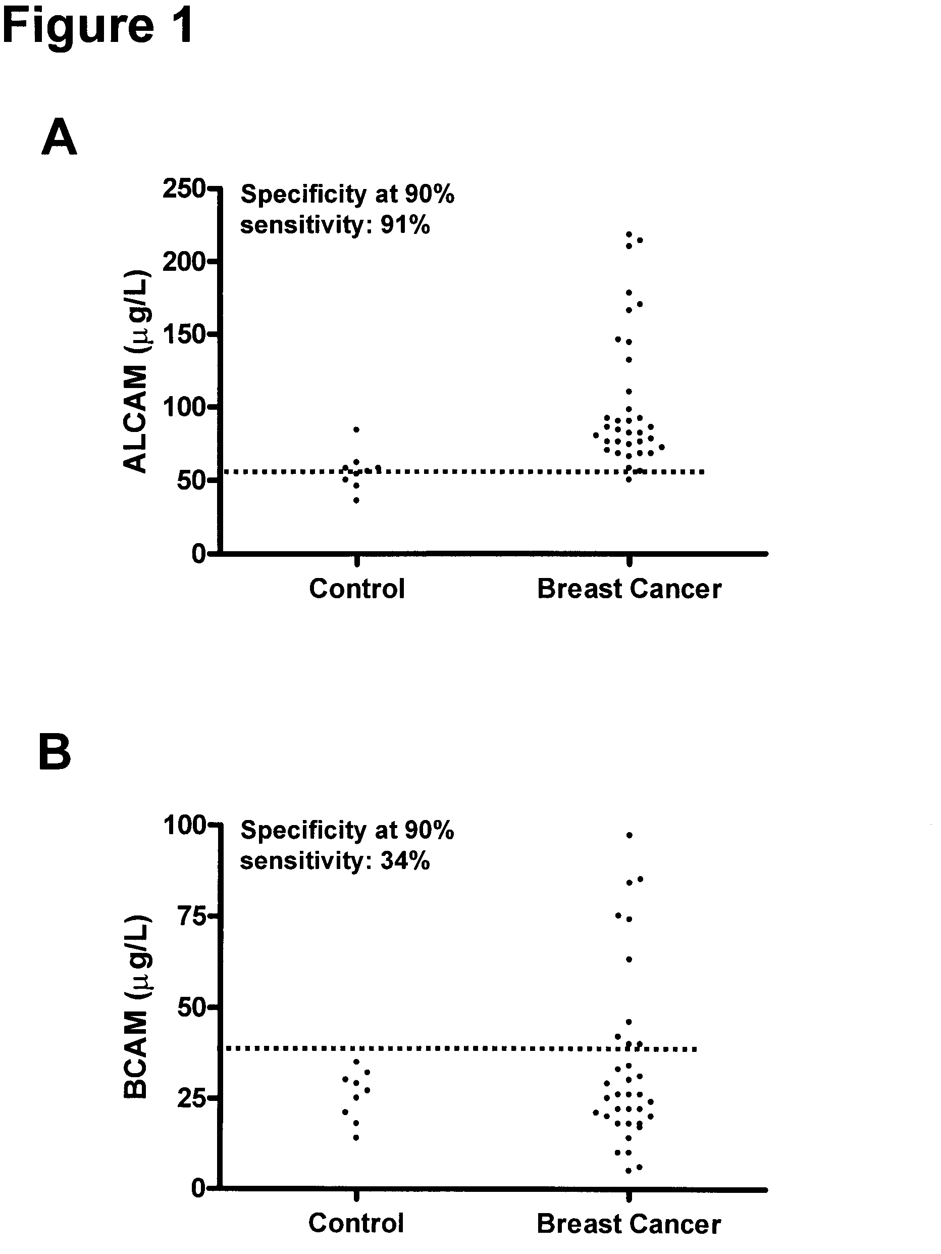
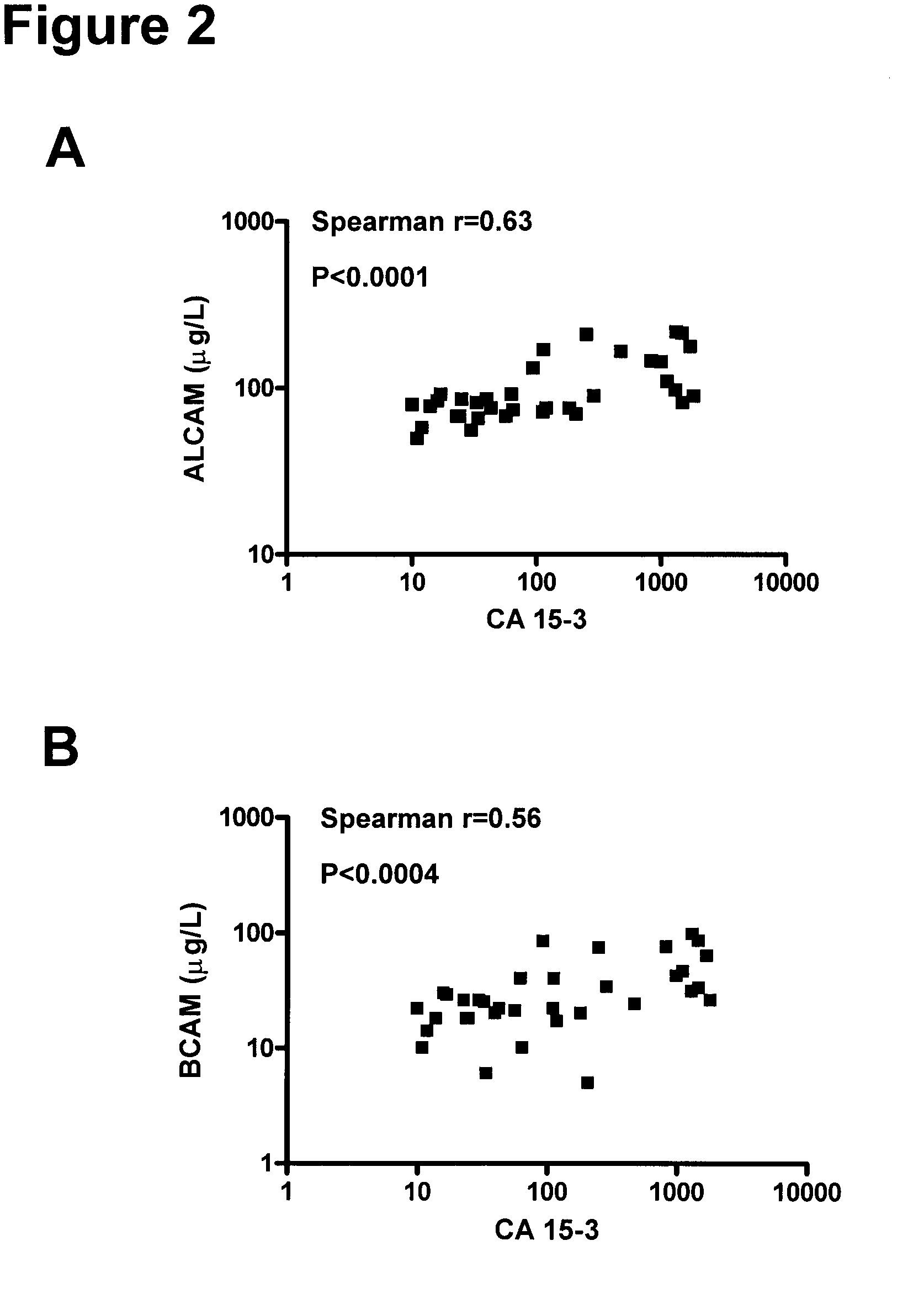
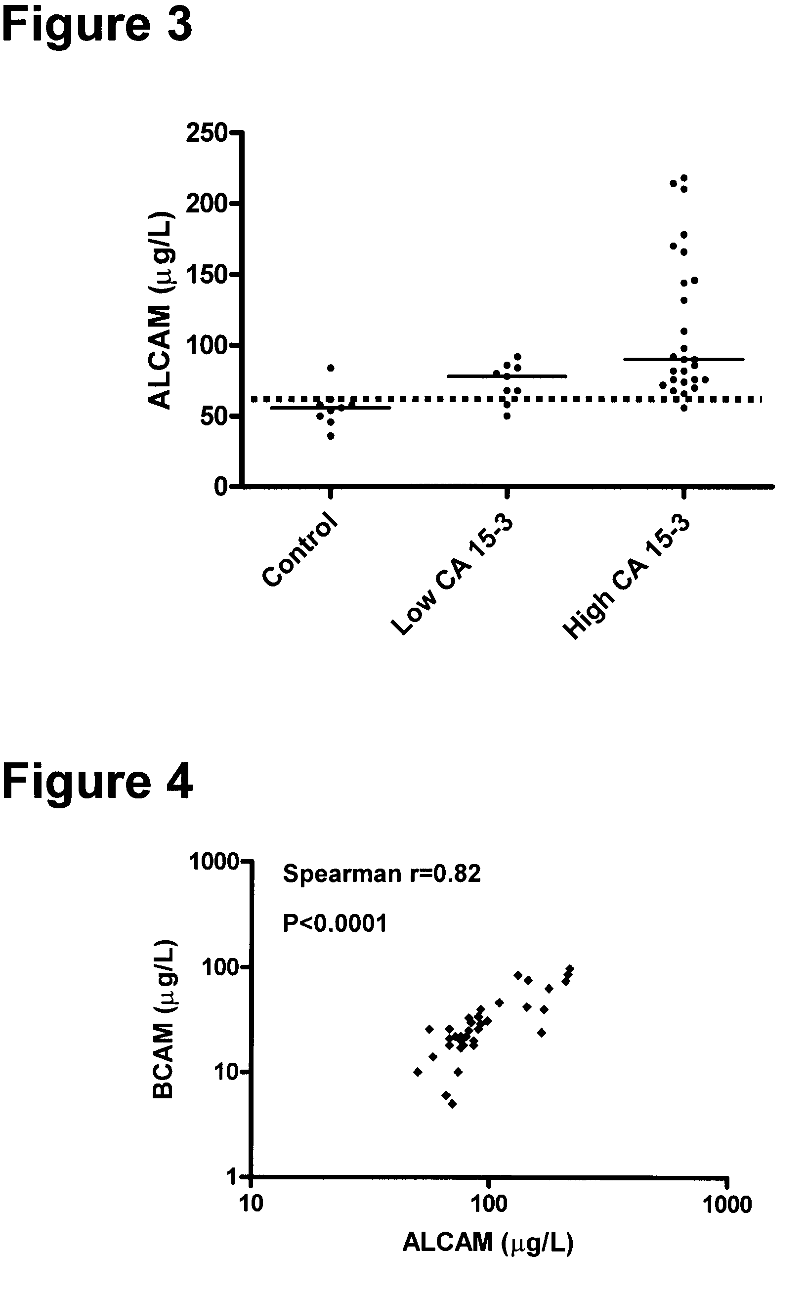
[0165]The clinical material used consisted of 150 serum samples from primary breast cancer patients (ages 34 to 82 years; median, 62 years), 100 serum samples from normal, apparently healthy women (ages 24 to 56 years; median, 40 years), and as an additional control, 50 serum samples from normal healthy men (ages 23 to 61 years; median, 48 years). The samples from primary breast cancer patients were from untreated individuals collected prior to surgery. Histologically, 94 were classified as invasive ductal carcinoma and / or multifocal invasive ductal carcinoma, 24 as invasive lobular carcinoma and / or multifocal invasive lobular carcinoma and 32 as either invasive ductal carcinoma+invasive lobular carcinoma, invasive ductal carcinoma with various aspects, lobular carcinoma in situ, medullary carcinoma or other. Histologic classification was based on the World Health Or...


PUM


Abstract

The present application describes biomarkers and methods useful for screening for, diagnosing or detecting the presence and severity of breast cancer in a subject. The present application also provides methods for determining the prognosis of a subject with breast cancer as well as methods for monitoring the therapeutic response to a breast cancer treatment or therapy.

Description

FIELD OF THE INVENTION[0001]The present application relates to methods and compositions for screening for, detecting or diagnosing likelihood and severity of breast cancer.BACKGROUND OF THE INVENTION[0002]Breast cancer is by far the most common cancer affecting women worldwide with approximately one million new cases diagnosed each year. It is a leading cause of death among women with solid tumors in North America1. It is a disease of the middle and late ages of life, as 75% of breast cancer is diagnosed in women over the age of 50. While breast cancer is less common at a young age, younger women tend to have a more aggressive form of the disease than older women. The five-year survival rate is close to 97% when the cancer is confined to the breast2. However, when breast cancer has metastasized at the time of diagnosis, the five-year survival rate is ˜23%. Gene expression patterns have been used to classify breast tumors into clinically relevant subgroups (luminal A, luminal B, basa...

Claims


Application Information

Patent Timeline
IPC IPC(8): G01N33/566G01N30/96
CPCG01N33/57415G01N2800/56G01N2333/70596
Inventor DIAMANDIS, ELEFTHERIOS P.KULASINGAM, VATHANY
Owner UNIV HEALTH NETWORK
Why Eureka
Try Eureka