Vessel segmentation in dce mr imaging

a dce and mr imaging technology, applied in image data processing, diagnostics, applications, etc., can solve the problem of misleading shift of bv frequency distribution towards higher bv values, and achieve the effect of improving the diagnostic efficacy of dce imaging

Active Publication Date: 2011-07-14
UNIV OSLO HF
View PDF5 Cites 34 Cited by
  • Summary
  • Abstract
  • Description
  • Claims
  • Application Information

AI Technical Summary

Benefits of technology

[0007]It is another object of the invention to improve the diagnostic accuracy of automated tumor grading by providing a method and a system for automatically segmenting and removing blood vessels in the perfusion related maps used in the tumor grading.
[0015]k-class cluster analysis splits n objects (here pixels) based on features (here for example rBV, ΔR2p, ΔR2max, T0, fmAUC) into k clusters, k<n, by maximizing variation between clusters relative to variation within each cluster. In other words, the analysis attempts to find the centers of natural clusters in the value space of the features, and group the objects according to these centers.
[0021]The features, or curve parameters, rBV, rBF, ΔR2p, and ΔR2max represent different measures of the degree the pixel / voxel entirely consist of blood. Here, rBV represents a measure of blood volume per voxel volume, which is largest (here typically equal to 1) if the voxel is entirely within a vessel. rBF represents tissue perfusion (flow) (vol / time) which is largest if entire volume is perfused, which is the case if voxel is entirely within vessel. ΔR2p which represents the ‘steady state’ΔR2 level following the first-passage of the contrast agent and is largest if the entire voxel contains blood. Similarly, the features, or curve parameters, T0 and fmAUC represent different measures of contrast arrival time at the tissue in a pixel, i.e. is tissue early (artery) or late (vein) in blood circulation. As the preferred specified features / parameters represent different measures of similar physiological situations, they may be used as substituent features (the ‘or’s) which may provide the advantage of improving simplicity and computation speed and reducing the required MRI acquisitions. In the alternative, the specified pair-wise features may be used as complimenting features (the ‘and’s) which may provide the advantage of improving the diagnostic accuracy (sensitivity, specificity, NPV, PPV) of the tumor grading.
[0026]The invention provides the advantage of removing false positives without introducing false negatives in a subsequent automated tumor grading.
[0033]It is a basic idea of the invention to apply a cluster analysis algorithm to automatically identify and remove image pixels representative of large vessels (arteries and veins), based on the shape and characteristics of the AIF for each pixel. Here, the AIF may be identified and separated from tissue by using parameters such as contrast arrival time (T0), Blood volume values (BV), peak enhancement values (ΔR2max) post first-pass enhancement level (ΔR2p), first moment of the area (fmAUC), relative mean transit time (rMTT). Vessel segmentation based on such dynamic first-pass parameters can improve the diagnostic efficacy of DCE imaging for tumor grading. The proposed method is attractive in that it provides a mask which covers all pixels affected by the intravascular susceptibility effect.

Problems solved by technology

Due to relatively small voxel sizes (typically tens of mm2) of the perfusion imaging technique, large vessels in the region could result in a misleading shift of the BV frequency distribution towards higher BV values.
Therefore, inclusion of BV values from vessels having dimensions of the order of or larger than the typical spatial resolution of the applied imaging technique is a confounding factor in the grading, as these are not separable from values from malicious tissue in the automated tumor segmentation.

Method used


Image

  • Vessel segmentation in dce mr imaging
  • Vessel segmentation in dce mr imaging
  • Vessel segmentation in dce mr imaging

Examples


Embodiment Construction

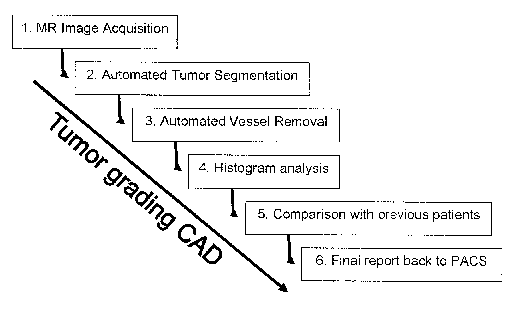
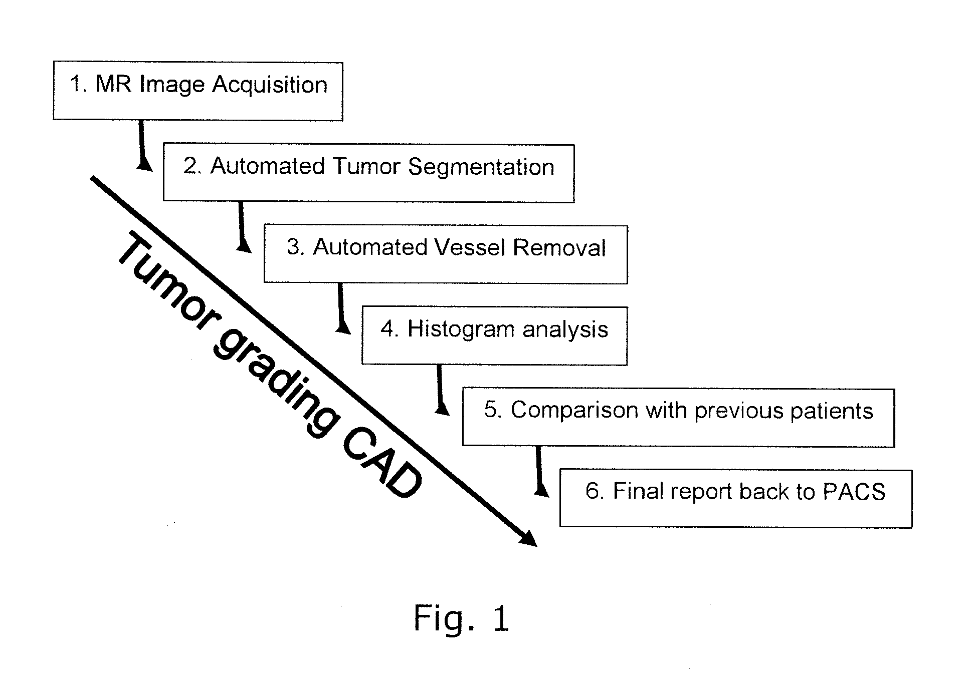
[0053]FIG. 1 illustrates the overall structure of a system or a method for computer aided diagnosis of tumors, comprising steps 1-6 as indicated.

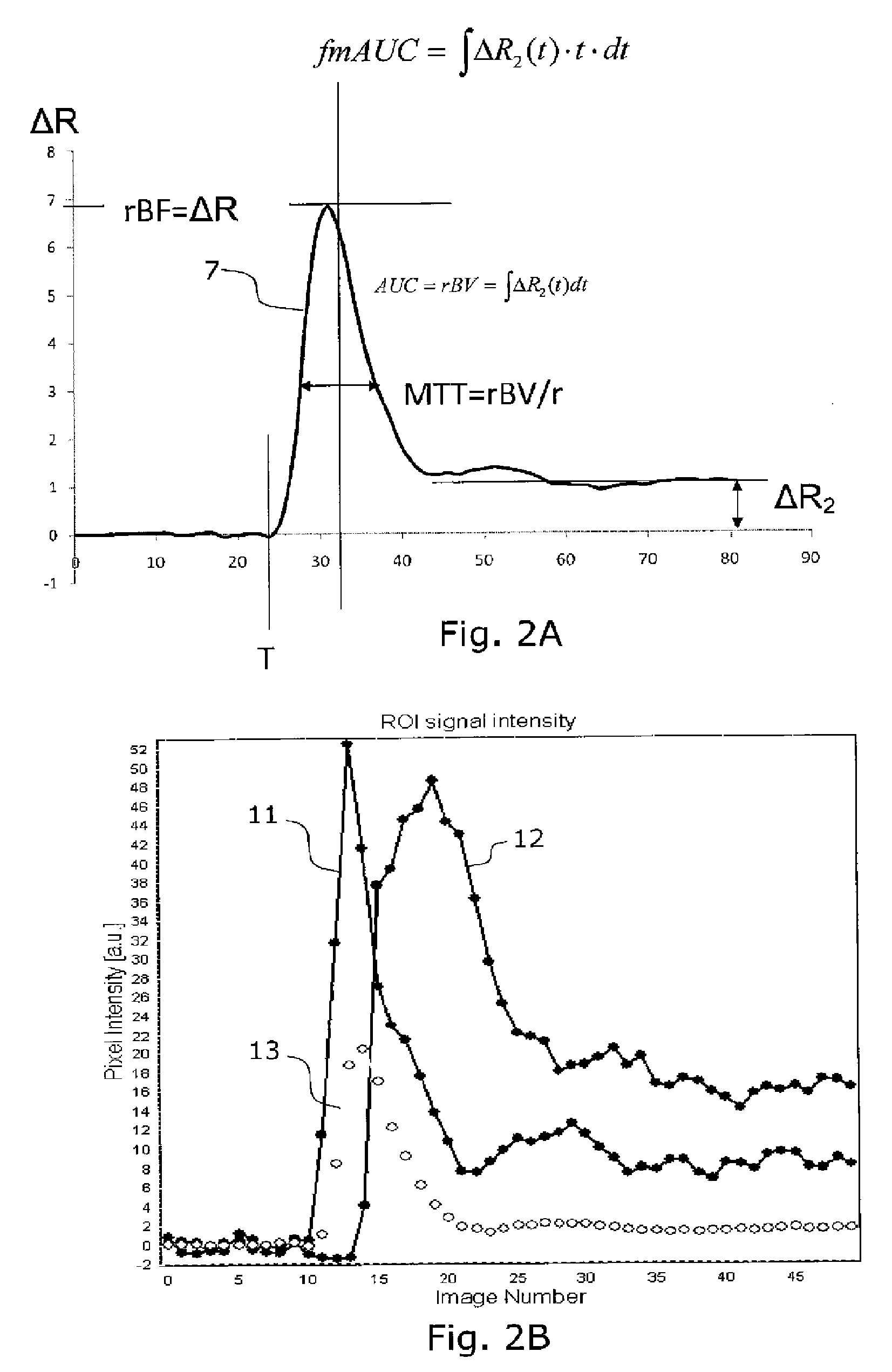
[0054]Some common abbreviations used in the present description are:[0055]MR: magnetic resonance[0056]AIF: arterial input function[0057]ΔR2: change in transverse relaxation rate (1 / T2 or 1 / T2*) due to the presence of an MR contrast agent in the tissue of interest[0058]DCE: 0dynamic contract enhanced[0059]DSC: dynamic susceptibility contrast[0060]rCBV: relative (cerebral) blood volume[0061]nCBV: normalized (cerebral) blood volume[0062]rMTT: relative mean transit time[0063]T0: contrast arrival time[0064]ΔR2max: maximum first-pass change in T2 relaxation rate, related to tissue perfusion (rBF)[0065]fmAUC: first moment of the area under curve[0066]ΔR2p: post first-pass enhancement level

[0067]FIG. 2A illustrates definitions of different parameters or features used in the cluster analysis on a curve 7 showing an arterial input function. Such curv...


PUM


Abstract

The invention relates to segmenting blood vessels in perfusion MR images, more particularly, the invention relates to automated vessel removal in identified tumor regions prior to tumor grading. Pixels from a perfusion related map from dynamic contrast enhancement (DCE) MR images are clustered into arterial pixels and venous pixels by e.g. a k-means class cluster analysis. The analysis applies parameters representing the degree to which the tissue entirely consists of blood (such as relative blood volume (rBV), peak enhancement (ΔR2max) and / or post first-pass enhancement level (ΔR2p)) and parameters representing contrast arrival time at the tissue (such as first moment of the area (fmAUC) and / or contrast arrival time (T0)). The artery and venous pixels are used to form a vessel mask. The invention also relates to a computer aided diagnostic (CAD) system for tumor grading, comprising automated tumor segmentation, vessel segmentation, and tumor grading.

Description

FIELD OF THE INVENTION[0001]The present invention relates to segmenting blood vessels in MR images, more particularly, the invention relates to automated vessel removal in identified tumor regions prior to tumor grading.BACKGROUND OF THE INVENTION[0002]Perfusion imaging based on dynamic contrast enhanced (DCE) MRI is a widely used technique for grading tumors, especially for brain tumors (gliomas) due to their inherent inaccessibility. Several studies have shown a good correlation between histopathological glioma grade and blood volume (BV) derived from perfusion imaging.[0003]Traditionally, an experienced neuroradiologist identifies the tumor region in relevant slices, and the tumor can be graded. however, there is a movement towards computer aided diagnostics (CAD) within tumor grading, and automated methods for segmenting (identifying) tumors from MR images exists, as well as automated methods for grading the identified tumors in BV maps (hot-spot or histogram methods).[0004]Bloo...

Claims


Application Information

Patent Timeline
Patent Type & Authority Applications(United States)
IPC IPC(8): G06K9/00
CPCG06T7/0012G06T7/0081G06T2207/30101G06T2207/10088G06T2207/30096G06T7/401G06T7/11G06T7/41
Inventor BJORNERUD, ATLEEMBLEM, KYRRE EEG
Owner UNIV OSLO HF
Why Eureka
Try Eureka