Ultrasonic image processing method and device, and ultrasonic image processing program

a technology of ultrasonic image processing and ultrasonic image, applied in image enhancement, instruments, applications, etc., can solve problems such as degrading sharpness

Active Publication Date: 2011-09-08
FUJIFILM HEALTHCARE CORP
View PDF4 Cites 14 Cited by
  • Summary
  • Abstract
  • Description
  • Claims
  • Application Information

AI Technical Summary

Benefits of technology

According to the present invention, an ultrasonic image processing method and device and an ultrasonic image processing program which are compatible with an analysis technique that varies depending on a region or a purpose of diagnosis or treatment can be provided.

Problems solved by technology

However, when the tissue border is noted in order to perform modulus-of-elasticity processing, sharpness is found to be degraded.

Method used


Image

  • Ultrasonic image processing method and device, and ultrasonic image processing program
  • Ultrasonic image processing method and device, and ultrasonic image processing program
  • Ultrasonic image processing method and device, and ultrasonic image processing program

Examples


Embodiment Construction

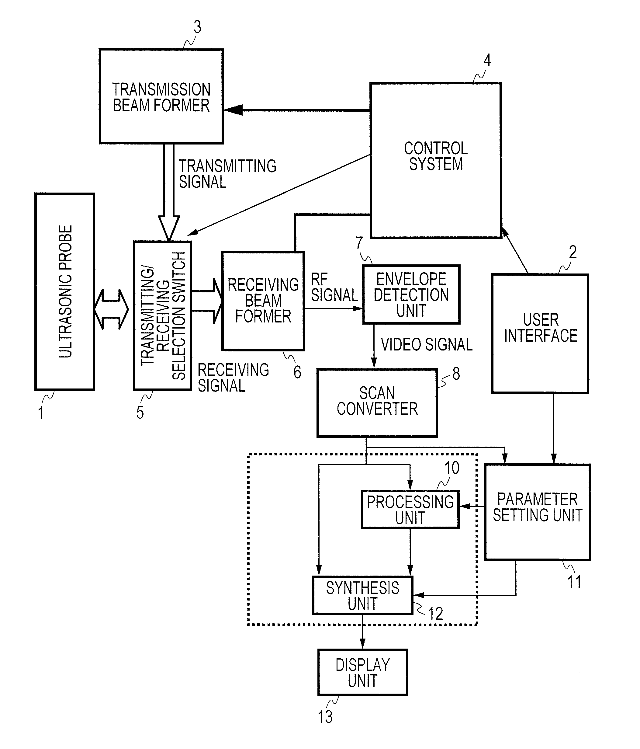
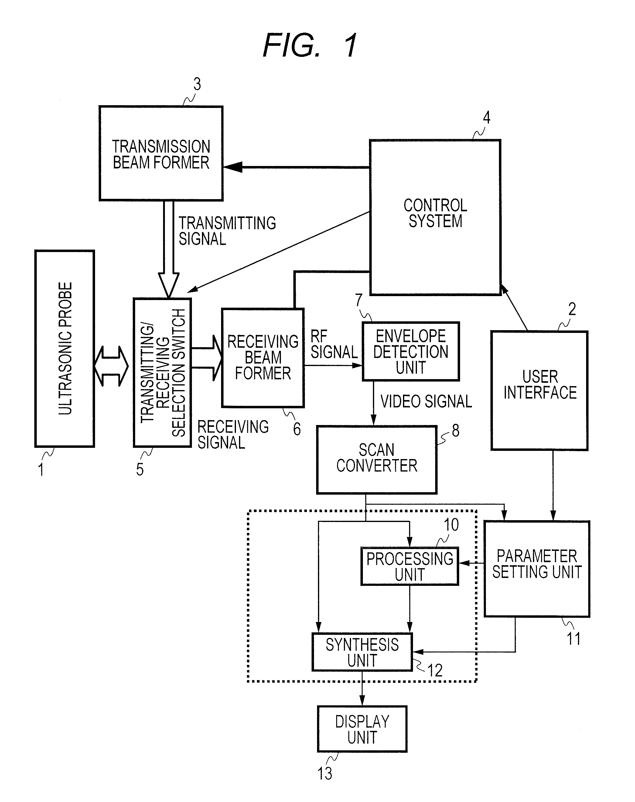
FIG. 1 shows an example of a system configuration for an ultrasonic border detection method of the present invention. An ultrasonic probe 1 having ultrasonic elements arrayed one-dimensionally transmits an ultrasonic beam (ultrasonic pulses) to a living body, and receives an echo signal (receiving signal) reflected from the living body. Under the control of a control system 4, a transmission signal having a delay time matched with a transmission focus is outputted from a transmission beam former 3, and sent to the ultrasonic prove 1 via a transmitting / receiving selection switch 5. An ultrasonic beam reflected or scattered inside the living body and returned to the ultrasonic probe 1 is converted into an electric signal by the ultrasonic probe 1, and sent as a receiving signal to a receiving beam former 6 via the transmitting / receiving selection switch 5. The receiving beam former 6 is a complex beam former that mixes two receiving signals that are 90° out of phase. The receiving bea...


PUM


Abstract

An ultrasonic image processing method and device, and an ultrasonic image processing program which can correspond to analytical methods different depending on a region or the purpose of a diagnosis or treatment. The ultrasonic image processing method comprises an image data creation step which stores a detection result obtained by irradiating a subject with ultrasonic waves by an irradiating section and detecting an ultrasonic signal from the subject by a detecting section and creates at least two-frame image data different in detection timing on the basis of the stored detection result, a motion vector distribution image creation step which creates a motion vector distribution image on the basis of a predetermined motion vector analysis through the use of a plurality frames of the image data, and a conversion step which converts a vector distribution image to a scalar distribution image on the basis of a plurality of established regions of interest (ROI).

Description

TECHNICAL FIELDThe present invention is a technology relating to an ultrasonic image processing method and device and an ultrasonic image processing program capable of clearly distinguishing a tissue border at the time of imaging a living body with ultrasonic waves.BACKGROUND ARTFor an ultrasonic image processing device employed in medical diagnostic imaging, there is disclosed a method in which, as described in, for example, patent literature 1, a modulus-of-elasticity distribution in a tissue is estimated based on a magnitude of a change in a small area in a diagnostic motion picture, and hardness is converted into a color map and then displayed. However, when the tissue border is noted in order to perform modulus-of-elasticity processing, sharpness is found to be degraded. As described in patent literature 2, a technique of producing a scalar distribution image directly from motion vectors of a diagnostic motion picture so as to improve the degree of distinction of the tissue bor...

Claims


Application Information

Patent Timeline
Patent Type & Authority Applications(United States)
IPC IPC(8): A61B8/00
CPCA61B8/08A61B8/469G06T7/2006A61B8/483A61B8/463A61B8/5246A61B8/5276G06T2207/10132G06T7/215
Inventor MASUI, HIRONARIAZUMA, TAKASHI
Owner FUJIFILM HEALTHCARE CORP
Why Eureka
Try Eureka