Electronic endoscope system

a technology of endoscopy and endoscope, which is applied in the field of electronic endoscopy systems, can solve the problems that have never been provided diagnoses, and achieve the effect of enhancing the oxygen saturation level and increasing the color saturation of the oxygen saturation level

Inactive Publication Date: 2011-09-29
FUJIFILM CORP
View PDF13 Cites 90 Cited by
  • Summary
  • Abstract
  • Description
  • Claims
  • Application Information

AI Technical Summary

Benefits of technology

[0009]An object of the present invention is to provide an electronic endoscope system that enables simultaneous acquisition of both blood vessel depth information and oxygen saturation level information and selective display of an oxygen saturation level of a characteristic blood vessel portion that is of interest to an observer in making a diagnosis.
[0016]Preferably, the second image production means increases a color saturation of the oxygen saturation level in the simulated color within the region of interest to selectively enhance the oxygen saturation level within the region of interest in the second oxygen saturation level image.

Problems solved by technology

However, there have never been provided diagnoses where the observers selectively displayed a portion that is of diagnostic interest.

Method used


Image

  • Electronic endoscope system
  • Electronic endoscope system
  • Electronic endoscope system

Examples


first embodiment

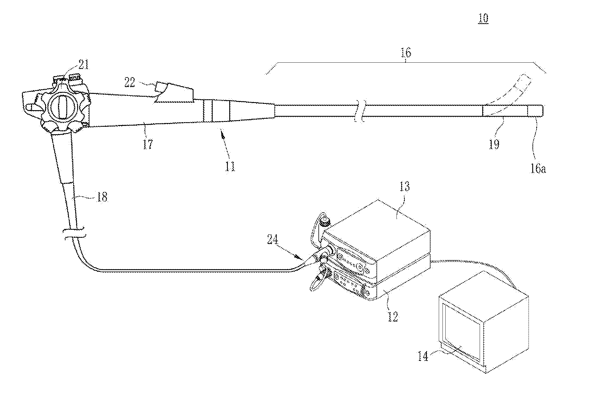
[0028]As illustrated in FIG. 1, an electronic endoscope system 10 according to the invention comprises an electronic endoscope 11 for imaging the inside of a subject's body cavity, a processor 12 for producing an image of a subject tissue in the body cavity based on a signal acquired by imaging, a light source device 13 for supplying light used to illuminate the inside of the body cavity, and a monitor (image display means) 14 for displaying the image of the inside of the body cavity. The electronic endoscope 11 comprises a flexible insertion section 16 that is inserted into a body cavity, an operating section 17 provided at the base of the insertion section 16, and a universal cord 18 for connecting the operating section 17 to the processor 12 and the light source device 13.

[0029]The insertion section 16 has a bending portion 19 at the tip thereof comprising connected bending pieces. The bending portion 19 bends up and down, left and right in response to the operation of an angle k...

second embodiment

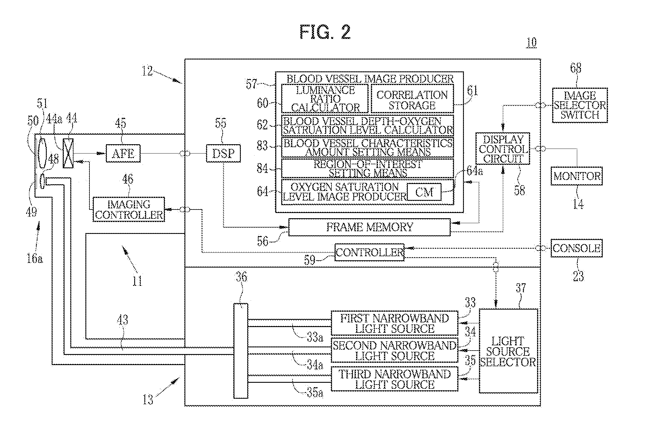
[0085]According to the invention, the blood vessel characteristics amount setting means 83 sets a blood vessel density as the blood vessel characteristics amount in the acquired image.

[0086]According to this embodiment, where a blood vessel density is set as the blood vessel characteristics amount, the region-of-interest setting means 84 sets a region of interest based on the blood vessel density.

[0087]First, the region of interest setting means 84 acquires the first narrowband image data stored in the frame memory 56, and then extracts micro blood vessels having a diameter of about 10 μm to 50 μm from the first narrowband image data. Then, the region of interest setting means 84 extracts, in particular, a portion having a high blood vessel density from the blood vessel region containing the thus extracted micro-blood vessels. The extraction of the portion having a high blood vessel density is achieved by binarizing the first narrowband image where the micro-blood vessels were extra...

third embodiment

[0093]According to the invention, the blood vessel characteristics amount setting means 83 sets a blood vessel branch point density as the blood vessel characteristics amount in the acquired image.

[0094]According to this embodiment, where a blood vessel branch point density is set as the blood vessel characteristics amount, the region-of-interest setting means 84 sets a region of interest based on the blood vessel branch point density.

[0095]First, the region of interest setting means 84 acquires the first narrowband image data stored in the frame memory 56, and then extracts micro-blood vessels having a diameter of about 10 μm to 50 μm from the first narrowband image data. Then, the region of interest setting means 84 extracts, in particular, a portion having a high blood vessel branch point density from the blood vessel region containing the thus extracted micro-blood vessels. The extraction of the portion having a high blood vessel branch point density may be achieved by binarizin...


PUM


Abstract

An electronic endoscope system includes a light source device for sequentially emitting plural kinds of light having different wavelength bands, an electronic endoscope for outputting image data of acquired images corresponding to the plural kinds of light sequentially illuminated to a subject tissue containing blood vessels, a setter for setting a blood vessel characteristics amount from the image data, a setter for setting a region of interest in the acquired image based on the amount, an image producer for producing a first oxygen saturation level image representing a distribution of an oxygen saturation level in the blood vessel from the image data, and an image display for displaying in simulated color a second oxygen saturation level image in which the oxygen saturation level within the region of interest is selectively enhanced.

Description

BACKGROUND OF INVENTION[0001]The present invention relates to an electronic endoscope system for acquiring information on a blood vessel from an image acquired by an electronic endoscope and producing an image from the acquired information.[0002]In recent years, a number of diagnoses and treatments using electronic endoscopes have been made in the field of medicine. A typical electronic endoscope is equipped with an elongated insertion section that is inserted into a subject's body cavity. The insertion section has therein incorporated an imager such as a CCD at the tip thereof. The electronic endoscope is connected to a light source device, which emits light from the tip of the insertion section to illuminate the inside of a body cavity. With the inside of the body cavity illuminated by light, the subject tissue inside the body cavity is imaged by an imager provided at the tip of the insertion section. Images acquired by imaging undergoes various kinds of processing by a processor ...

Claims


Application Information

Patent Timeline
Patent Type & Authority Applications(United States)
IPC IPC(8): A61B1/04
CPCA61B1/00009A61B5/7485A61B1/0005A61B1/05A61B1/0638A61B5/0084A61B5/02007A61B5/14551A61B5/1459A61B5/489G06T7/0012G06T2207/10024G06T2207/10068G06T2207/10152G06T2207/30101H04N5/2256H04N2005/2255A61B5/7425A61B1/063A61B1/00045A61B1/000094A61B1/0655H04N23/555H04N23/56
Inventor SAITO, TAKAAKI
Owner FUJIFILM CORP
Why Eureka
Try Eureka