Methods and apparatus for ultrasound strain imaging

a strain imaging and ultrasound technology, applied in the field of ultrasound strain imaging, can solve the problems of difficult control of compression rate, compromising image quality, and difficult to generate reliable, high-quality ultrasound strain images from freehand palpations

Active Publication Date: 2011-09-29
THE JOHN HOPKINS UNIV SCHOOL OF MEDICINE
View PDF12 Cites 35 Cited by
  • Summary
  • Abstract
  • Description
  • Claims
  • Application Information

AI Technical Summary

Benefits of technology

[0010]According to a first aspect of the present invention, a method for providing ultrasonic strain imaging comprises placing an ultrasound probe onto a target area to be imaged, applying palpations to the target area, acquiring a sequence of RF frames during palpations of the target area, measuring positions and orientations of the ultrasound probe at each the RF frame in the sequence of the RF frames, synchronizing each of the positions and orientations of the ultrasound probe to a respective RF frame in the sequence of RF frames, automatically selecting multiple pairs of RF frames from an approximately same location and orientation from the synchron

Problems solved by technology

However, generating reliable, high-quality ultrasound strain images from freehand palpations is a challenging task.
However, it is difficult to control the compression rate as it is governed by hand motion and the frame rate of RF data.
Also, small lateral or out-of-plane motions can compromise the quality of images.
However, it is difficult to induce pure axial motion with freehand compression.
However, these algorithms only partially address the problem by compensating for in-plane motions and applying smoothness constraints.
The images ar

Method used


Image

  • Methods and apparatus for ultrasound strain imaging
  • Methods and apparatus for ultrasound strain imaging
  • Methods and apparatus for ultrasound strain imaging

Examples


example

[0075]The following Example has been included to provide guidance to one of ordinary skill in the art for practicing representative embodiments of the presently disclosed subject matter. In light of the present disclosure and the general level of skill in the art, those of skill can appreciate that the following Example is intended to be exemplary only and that numerous changes, modifications, and alterations can be employed without departing from the scope of the presently disclosed subject matter. The following Example is offered by way of illustration and not by way of limitation.

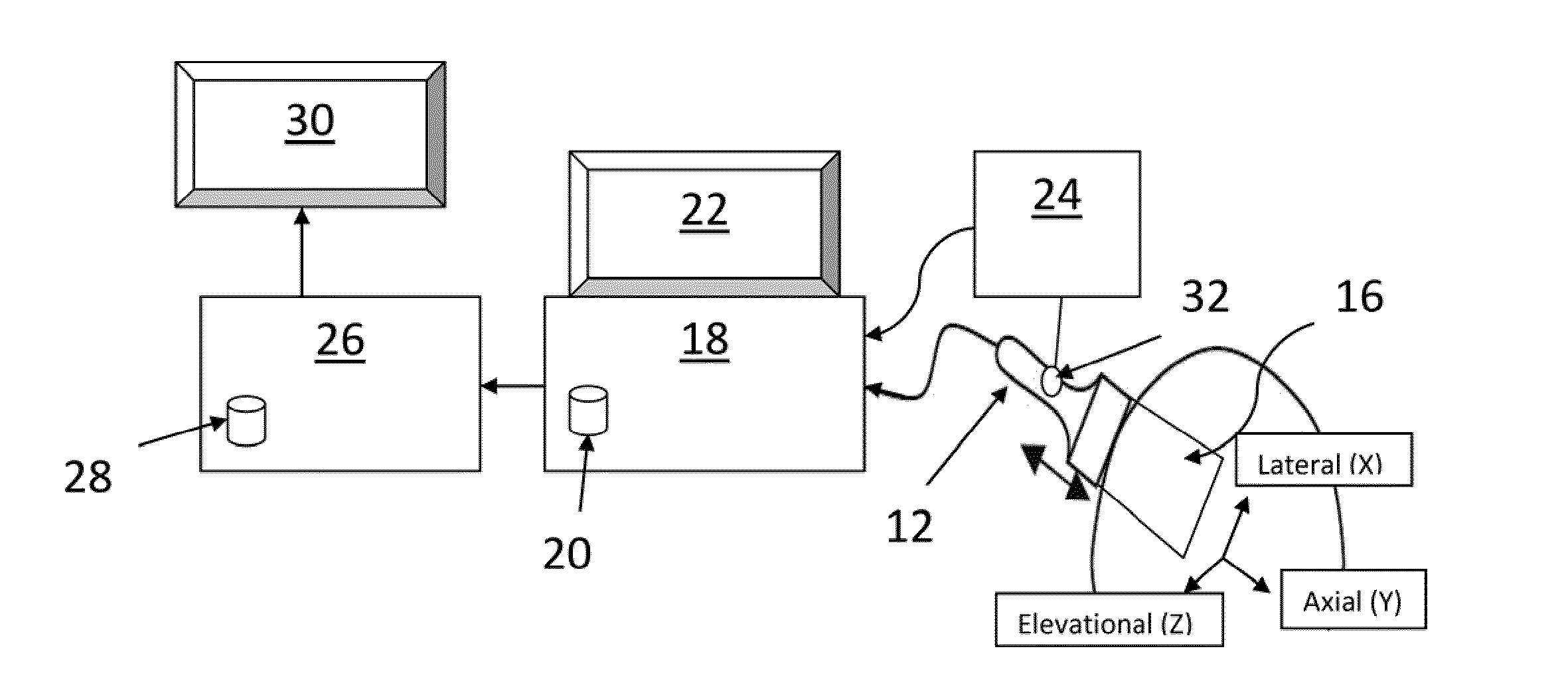
[0076]Ultrasound data was acquired using a SONOLINE Antares™ ultrasound system (Siemens Medical Solutions USA, Inc.) with a high-frequency ultrasound transducer (VF10-5) at center frequency of 6-8 MHz. RF data was accessed through the Axius Direct™ Ultrasound Research Interface provided by Siemens. A data acquisition program was connected to this interface to send the command for capturing RF data. At th...


PUM


Abstract

A system and method for improved ultrasound strain imaging includes using data from a tracking system to enhance the quality of ultrasound strain image and to reduce the dependency of image quality of the user's expertise. The tracking information is synchronized with the RF frames and interpolated to find the transformation corresponding to each frame. The RF frames with their transformations are incorporated into calculation of ultrasound strain images. The tracking system may be an optical tracker, electromagnetic tracker, accelerometer, or a structured light system. The structured light system may also be used for probe calibration, by calibrating the surface of the probe pre-operatively. In addition, a relative Young's Modulus may be calculated using tracking information that is independent from the distribution of input force.

Description

REFERENCE TO RELATED APPLICATIONS[0001]This application claims the benefit of U.S. Provisional Patent Application No. 61 / 318,040, filed on Mar. 26, 2010, which is hereby incorporated by reference for all purposes as if fully set forth herein.FIELD OF THE INVENTION[0002]The present invention pertains to a method and system for ultrasound strain imaging. More particularly, the present invention pertains to a method and system for ultrasound strain imaging using an external tracker.BACKGROUND OF THE INVENTION[0003]Ultrasound imaging has become a widely used medical imaging modality, due in part to its effectiveness in safely imaging tissue, its ease of use, and lower cost. Ultrasound has become an essential imaging tool in applications such as identifying tissue anomalies, monitoring fetal development, and assisting in guiding surgical devices in invasive treatments.[0004]More recently, strain imaging has been shown helpful in numerous medical applications, especially in the detection ...

Claims


Application Information

Patent Timeline
IPC IPC(8): A61B8/14
CPCA61B8/4245A61B8/58A61B8/5215A61B8/485A61B8/4254G16H50/20A61B8/4263
Inventor FOROUGHI, PEZHMANBOCTOR, EMADHAGER, GREGORY
Owner THE JOHN HOPKINS UNIV SCHOOL OF MEDICINE
Why Eureka
Try Eureka