Methods and systems of combining magnetic resonance and nuclear imaging

a nuclear imaging and magnetic resonance technology, applied in the field of multimodality medical imaging, can solve the problems of increasing noise and signal distortion, mostly dismissing the possibility of combining spect and mri within a single system, etc., and achieve the effect of reducing or minimizing mis-registration artifacts

Inactive Publication Date: 2011-11-03
GAMMA MEDICA
View PDF6 Cites 58 Cited by
  • Summary
  • Abstract
  • Description
  • Claims
  • Application Information

AI Technical Summary

Benefits of technology

An aspect of the present invention provides a dual-modality, co-registered, and optionally fused image dataset from MRI and single-photon emission (SPE) nuclear medicine imaging modalities in a single imaging session. The single dual-modality imaging session allows a body (e.g., a human or animal body), body part (e.g., head, knee, breast), organ (e.g., heart, prostate, thyroid) or other object being scanned to remain essentially motionless relative to the two imaging systems for sequential scanning while using the same body position on the same bed, thereby reducing or minimizing mis-registration artifacts from changes in body orientation between the imaging studies. In this case, the SPE system could be located adjacent to the MRI system and in its fringe magnetic field and the two fields of view would not coincide. It is also possible for the SPE system to be located inside the MRI system, but not at the center of the MRI field of view.

Problems solved by technology

Although there may be clinical benefits to combine SPECT and MRI images, any prospect of combining SPECT and MRI within a single system has been mostly dismissed because the functions of the PMTs in a typical SPECT system are severely compromised by the high magnetic fields needed for MRI and because magnetic field uniformity needed for MRI is distorted by the electrically conducting components in the typical SPECT system.
This large volume occupied by the typical Anger camera used in SPECT is another impediment to its use within the bore of an MRI imaging system.
However, locating the electronics away from the magnetic field requires that they be connected via relatively long cables that result in an increased noise and signal distortion.

Method used


Image

  • Methods and systems of combining magnetic resonance and nuclear imaging
  • Methods and systems of combining magnetic resonance and nuclear imaging
  • Methods and systems of combining magnetic resonance and nuclear imaging

Examples


Embodiment Construction

In the following detailed description, only certain exemplary embodiments of the present invention are shown and described, by way of illustration. As those skilled in the art will recognize, the described exemplary embodiments may be modified in various ways, all without departing from the spirit or scope of the present invention. Accordingly, the drawings and description are to be regarded as illustrative in nature, and not restrictive.

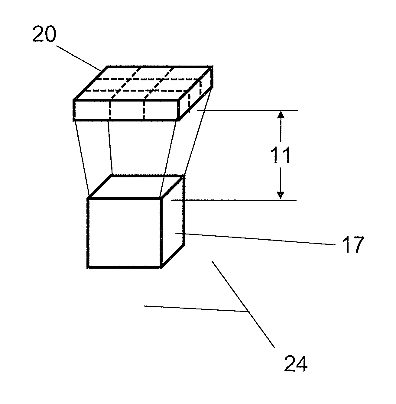
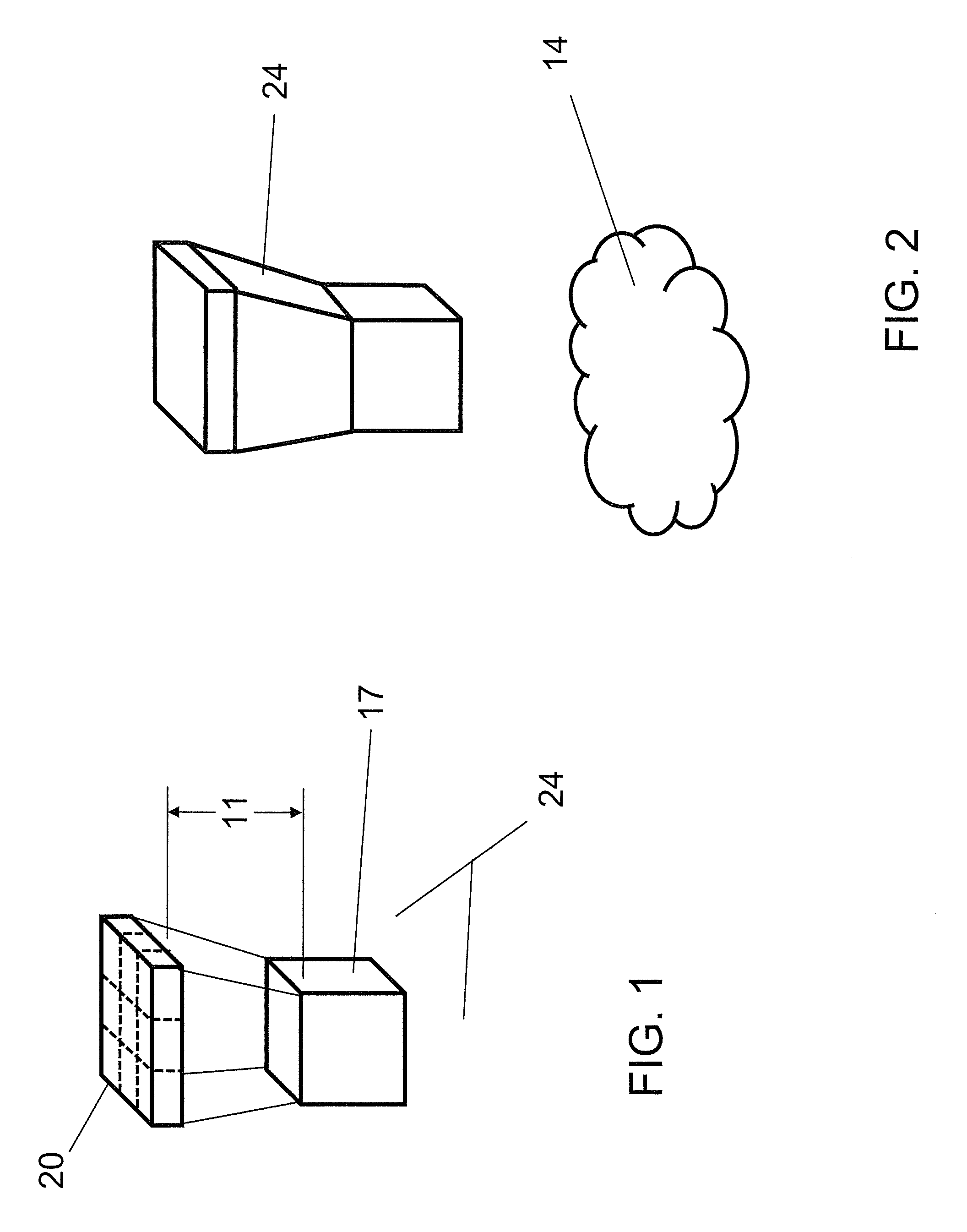
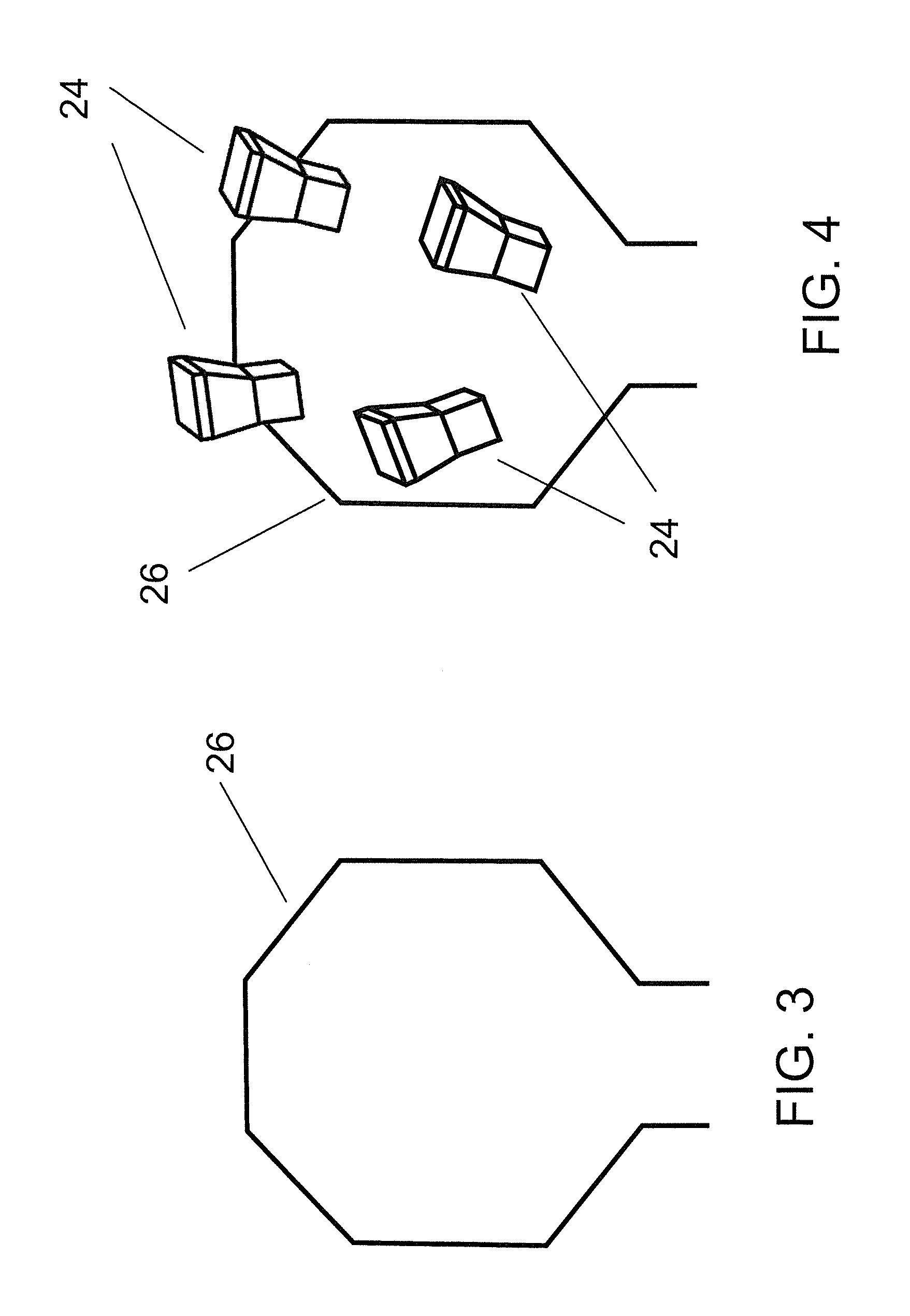
An embodiment of the present invention is designed to enhance or augment MRI imaging by incorporating an additional modality within an RF coil for sequential or simultaneous operation of the modality with an MRI system. The added modality is tomographic SPECT or planar imaging based on the single-photon emission (SPE) radiotracer principle.

In one embodiment of the present invention, the SPE imaging system is based on a semiconductor direct conversion detector, such as a cadmium zinc telluride (CZT) detector. The embodiment of the present invention r...


PUM


Abstract

An multi-modality imaging system for imaging of an object under study that includes a magnetic resonance imaging (MRI) apparatus and an MRI-compatible single-photon nuclear imaging apparatus imbedded within the RF coil of the MRI system such that sequential or simultaneous imaging can be done with the two modalities using the same support bed of the object under study during the imaging session.

Description

FIELD OF THE INVENTIONThe invention relates generally to multi-modality medical imaging. More particularly, the invention relates to methods and systems for combining magnetic resonance imaging (MRI) with single photon emission (SPE) imaging, such as single photon emission computed tomography (SPECT).BACKGROUND OF THE INVENTIONMagnetic resonance imaging is a technique used to visualize the inner volume of an object (e.g., a human or animal body or a body part or tissue specimen or a test phantom). Magnetic field strengths for MRI studies of humans typically require 1.5 or 3.0 Tesla (T) and studies of animals typically require 4.7 or 7.0 T, although magnets up to 17 T have been reported. Organ-specific radio frequency (RF) coils are routinely used in neurology, mammography, cardiology, and urology applications. Additionally, RF coils for specific orthopaedic imaging applications such as shoulder or knee evaluation are used in clinical radiology. In more general applications, a whole-...

Claims


Application Information

Patent Timeline
Patent Type & Authority Applications(United States)
IPC IPC(8): A61B5/055
CPCG01R33/481A61B5/055A61B5/0035
Inventor WAGENAAR, DOUGLAS J.PATT, BRADLEY E.MEIER, DIRKHUGG, JAMES W.
Owner GAMMA MEDICA
Why Eureka
Try Eureka