Apparatus, systems, and methods for localizing markers or tissue structures within a body

a technology of tissue structure and apparatus, applied in the field of apparatus and methods for performing surgical procedures, can solve the problems of limited guidance for localization, limited guidance in determining, and wires may move, and achieve the effects of facilitating identification of markers, enhancing reflection of ultrasound waves, and enhancing electromagnetic signals

Active Publication Date: 2011-12-22
CIANNA MEDICAL INC
View PDF11 Cites 117 Cited by
  • Summary
  • Abstract
  • Description
  • Claims
  • Application Information

AI Technical Summary

Benefits of technology

[0021]In accordance with yet another embodiment, an implantable marker is provided for localization of a target tissue region within a patient's body that includes an elongate core member, and a plurality of beads carried by the core member. Optionally, the beads may include a plurality of surfaces and / or edges, e.g., defining a plurality of dihedral or trihedral corners, to enhance reflection of electromagnetic signals to facilitate identification of the marker. In addition or alternatively, the marker may include an electronic circuit, e.g., embedded in or otherwise carried by one of the beads or the core member, that may provide one of an active reflector and an active transponder. In addition, the marker may include echogenic features and / or the plurality of surfaces and / or edges may enhance reflection of ultrasound waves, e.g., to enhance or otherwise facilitate imaging and / or locating the marker using external ultrasound imaging and the like.
[0022]In accordance with still another embodiment, a method is provided for identifying a lesion within a target tissue region of a patient's breast that includes introducing a marker through breast tissue into the target tissue region, and imaging the breast using ultrasound. The marker may include one or more beads and / or a plurality of surfaces and / or edges, e.g., defining dihedreal or trihedral corners, that enhance identifying the marker in ultrasound images to facilitate identifying the lesion. In addition or alternatively, the marker may include echogenic materials and / or features for enhancing detection using ultrasound. For example, the target may be an elongate marker including a plurality of beads coupled to a core element, the beads including angled surfaces and / or edges to enhance detection using ultrasound imaging. The core element may be biased to one or more predetermined shapes, e.g., a wave shape, a tapered helix, a cylindrical helix, and the like, yet may be sufficiently resilient to be straightened, e.g., to facilitate loading the marker into a delivery device. In another embodiment, the target may include a spherical, elliptical, discus, or other shape, e.g., including one or more surface features to enhance reflection of ultrasound signals.
[0023]Optionally, the method may include removing a tissue specimen from the target tissue region, the tissue specimen including the lesion and the target. The target may enhance locating and / or identifying the lesion and / or desired specimen being removed, e.g., using ultrasound imaging.

Problems solved by technology

However, such images are generally two dimensional and therefore provide only limited guidance for localization of the lesion since the breast and any lesion to be removed are three-dimensional structures.
Further, such images may provide only limited guidance in determining a proper margin around the lesion, i.e., defining a desired specimen volume to be removed.
One problem with using a wire for localization is that the wire may move between the time of placement and the surgical procedure.
For example, if the wire is not secured sufficiently, the wire may move relative to the tract used to access the lesion and consequently the tip may misrepresent the location of the lesion.
If this occurs, when the location is accessed and tissue removed, the lesion may not be fully removed and / or healthy tissue may be unnecessarily removed.
Again, since such images are two dimensional, they may provide limited guidance to localize the lesion being treated or removed.
Because the seed is delivered through a needle that is immediately removed, there is risk that the seed may migrate within the patient's body between the time of placement and the surgical procedure.
Thus, similar to using a localization wire, the seed may not accurately identify the location of the lesion, particularly, since there is no external way to stabilize the seed once placed.
Further, such gamma probes may not provide desired precision in identifying the location of the seed, e.g., in three dimensions, and therefore may only provide limited guidance in localizing a lesion.

Method used


Image

  • Apparatus, systems, and methods for localizing markers or tissue structures within a body
  • Apparatus, systems, and methods for localizing markers or tissue structures within a body
  • Apparatus, systems, and methods for localizing markers or tissue structures within a body

Examples


Embodiment Construction

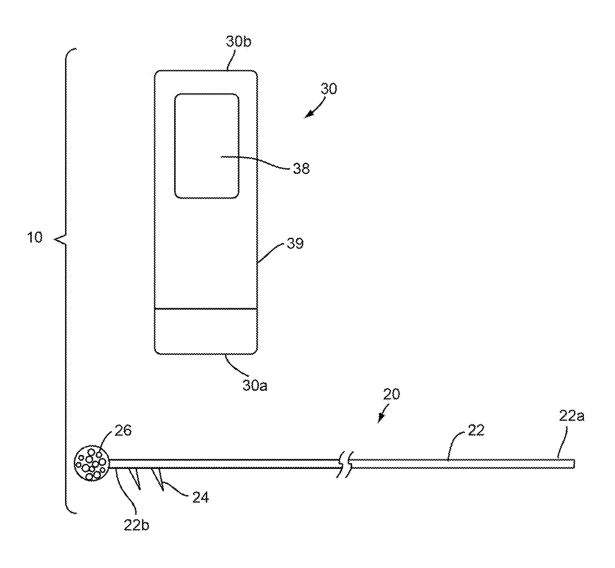
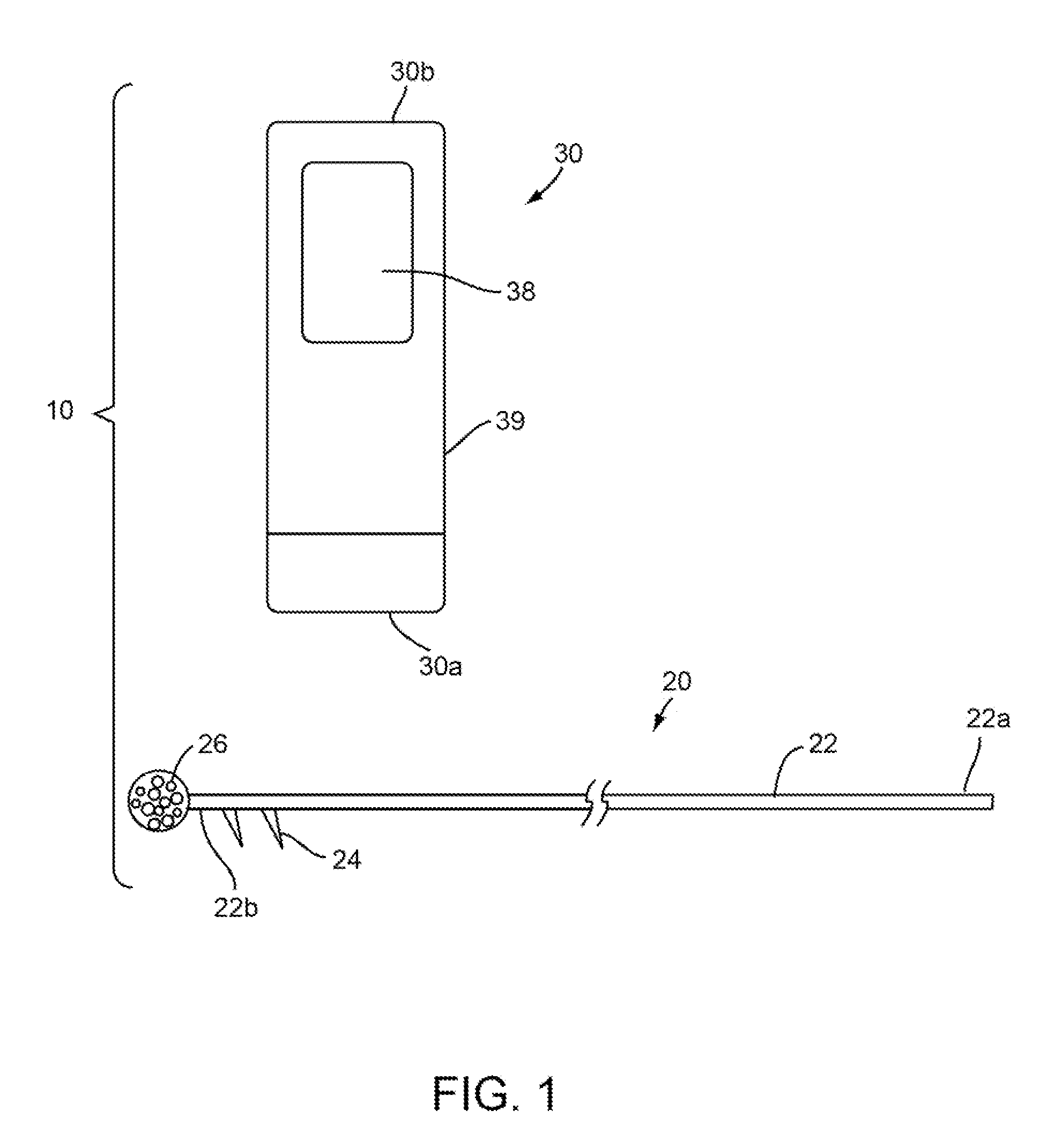
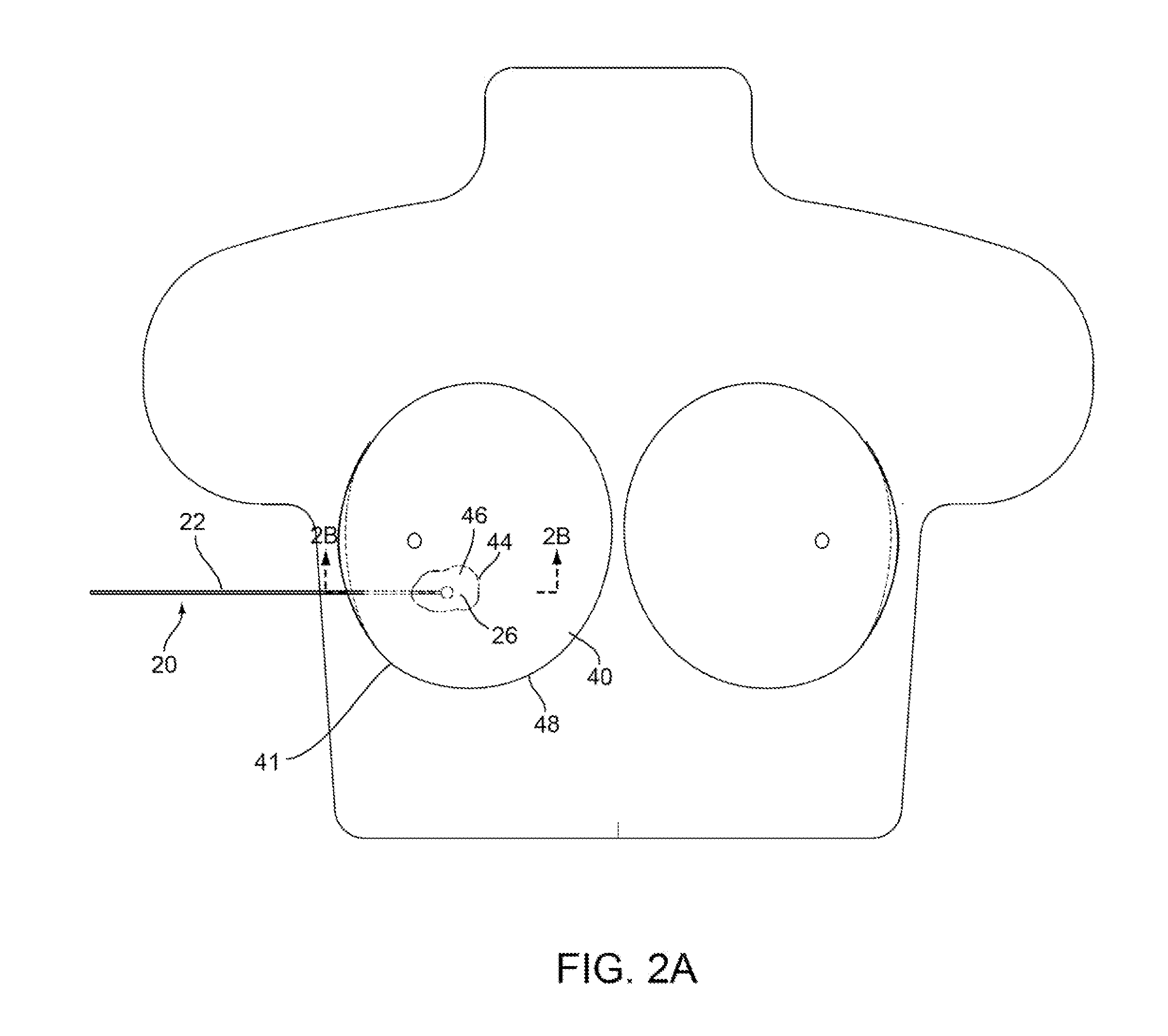
[0081]Turning to the drawings, FIG. 1 shows an exemplary embodiment of a system 10 for localization of a target tissue region within a patient's body, such as a tumor, lesion, or other tissue structure within a breast or other location within a body. The system 10 generally includes a marker device or localization wire 20 and a probe 30 for detecting at least a portion of the localization wire 20 using electromagnetic pulses, waves, or other signals, such as radar. The localization wire 20 may include an elongated member or shaft 22 including a proximal end 22a, a distal end 22b, and a target 26 on the distal end 22b. Optionally, the system 10 may include one or more additional localization wires and / or targets (not shown) in addition to localization wire 20.

[0082]The shaft 22 may be formed from a relatively rigid material, e.g., a solid rod or hollow tubular body, having sufficient column strength to facilitate percutaneous introduction of the localization wire 20 through tissue. T...


PUM


Abstract

Apparatus, systems, and methods are provided for localizing lesions within a patient's body, e.g., within a breast. The system may include one or more markers implantable within or around the target tissue region, and a probe for transmitting and receiving electromagnetic signals to detect the one or more markers. During use, the marker(s) are into a target tissue region, and the probe is placed against the patient's skin to detect and localize the marker(s). A tissue specimen, including the lesion and the marker(s), is then removed from the target tissue region based at least in part on the localization information from the probe.

Description

RELATED APPLICATION DATA[0001]This application is a continuation-in-part of co-pending application Ser. No. 12 / 824,139, filed Jun. 25, 2010, which claims benefit of co-pending provisional application Ser. Nos. 61 / 220,900, filed Jun. 26, 2009, 61 / 255,469, filed Oct. 27, 2009, and 61 / 297,694, filed Jan. 22, 2010, the entire disclosures of which are expressly incorporated by reference herein.FIELD OF THE INVENTION[0002]The present invention relates generally to apparatus and methods for performing surgical procedures, and more particularly to apparatus and methods for localizing targets, markers, lesions, and / or other body structures within a patient's body, e.g., during surgical or other procedures, such as during lumpectomy procedures.BACKGROUND[0003]Before a biopsy or surgical procedure to remove a lesion within a breast, such as a lumpectomy procedure, the location of the lesion must be identified. For example, mammography or ultrasound imaging may be used to identify and / or confir...

Claims


Application Information

Patent Timeline
Patent Type & Authority Applications(United States)
IPC IPC(8): A61B8/00
CPCA61B5/0507A61B2019/5487A61B5/064A61B5/4312A61B8/0825A61B8/0841A61B19/5225A61B19/54A61B2017/00438A61B2017/00442A61B2019/5251A61B2019/5272A61B2019/5408A61B2019/547A61B2019/5475A61B5/06A61B2034/2072A61B2090/3975A61B90/37A61B2034/2051A61B2090/3908A61B2090/397A61B2090/3987A61B2090/3925A61B10/0041A61B90/39A61B2090/3983A61B8/481A61B17/3468
Inventor CHI SING, EDUARDONGUYEN, TOMMY G.URQUIDI, LUIS
Owner CIANNA MEDICAL INC
Why Eureka
Try Eureka