

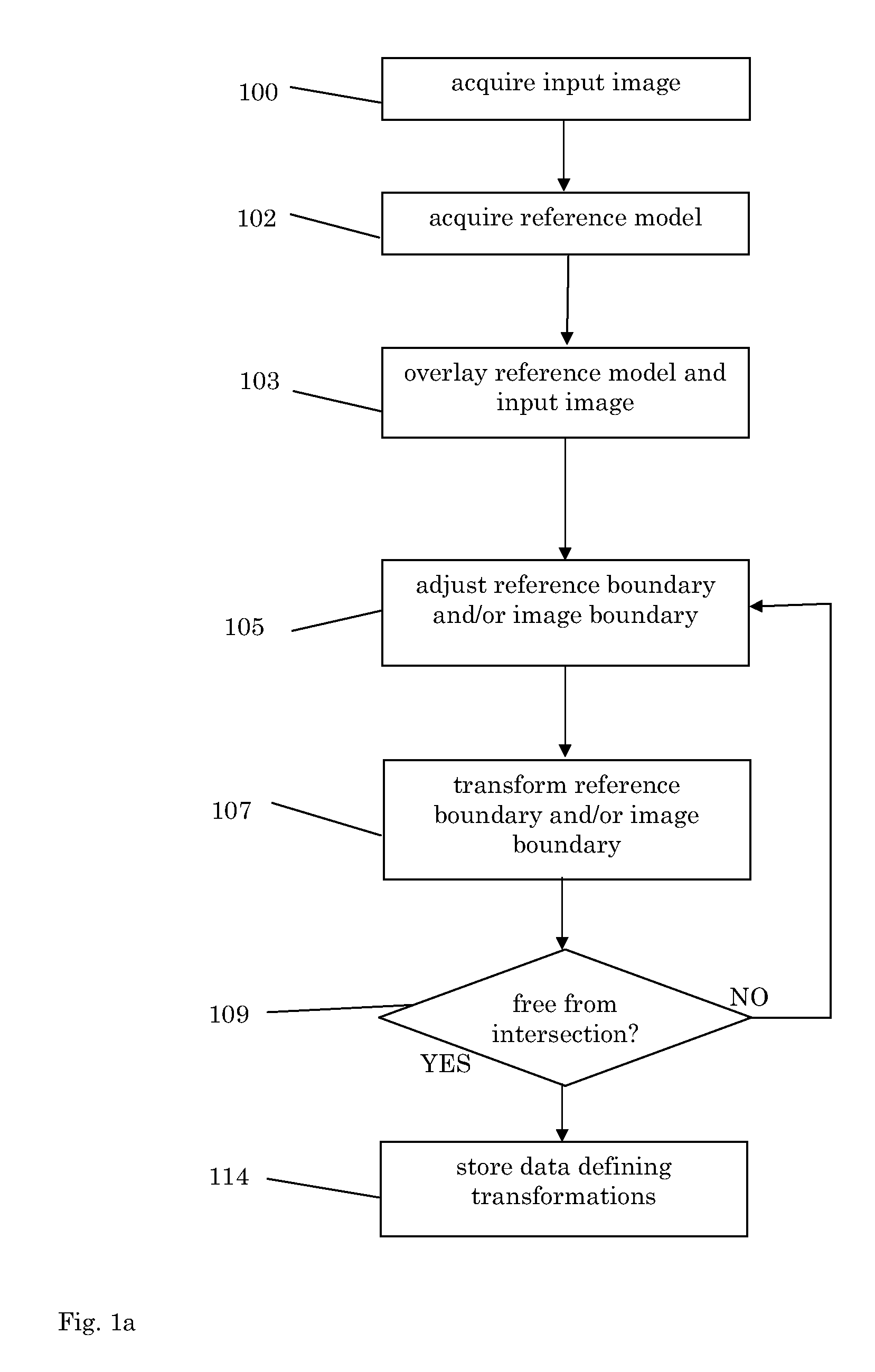
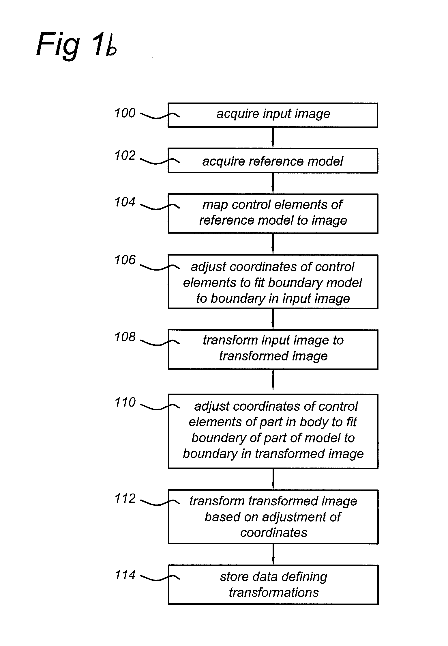
Method of and arrangement for linking image coordinates to coordinates of reference model
a reference model and coordinate system technology, applied in image analysis, instruments, computing, etc., can solve the problems of inability to achieve a sufficiently detailed reconstruction of the lungs and thorax, no standardized heart geometries, and individual models that incorporate the realistic geometry of all organs may take weeks or even months to crea
- Summary
- Abstract
- Description
- Claims
- Application Information
AI Technical Summary
Benefits of technology
Problems solved by technology
Method used
Image

Examples
Embodiment Construction
[0070]Before describing the invention in more detail a definition of terms will be given:
[0071]inverse computation: any technique to estimate electrical properties of an internal organ such as the heart or brain from surface recordings using volume conduction models;
[0072]mesh: any set of points and their connections used to describe either a surface or a volume in 3D;
[0073]imaging modality: a technique to measure internal structure like MRI, CT or echo;
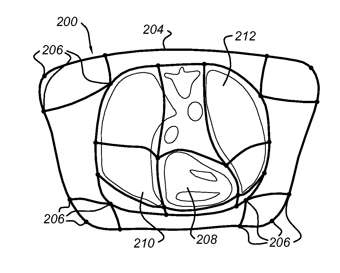
[0074]structural element: part of a patient or associated part of reference model in patient coordinates and / or abstract space coordinates. The largest structural element corresponds to the physical structure, i.e. entire body. A structural element can be subdivided in smaller structural elements (e.g. rectangular blocks);
[0075]transformation: (possibly non-linear) mapping of relative coordinates to patient coordinates or deformation of image defined in a coordinate system;
[0076]patient coordinates: coordinate system that was used to...
PUM
 Abstract
Description
Claims
Application Information