
Visualization of the coronary artery tree

a visualization and coronary artery technology, applied in the field of coronary artery reconstruction and examination apparatus, can solve problems such as providing inadequate information to users

Active Publication Date: 2012-01-26
KONINKLIJKE PHILIPS ELECTRONICS NV
View PDF13 Cites 4 Cited by
  • Summary
  • Abstract
  • Description
  • Claims
  • Application Information

AI Technical Summary

Benefits of technology

[0028]The examination apparatus provides the possibility of supplying a user with improved information about the current coronary artery situation, for example, of a patient. As the examination apparatus, according the invention, is capable of visualising a combined coronary tree volume data set image, the assessment of the coronary artery geometry is facilitated. This allows a quick and reliable diagnosis of the coronary situation. The combined coronary tree volume data set image, e.g. the model image, shows both sides of the artery tree at the same time of the cardiac cycle. In other words, as an effect of the invention, the artery branches shown on the generated image have the same relation as the investigated artery trees in reality.
[0029]Further exemplary embodiments of the apparatus are set forth in the dependent claims. The aspects and advantages discussed in relation to the exemplary embodiments of the method described above also apply to the exemplary embodiments of the apparatus, according to the invention, and vice versa.
[0030]The invention also provides an X-ray imaging system with an examination apparatus, according to the above mentioned embodiment, and according to the embodiments defined in the respective sub claims.
[0031]Thus, a system can be provided that supplies the user with improved information about the patient and that can also be used for other examination aspects related to the X-ray imaging system.
[0032]According to another exemplary embodiment of the present invention, a computer readable medium is provided, in which a computer program for reconstruction of the coronary arteries is stored which, when being executed by a processor, causes the processor to carry out the above mentioned method steps.
[0033]Furthermore, according to another exemplary embodiment of the present invention, a computer program element for reconstruction of the coronary arteries is provided which, when being executed by a processor, causes the processor to carry out the above mentioned method steps.

Problems solved by technology

But it has shown that this provides inadequate information to the user, i.e., for example, a surgeon or cardiologist.

Method used


Image

  • Visualization of the coronary artery tree
  • Visualization of the coronary artery tree
  • Visualization of the coronary artery tree

Examples


Embodiment Construction

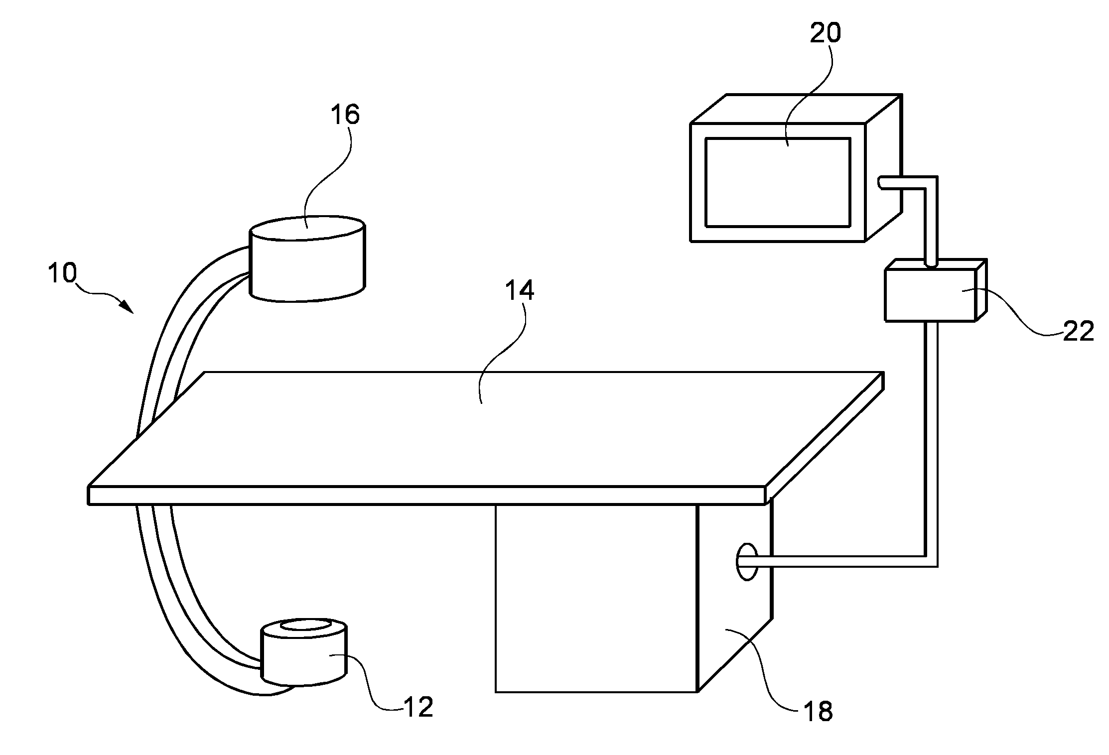
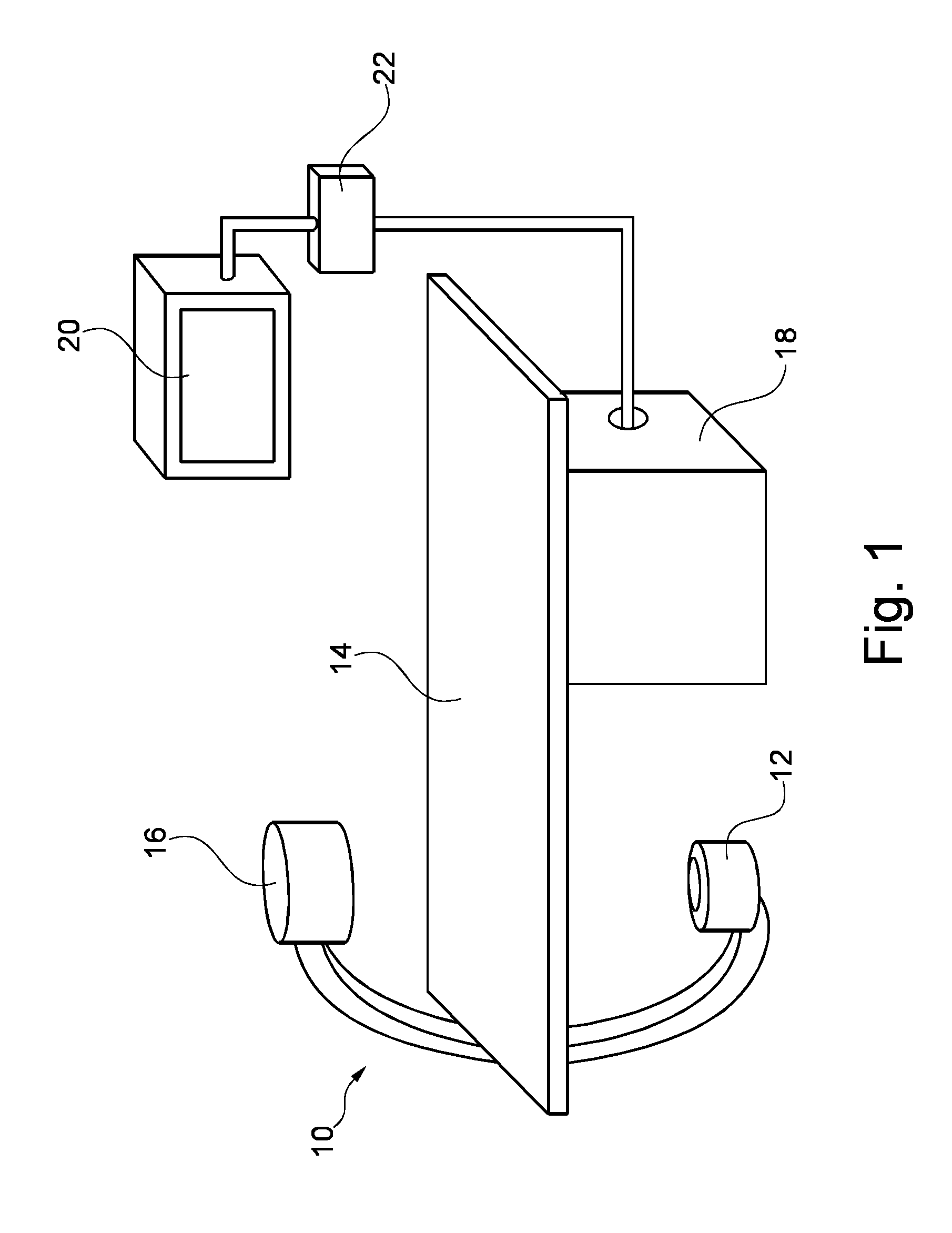
[0047]FIG. 1 schematically shows an X-ray imaging system 10 with an examination apparatus for reconstruction of the coronary arteries. The examination apparatus comprises an X-ray image acquisition device with a source of X-ray radiation 12 provided to generate X-ray radiation. A table 14 is provided to receive a subject to be examined. Further, an X-ray image detection module 16 is located opposite the source of X-ray radiation 12, i.e. during the radiation procedure the subject is located between the source of X-ray radiation 12 and the detection module 16. The latter is sending data to a data processing unit or calculation unit 18, which is connected to both the detection module 16 and the radiation source 12. The calculation unit 18 is located underneath the table 14 to save space within the examination room. Of course, it could also be located at a different place, such as a different room or laboratory. Furthermore, a display device 20 is arranged in the vicinity of the table ...


PUM


Abstract

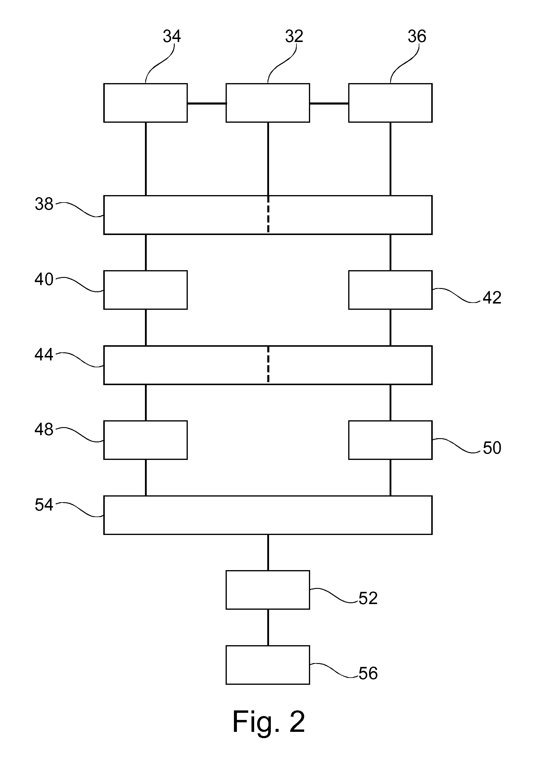
The present invention is related to a method for reconstruction of the coronary arteries and an examination apparatus for reconstruction of the coronary arteries. To provide improved coronary artery information, an apparatus and a method are provided where a gating signal is provided (32) and a first gated X-ray image sequence of one of the left or right branches of the coronary arteries is acquired (34) with injected contrast agent into the one of the left or right branches of the coronary arteries. Further, a second gated X-ray image sequence of the other branch of the coronary arteries is acquired (36) with injected contrast agent into said other branch. Then, a gated reconstructing (38) of the left and the right coronary artery is suggested and a volume data (40, 42) of the coronary arteries is generated. The volume data of the left and right coronary arteries is registered (44) in relation to time and space. Further; the registered volume data (48, 50) of the left and the right coronary arteries is combined and a combined coronary artery tree volume data set (52) is generated (54). Finally, the combined coronary tree volume data set is visualized (56).

Description

FIELD OF THE INVENTION[0001]The present invention is related to a method for reconstruction of the coronary arteries and an examination apparatus for reconstruction of the coronary arteries.BACKGROUND OF THE INVENTION[0002]During interventions, for diagnosis purposes, treatment planning or outcome control of an intervention, it is known to visualise the left and right coronary arteries partly surrounding the heart by using X-ray image generating devices of the so-called C-type. The visualisation is usually based on a reconstruction of the coronary arteries by the means of at least two acquired images. For acquisition, the left or the right branch of the coronary arteries (LCA or RCA) are selectively injected with a contrast agent. In other words, images are acquired for the left branch separately from the acquisition of the images of the right branch. Usually the acquisitions are analysed independently for a diagnosis on the whole coronary artery tree. But it has shown that this pro...

Claims


Application Information

Patent Timeline
IPC IPC(8): H05G1/10G06K9/60
CPCA61B6/504A61B6/541G06T11/006A61B6/466G06T2211/404A61B6/481A61B6/4441
Inventor HANSIS, EBERHARD SEBASTIANGRASS, MICHAEL
Owner KONINKLIJKE PHILIPS ELECTRONICS NV
Learn More