Method and apparatus for automated whole blood sample analyses from microscopy images

a whole blood sample and microscopy image technology, applied in the field of methods and apparatuses for performing analyses on whole blood sample from microscopy images, can solve the problems of laborious blood smears, cost prohibitive, time-consuming,

Active Publication Date: 2012-02-09
ABBOTT POINT CARE
View PDF4 Cites 88 Cited by
  • Summary
  • Abstract
  • Description
  • Claims
  • Application Information

AI Technical Summary

Benefits of technology

[0009]According to an aspect of the present invention, a method for identifying at least one type of white blood cell (WBC) within a whole blood sample is provided, comprising the steps of: a) adding at least one colorant to the whole blood sample, which colorant is operable to differentially identify at least one WBC type from another WBC type; b) disposing the blood sample into a chamber defined by at least one transparent panel; c) creating at least one image of the sample quiescently residing within the chamber; d) identifying WBCs within the sample image; e) quantitatively analyzing at least some of the identified WBCs within the image relative to one or more predetermined quantitatively determinable features; and f) identifying at least one type of WBC from the identified WBCs using the quantitatively determinable features.
[0010]According to another aspect of the present invention, an apparatus for analyzing a whole blood sample quiescently residing within a chamber is provided. The apparatus includes an objective lens, a sample illuminator, an image dissector, and a programmable analyzer. The sample illuminator is operable to provide a fluorescent excitation light and one or more transmission lights. The image dissector is adapted to receive one or both of light fluorescing from the sample and lights transmitted through the sample, and produce signals representative of such light. The programmable analyzer is adap

Problems solved by technology

Blood smears are problematic for several reasons; e.g., the cells must be killed and fixed, which process precludes many types of supravital stains and analyses whose'results depend upon living cells, and blood smears are labor intensive, cost

Method used


Image

  • Method and apparatus for automated whole blood sample analyses from microscopy images
  • Method and apparatus for automated whole blood sample analyses from microscopy images
  • Method and apparatus for automated whole blood sample analyses from microscopy images

Examples


Embodiment Construction

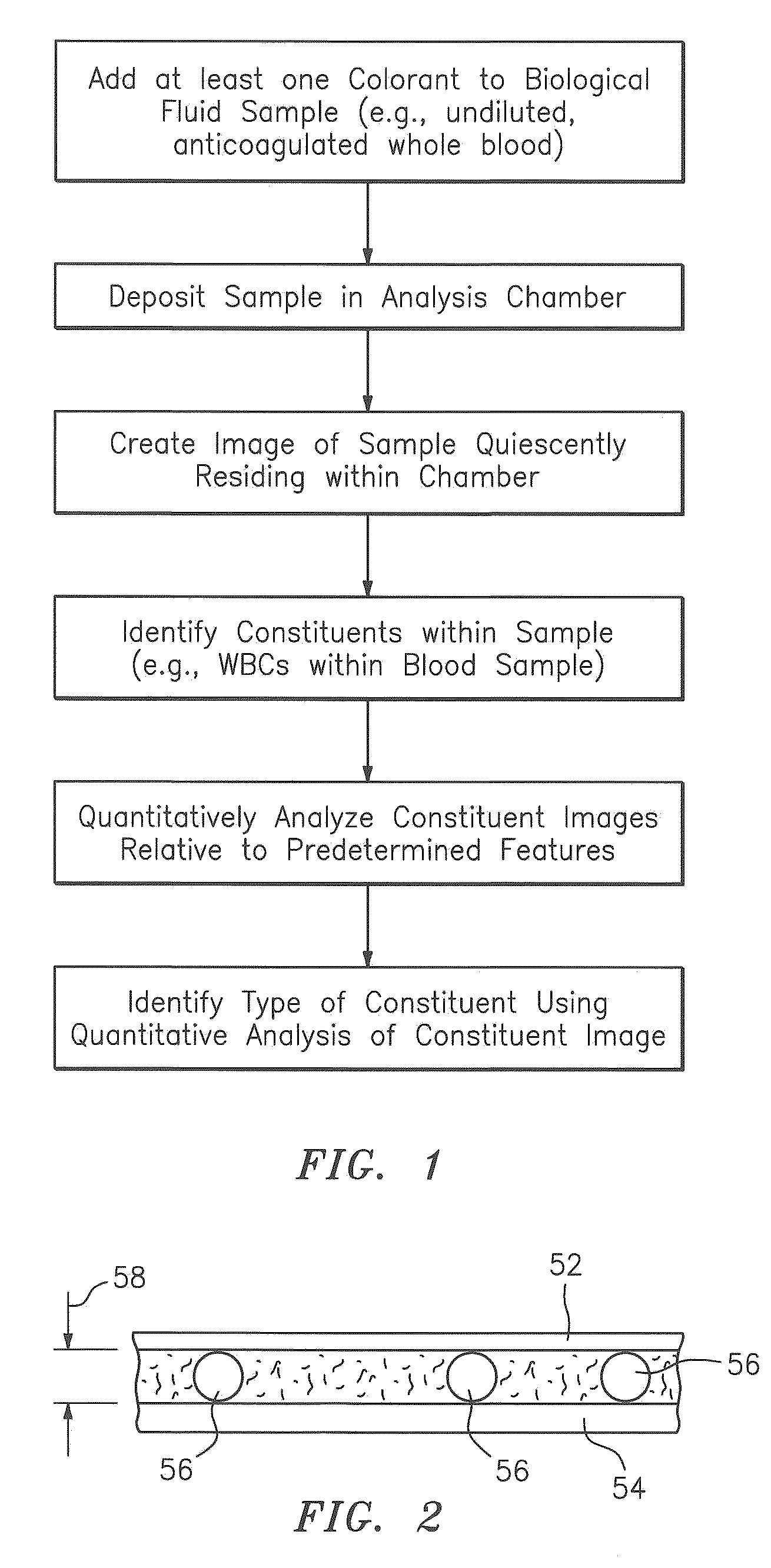
[0050]Now referring to FIG. 1, as will be described in greater detail below, the present invention includes a method and an apparatus for identifying constituents within a biological fluid sample quiescently residing within an analysis chamber. Typically, at least one colorant is added to the sample to facilitate distinguishing one constituent from another within the sample. The sample quiescently residing within the chamber is imaged, and constituents within the image of the sample are located. At least some of the constituents located within the image are analyzed to determine the presence of one or more features (e.g., to determine the extent to which a constituent possesses a particular characteristic) of each particular constituent analyzed. Each feature can be quantitatively evaluated from the image. At least one type of constituent is determined from the located constituents using the features.

[0051]The present invention has particular utility when applied to perform a leukoc...


PUM


Abstract

A method and apparatus for identifying one or more target constituents (e.g., white blood cells) within a biological sample is provided. The method includes the steps of: a) adding at least one colorant to the sample; b) disposing the sample into a chamber defined by at least one transparent panel; c) creating at least one image of the sample quiescently residing within the chamber; d) identifying target constituents within the sample image; e) quantitatively analyzing at least some of the identified target constituents within the image relative to one or more predetermined quantitatively determinable features; and f) identifying at least one type of target constituent within the identified target constituents using the quantitatively determinable features.

Description

[0001]The present application is entitled to the benefit of and incorporates by reference essential subject matter disclosed in the U.S. Provisional Patent Application Ser. No. 61 / 371,020, filed Aug. 5, 2010.BACKGROUND OF THE INVENTION[0002]1. Technical Field[0003]The present invention relates to methods and apparatus for performing analyses on whole blood samples from microscopy images in general, and to automated version of the same in particular.[0004]2. Background Information[0005]Medical diagnostics often include analyses of a whole blood sample from a patient. One of the more popular diagnostics is a complete blood count (referred to as a “CBC”), which is a suite of tests that may include, in addition to the enumeration of the cellular components, red blood cell metrics, reticulocyte counts, and a leukocyte differential count (“LDC”; sometimes referred to as a “white blood cell differential”), which is the identification and enumeration of the types of white blood cells (WBCs)...

Claims


Application Information

Patent Timeline
IPC IPC(8): C12Q1/04C12M1/34
CPCG01N15/1475G01N21/5907G01N2015/008G01N33/5094G01N21/6458G01N33/49
Inventor HERZOG, DAVIDWANG, ZHIZHOUWARDLAW, STEPHEN C.XIE, MIN
Owner ABBOTT POINT CARE
Why Eureka
Try Eureka