Markers for cancer detection

a cancer and marker technology, applied in the field of cancer detection markers, can solve the problems of poor patient prognosis, uncomfortable, expensive,

Inactive Publication Date: 2012-02-16
TECHNION RES & DEV FOUND LTD
View PDF0 Cites 40 Cited by
  • Summary
  • Abstract
  • Description
  • Claims
  • Application Information

AI Technical Summary

Benefits of technology

[0035]In currently preferred embodiments, the biological sample comprises a tissue sample. In currently preferred embodiments, the tissue sample comprises gastrointestinal tissue, particularly colorectal tissue or pre-cancerous tissue such as polyps. In currently preferred embodiments, the biological sample is selected from a tumor within the gastrointestinal tract, particularly a colorectal tumor. In particular embodiments, the gastrointestinal tissue is from an area or organ selected from the group consisting of the esophagus, the stomach, the small intestine, the large intestine (colon), the rectum, the appendix and a combination thereof.
[0036]In particular embodiments, the cancer being detected is selected from the group consisting of adrenal cancer, bladder cancer, bone cancer, brain cancer, breast cancer, cervical cancer, colorectal cancer, fallopian tube cancer, gastric cancer, head and neck cancer, hepatic cancer, lung cancer including small cell lung cancer and non-small cell lung cancer, melanoma, neuroblastoma, oral cancer, ovarian cancer, pancreatic cancer, prostate cancer, thyroid and parathyroid cancer, renal cancer, sarcoma, thymoma, hematological malignancies and germ cell tumors. In a currently preferred embodiment, the cancer is colorectal cancer. Each of the aforementioned cancers represents a separate embodiment of the invention and may be used independently from any of the others.

Problems solved by technology

However, colorectal cancer is often detected only upon manifestation of clinical symptoms, such as pain and black tarry stool.
Generally, such symptoms are present only when the disease is well established, often after metastasis has occurred, and the prognosis for the patient is poor, even after surgical resection of the cancerous tissue.
While this technique allows for identification, removal, and biopsy of potentially cancerous growths such as polyps, its use is associated with certain disadvantages, such as being expensive, uncomfortable, inherently risky due to its invasive nature, and the inability to access some portions of the large intestine and most of the small intestine.
The transit of endoscopy capsules through the small intestine is dependent on peristalsis, meaning that some areas with lesions may be missed if the capsule is not retained in that area for a sufficient amount of time.
Further, the endoscopy capsules in current use are not capable of identifying molecular markers, which may be early indicators of colorectal cancer, even prior to the development of pre-cancerous polyps.
U.S. Pat. No. 7,452,727 discloses a automatable method for identifying cancer cells and their precursor cells that is based on detecting at least two molecular markers, wherein the detection of each marker alone is not a reliable indicator of the presence of cancer cells and their precursor cells.

Method used


Image

  • Markers for cancer detection
  • Markers for cancer detection
  • Markers for cancer detection

Examples


example 1

Clinical Characteristics of Patients Undergoing Surgical Gastrointestinal Re-Sectioning

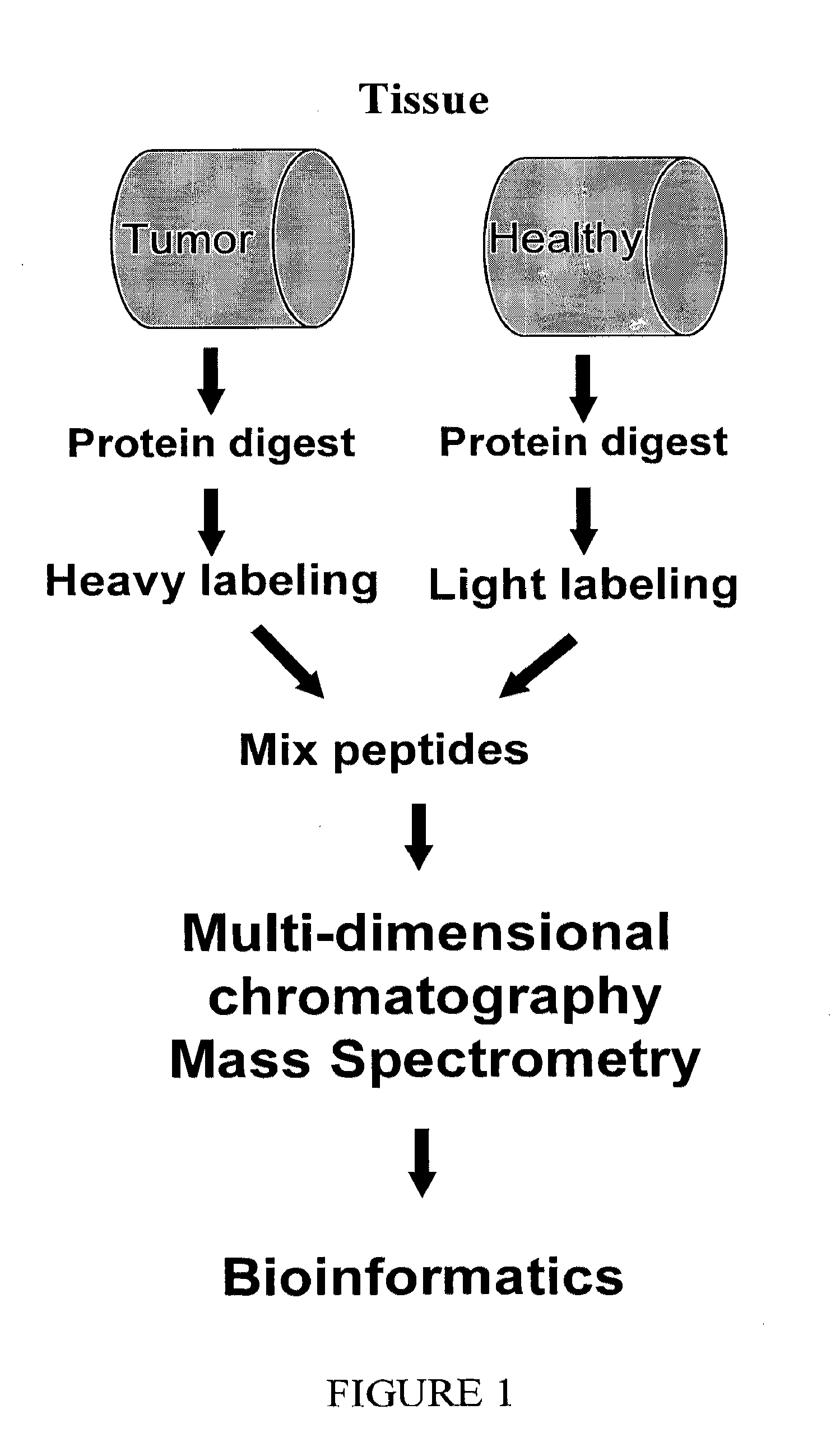
[0203]The entire protein repertoires of healthy and diseased gastrointestinal tissues were analyzed from samples obtained from greater than 50 patients undergoing surgery for colorectal cancer.

[0204]The clinical characteristics of the patients studied are provided in Table 2.

TABLE 2PatientAge atNo.DiagnosisGenderAppearanceDiagnosisTNMStageGrade20178MprimaryAdeno-T3N1M0IIIhighcarcinoma20281FprimaryMucinousT3N1IIIlowadeno-carcinoma20385MprimaryAdeno-T3N0M0IIhighcarcinoma20571FprimaryAdeno-T3N1M0IIIhighcarcinoma20781FprimaryAdeno-T3N2IIIhighcarcinoma20878MprimaryAdeno-T3N2M1IVhighcarcinoma20971MprimaryAdeno-T3N0M1IIhighcarcinoma21157FprimaryAdeno-T2N0M0Iintermedcarcinoma21250FprimaryAdeno-T3N1M0IIIhighcarcinoma21454FprimaryAdeno-T3N1M0IIIhighcarcinoma21777FLocalAdeno-T3N0M0IIhighrecurrancecarcinoma21879MprimaryAdeno-T3N2M1IVlow(T + P + N)carcinoma21960MprimaryAdeno-T3N1M0IIIhighcarcinoma22073Mprimary...

example 2

Identification of a Group of Protein Markers that are Found in Polyps and in Advanced Stages of Colorectal Cancer

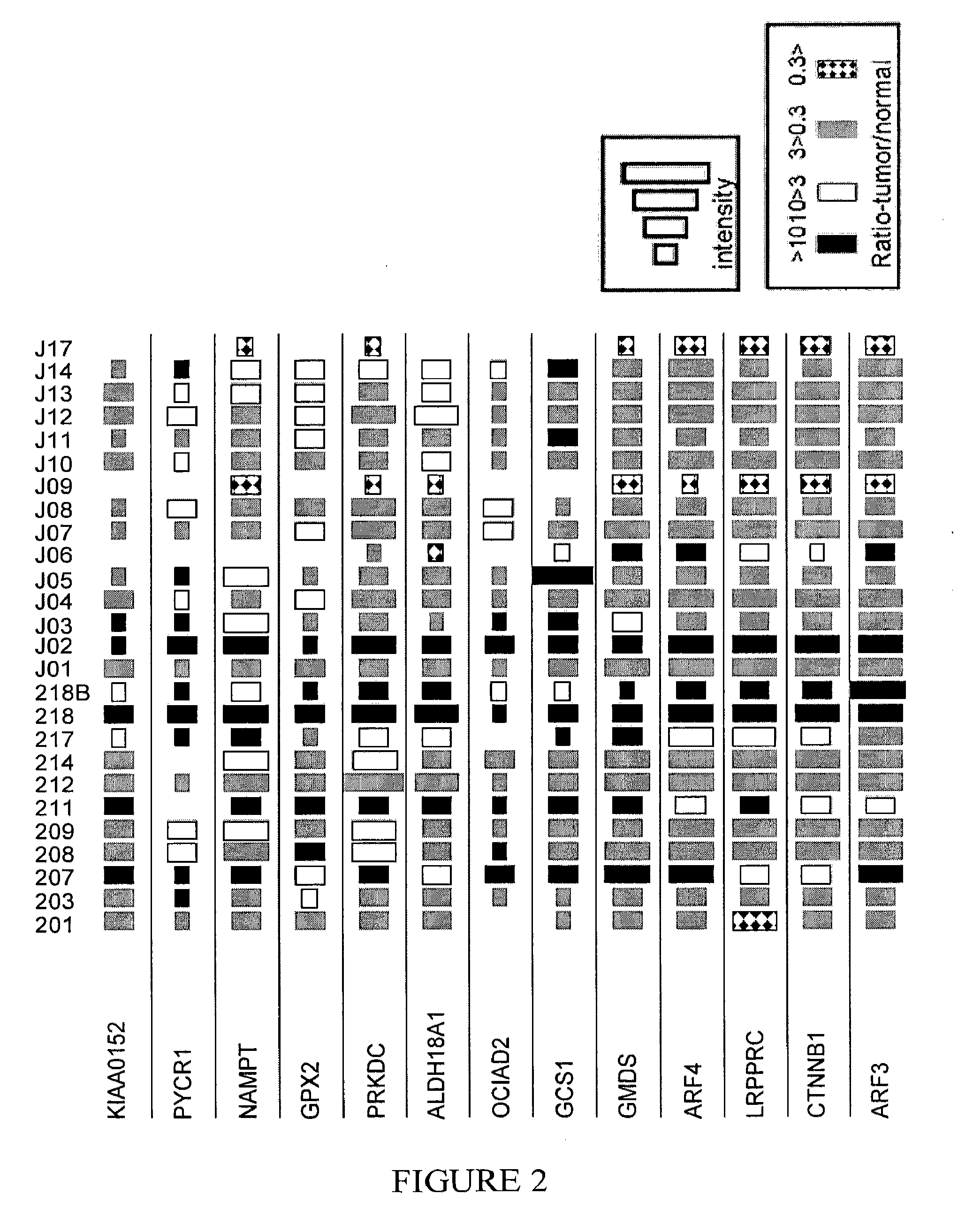
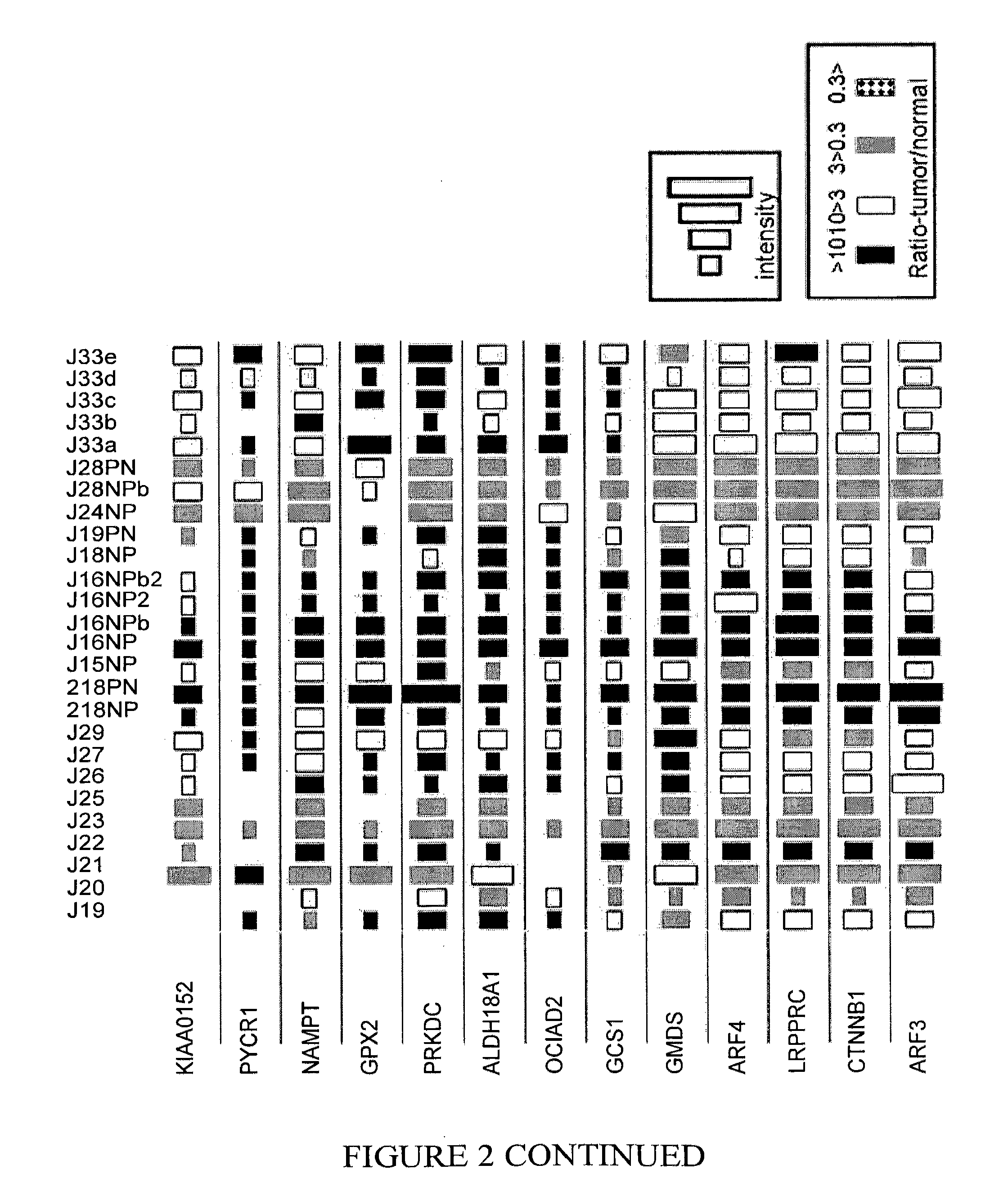
[0205]Table 3 lists proteins that were observed to be highly expressed in polyps, as well as in early and advanced stages of various colorectal cancers. As shown in Table 3 and in FIG. 2, in a large number of patients, the expression level of these proteins in polyps or cancerous tissue was at least 3 times greater than the expression level in healthy tissue from the same patient. Similarly, few if any, of the patients exhibited decreased expression levels of the same proteins in polyps or cancerous tissue as compared to the corresponding level in healthy tissue from the same patient. That is, a ratio of protein expression (polyps / tumor vs healthy tissue from the same patient), that was less than 1:3, was rarely observed. Accordingly, a diagnostic array of reagents directed to detection of at least some of the proteins in this group may be used as a general screening test...

example 3

Identification of a Group of Protein Markers that are More Highly Expressed in Polyps than in Advanced Stages of Colorectal Cancer

[0206]Table 4 lists proteins that were observed to be highly expressed in polyps, whereas in more advanced stages of colorectal cancer these proteins tended to have decreased levels of expression. Accordingly, a diagnostic array of reagents directed to detection of at least some of the proteins in this group may be used as a screening test for very early detection of colorectal cancer. Such a screening test could identify susceptible at-risk individuals, even prior to the stage at which polyp visualization is possible by endoscopic techniques.

TABLE 4Protein(IPI Acc. No.)nBigTumornSmallTumornBigPolypnSmallPolypCPT2 Carnitine O-65120palmitoyltransferase 2,mitochondrial precursor(IPI00012912)ARL1 ADP-ribosylation factor-61120like protein 1(IPI00219518)PFKL Isoform 1 of 6-62110phosphofructokinase, liver type(IPI00332371)GOT2 Aspartate63110aminotransferase, mi...


PUM

PropertyMeasurementUnit
nucleic acid hybridization assayaaaaaaaaaa
fluorescentaaaaaaaaaa

Abstract

The present invention relates to methods for detecting, prognosing and staging cancers, in particular cancers of the gastrointestinal tract. The methods of the invention comprise detecting specific protein markers in a tissue of interest, wherein the detected levels thereof may be indicative of pre-cancerous or cancerous tissue, or the stage or prognosis of a cancer. Further provided are methods of treating cancer, and cancer detection kits.

Description

FIELD OF THE INVENTION[0001]The present invention relates to methods for detecting, prognosing and staging cancers, in particular cancers of the gastrointestinal tract. The methods of the invention comprise detecting specific protein markers in a tissue of interest, wherein the detected levels thereof may be indicative of pre-cancerous or cancerous tissue, or the stage or prognosis of a cancer. Further provided are methods of using the markers for treating cancer, and cancer detection kits.BACKGROUND OF THE INVENTION[0002]Colorectal cancer, also referred to as colon cancer or large bowel cancer, is a malignant neoplastic disease associated with tumors in the colon, rectum and appendix. With 655,000 deaths worldwide per year, it is the third most common form of cancer and the second leading cause of cancer-related death in the Western world.[0003]Colorectal cancers originate in the colorectal epithelium and typically are not extensively vascularized (and therefore not invasive) durin...

Claims


Application Information

Patent Timeline
Patent Type & Authority Applications(United States)
IPC IPC(8): G01N33/574C40B30/06C12Q1/68C12Q1/02A61K49/00
CPCA61K39/0011G01N33/57419G01N2800/56
Inventor ADMON, ARIEBARNEA, EILONBEER, ILANZIV, TAMAR
Owner TECHNION RES & DEV FOUND LTD
Why Eureka
Try Eureka