Apparatus, method and medium storing program for reconstructing intra-tubular-structure image

a technology of intratubular structure and image, applied in image enhancement, tomography, instruments, etc., can solve the problems of difficult to recognize the correspondence between the position of the blood vessel in real three-dimensional space, and it is not easy to adopt the method. , to achieve the effect of intuitively and easily recognized

Inactive Publication Date: 2012-04-05
FUJIFILM CORP
View PDF3 Cites 109 Cited by
  • Summary
  • Abstract
  • Description
  • Claims
  • Application Information

AI Technical Summary

Benefits of technology

[0011]In view of the foregoing circumstances, it is an object of the present invention to provide an apparatus, a method and a program for reconstructing an image of the inside of a tubular structure that can make it possible to easily and intuitionally recognize, based on the morphology of the tubular structure in real space, a three-dimensional tomographic image of the inside of a tubular structure represented in a coordinate system along a center line of the tubular structure.

Problems solved by technology

Therefore, it has been difficult to recognize a correspondence between the position of the blood vessel in the 3D-IVUS image represented in a coordinate system along the center line of the blood vessel and the position of the blood vessel in real three-dimensional space.
Further, since the method disclosed in Patent Document 1 requires hardware, such as an MPS sensor and an system for analyzing signal values of the MPS sensor, it is not easy to adopt the method disclosed in Patent Document 1.

Method used


Image

  • Apparatus, method and medium storing program for reconstructing intra-tubular-structure image
  • Apparatus, method and medium storing program for reconstructing intra-tubular-structure image
  • Apparatus, method and medium storing program for reconstructing intra-tubular-structure image

Examples


Embodiment Construction

[0065]Hereinafter, embodiments of an apparatus, a method and a program for reconstructing an image of the inside of a tubular structure according to the present invention will be described in detail with reference to drawings.

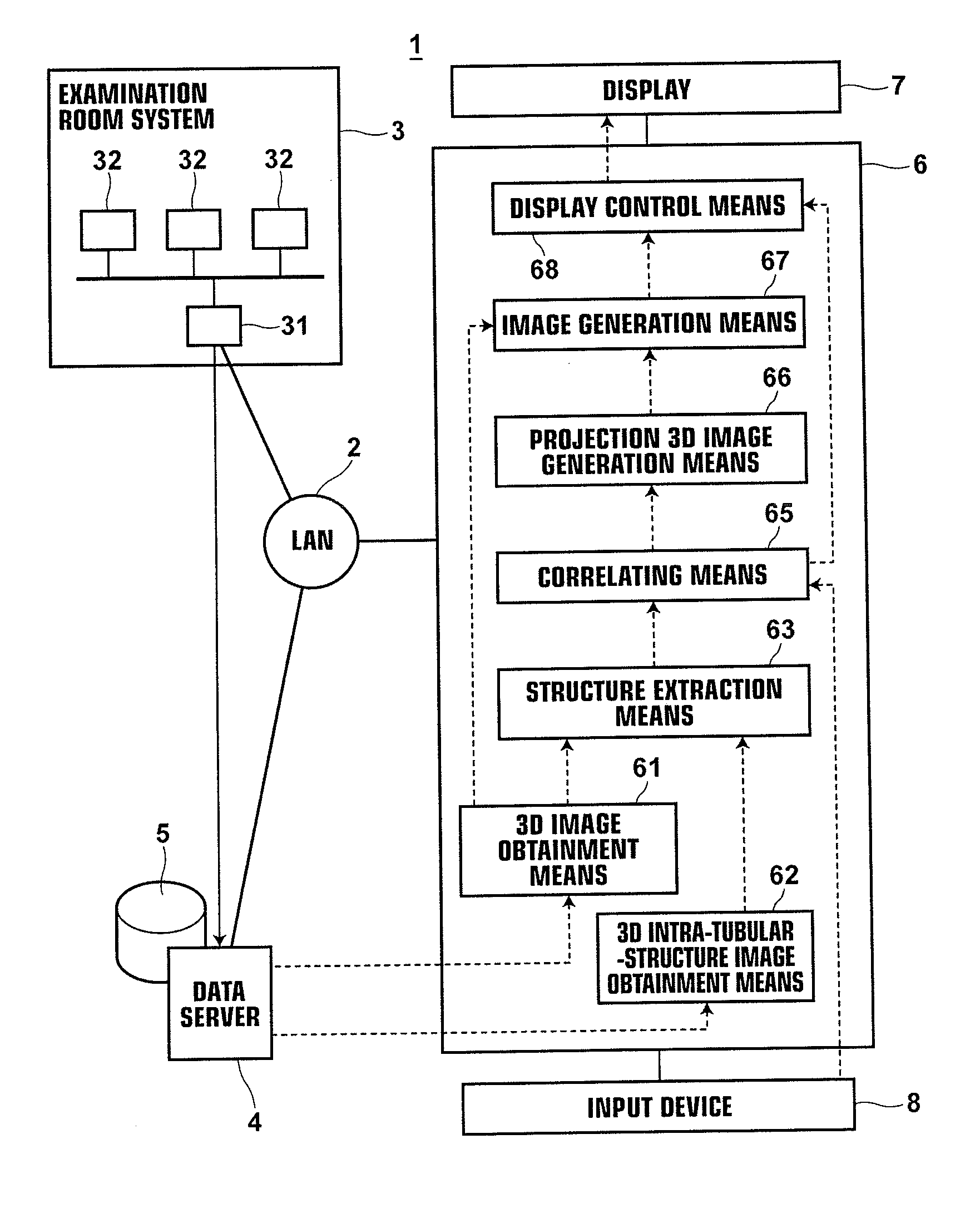
[0066]FIG. 1 is a schematic diagram illustrating the configuration of a hospital system 1 including an apparatus 6 for reconstructing an image of the inside of a tubular structure (an intra-tubular-structure image reconstruction apparatus) according to an embodiment of the present invention. The hospital system 1 includes an examination room system 3, a data server 4 and a workstation (WS) 6 for diagnosis, which are connected to each other through a local area network (LAN) 2.

[0067]The examination room system 3 includes various kinds of modality 32 for imaging a patient to be examined and an examination room workstation (WS) 31 for checking and adjusting images output from each of the modalities 32. An IVUS apparatus and a CT (Computed Tomography) apparatus, wh...


PUM


Abstract

A tubular structure, such as a blood vessel, of a subject is extracted from each of a three-dimensional image representing the tubular structure and a three-dimensional intra-tubular-structure image that has been generated from plural tomographic images of the tubular structure obtained by performing tomography on the tubular structure plural times from the inside of the tubular structure along a path in the tubular structure. Further, an arbitrary range in one of the tubular structure extracted from the three-dimensional image and the tubular structure extracted from the three-dimensional intra-tubular-structure image is correlated with a corresponding range in the other one of the tubular structures. Further, a projection three-dimensional image is generated by projecting an image of a specific structure included in the range in the three-dimensional intra-tubular-structure image into the correlated range in the three-dimensional image.

Description

BACKGROUND OF THE INVENTION[0001]1. Field of the Invention[0002]The present invention relates to an apparatus for reconstructing an image of the inside of a tubular structure derived from IntraVascular UltraSound (IVUS) diagnosis, optical coherence tomography (OCT), or the like. Further, the present invention relates to a method and a program for reconstructing an image of the inside of the tubular structure, and a medium storing the program.[0003]2. Description of the Related Art[0004]In recent years, use of a two-dimensional tomographic image of a tubular structure, such as a blood vessel, in image-based diagnosis of the tubular structure was known. The two-dimensional tomographic image is generated based on image signals of the tubular structure obtained by scanning the inside of the tubular structure while rotating a probe attached to a leading end of a catheter in the tubular structure. IntraVascular UltraSound (IVUS) diagnosis and optical coherence tomography (OCT) technique, ...

Claims


Application Information

Patent Timeline
Patent Type & Authority Applications(United States)
IPC IPC(8): A61B8/14
CPCG06T7/0028G06T2207/10081G06T2207/10088G06T2207/30101G06T2207/10136G06T2207/30048G06T2207/10101G06T7/33
Inventor KITAMURA, YOSHIRO
Owner FUJIFILM CORP
Why Eureka
Try Eureka