Erg monoclonal antibodies

a monoclonal antibody and antibody technology, applied in the field of erg monoclonal antibodies, can solve the problems of difficult pathology in diagnosing prostate cancer, limited clinical diagnosis of molecular markers, and no antibody for detecting erg in clinical specimens

Active Publication Date: 2012-05-31
THE HENRY M JACKSON FOUND FOR THE ADVANCEMENT OF MILITARY MEDICINE INC
View PDF1 Cites 5 Cited by
  • Summary
  • Abstract
  • Description
  • Claims
  • Application Information

AI Technical Summary

Problems solved by technology

If some of these features are not apparent, it can be difficult for pathologists to diagnose prostate cancer, especially in the case of prostate needle biopsies with very limited tumor content (Mostofi et al., 1992; Mostofi et al., 1993).
However, these molecular markers have marked limitations in routine diagnosis.
Although, the ERG proto-oncogene was initially characterized more than twenty years ago (Rao et al., 1987a; Rao et al., 1987b; Reddy et al., 1987), currently there is no available antibody for detecting ERG in clinical specimens.
The high degree of homology between members of this family presents a significant obstacle for raising an antibody against a specific member of this protein family.

Method used


Image

  • Erg monoclonal antibodies
  • Erg monoclonal antibodies
  • Erg monoclonal antibodies

Examples


example 1

9FY Antibody Generation

[0104]The hybridoma clone 9FY was obtained by immunizing Balb / C mice with a chemically synthesized polypeptide having amino acids 42-66 of SEQ ID NO:1 and a cysteine residue added at either the N- or C-terminus to keep the —COOH free (unconjugated). The immunizing polypeptide was conjugated to keyhole limpet hemocyanin (KLH) and injected into the mice with an adjuvant at three separate injection sites at three week intervals. The first injection used Freund's complete as the adjuvant. The second and third injection used Freund's incomplete adjuvant. Serum bleeds were assessed for binding to the immunizing polypeptide using a direct ELISA screen. Mice with the highest titer were chosen for the first hybridoma fusion step. Eight positives clones were confirmed by ELISA and supernatants were analyzed by immunoblot assays using ERG3 protein heterologously expressed in HEK-293 cells. Two of the clones were found to be positive in the immunoblot assay. One of the ne...

example 2

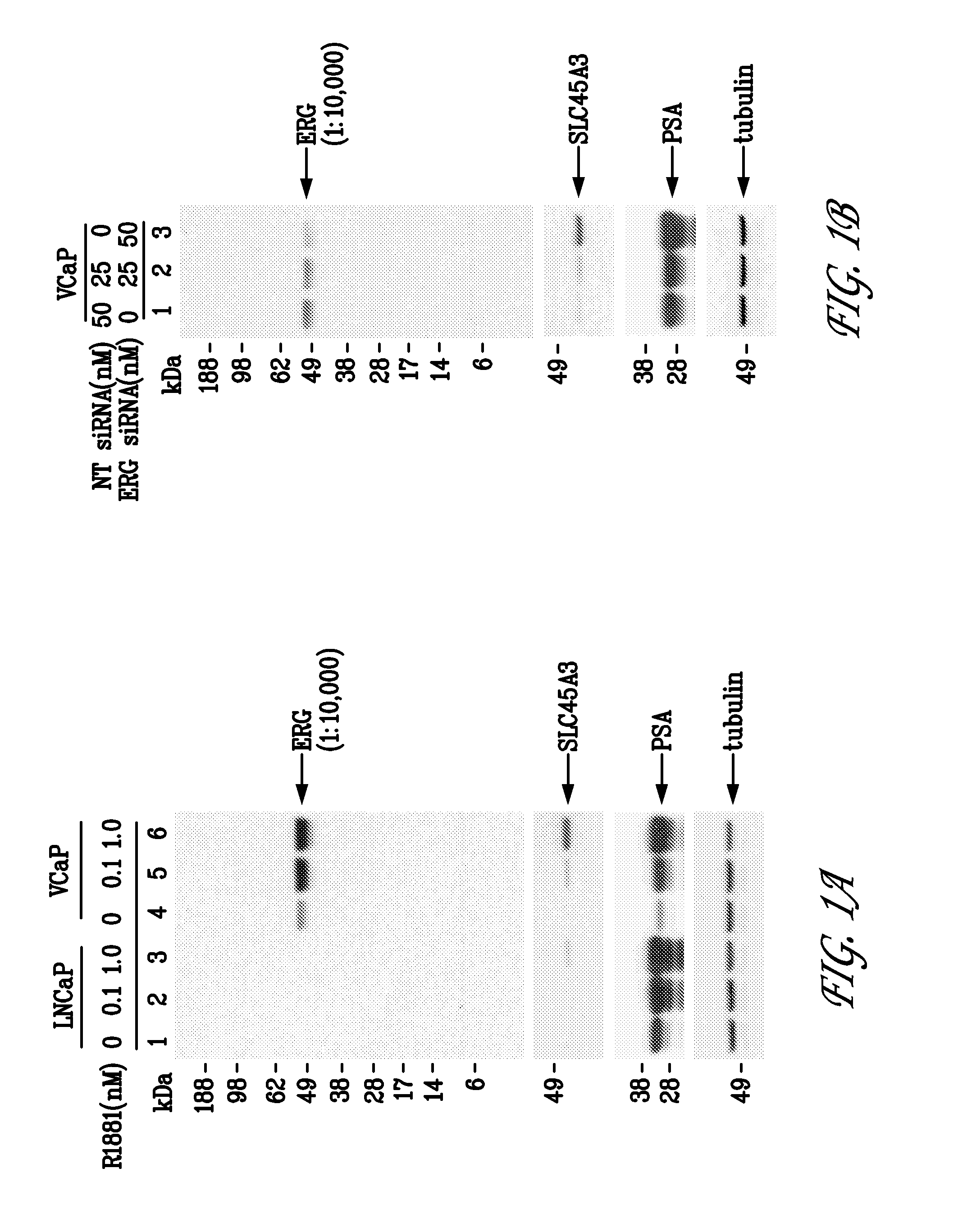
9FY Antibody Specifically Recognizes ERG Protein in Prostate Cancer Cells

[0105]VCaP cells (Korenchuck et al., 2001, In Vivo, 15:163-68) are a human prostate cancer cell line that over express a TMPRSS2 / ERG fusion frequently detected in human prostate tumors. In VCaP cells, endogenous ERG gene transcription is controlled by the androgen inducible TMPRSS2 promoter. LNCaP cells are a human prostate cancer cell line that do not harbor a TMPRSS2 / ERG fusion and do not express detectable levels of ERG.

[0106]1. Western Blots

[0107]LNCaP (ATCC #CRL-1740) and VCaP cells (ATCC, #CRL-2876) were grown in RPMI-1640 (ATCC; #30-2001) and DMEM medium (ATCC; #30-2002), respectively, supplemented with 10% fetal bovine serum (ATCC; #30-2020) and 2 mM glutamine. LNCaP and VCaP cells (2×10⁶) were seeded onto 10 cm dishes and maintained for five days and three days, respectively, in media with 10% charcoal-stripped fetal bovine serum (c-FBS; #100119 Gemini Bio-Products, Calabasas, Calif.). For androgen ind...

example 3

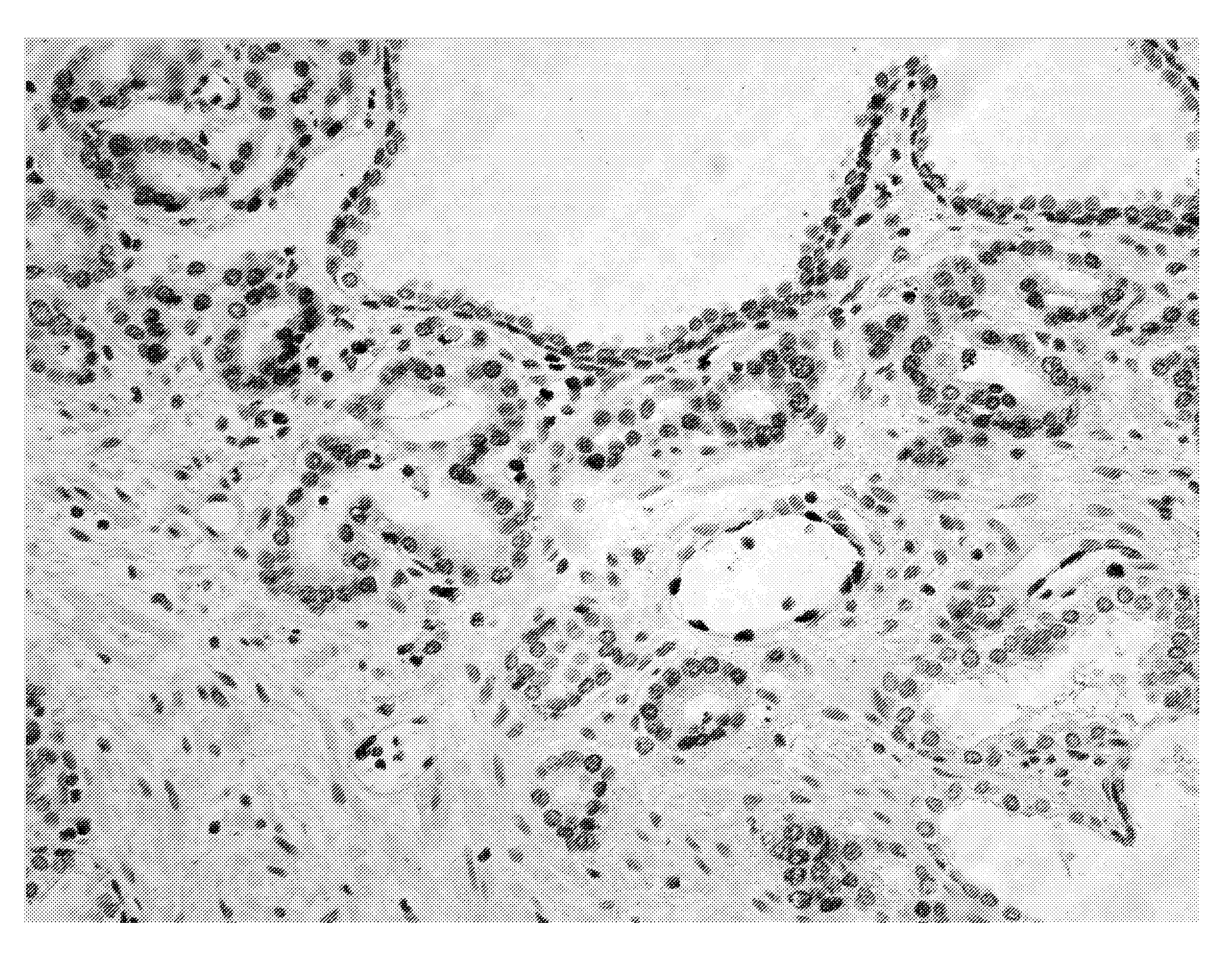
Immunohistochemical Staining in FFPE Specimens

[0122]The 9FY antibody was also tested in formalin-fixed paraffin embedded (FFPE) human prostate specimens. Radical prostatectomy specimens were fixed in formalin and embedded as whole mounts in paraffin. Each prostate was sectioned at 0.22 cm intervals in a transverse plane perpendicular to the long axis of the posterior surface of the prostate and completely embedded as whole mounts. The sections were analyzed for immunohistochemistry (IHC) staining on four-micron sections of the whole-mounted blocks.

[0123]The tissue sections for IHC were prepared as described previously (Furusato et al., 2007). Slides were incubated with 1:1200 9FY antibody. VECTOR® VIP (Vector Laboratories, Burlingame, Calif.) (purple) was used as chromogen substrate and the slides were counterstained with hematoxylin. IHC revealed nuclear staining of prostate tumor cells that is consistent with the sub-cellular localization of the ERG transcription factor (FIGS. 5A-...


PUM

PropertyMeasurementUnit
dissociation constantaaaaaaaaaa
temperatureaaaaaaaaaa
concentrationaaaaaaaaaa

Abstract

Monoclonal antibodies, or antigen-binding fragments thereof, that bind to ERG, and more specifically, to an epitope formed by amino acids 42-66 of ERG3 are disclosed. The monoclonal antibodies can be non-human antibodies (e.g., rabbit or mouse) or humanized monoclonal antibodies having the CDR regions derived from those non-human antibodies. In other embodiments, the monoclonal antibodies are chimeric, having the light and heavy chain variable regions of a non-human ERG anti-body. Methods of using the antibodies to detect ERG, or fusion proteins comprising all or part of an ERG polypeptide, such as an ERG polypeptide encoded by a TMPRSS2 / ERG, SLC45A3 / ERG, or NDRG1 / ERG fusion transcript, are also provided, including methods of detecting ERG or ERG fusion events in a clinical setting. The antibodies can also be used to inhibit the activity of ERG or fusion proteins comprising all or part of an ERG polypeptide, such as an ERG polypeptide encoded by a TMPRSS2 / ERG, SLC45A3 / ERG, or NDRG1 / ERG fusion transcript and to treat malignancies associated with overexpression of ERG or an ERG fusion event, such as prostate cancer, Ewing's sarcoma, acute myeloid leukemia, acute T-lymphoblastic leukemia, endothelial cancer, and colon cancer.

Description

CROSS REFERENCE TO RELATED APPLICATIONS[0001]This application claims the benefit of, and relies on the filing date of, U.S. provisional patent application No. 61 / 173,834, filed 29 Apr. 2009, the entire disclosure of which is incorporated herein by reference.STATEMENT OF GOVERNMENT INTEREST[0002]The present invention was made partially with U.S. Government support from research funded by contract number HU0001-04-C-1502 from the Uniformed Services University of the Health Sciences and grant number W81XWH-08-1-0532 from the U.S. Army Medical Research Acquisition Activity. The U.S. Government has certain rights in the invention.BACKGROUND[0003]The ETS-related gene (ERG) proto-oncogene is overexpressed in 60-70% of prostate tumors as a result of recurring gene fusions involving TMPRSS2 and the ETS family of genes (Petrovics et al., 2005; Tomlins et al., 2005; reviewed in Kumar-Sinha et al., 2008). Emerging studies on human prostate cancer specimens and various experimental models unders...

Claims


Application Information

Patent Timeline
Patent Type & Authority Applications(United States)
IPC IPC(8): A61K39/00A61P35/00C07K14/47C12P21/08C07K16/32G01N33/577
CPCC07K16/32G01N33/5748C07K2317/56A61P35/00
Inventor SRIVASTAVA, SHIVTAN, SHYH-HANDOBI, ALBERT
Owner THE HENRY M JACKSON FOUND FOR THE ADVANCEMENT OF MILITARY MEDICINE INC
Why Eureka
Try Eureka