Methods and System for Image Guided Cell Ablation with Microscopic Resolution

a technology of image guided cell and microscopic resolution, which is applied in the field of abnormal cell ablation, can solve the problems of not being able to remove all cancer cells, local recurrence, and poorer outcomes, and achieve the effect of preventing excess treatment to healthy tissu

Active Publication Date: 2012-06-14
LUMICELL
View PDF6 Cites 57 Cited by
  • Summary
  • Abstract
  • Description
  • Claims
  • Application Information

AI Technical Summary

Benefits of technology

[0036]Optionally the system further contains an additional feedback algorithm after the ablation of diseased cells to post-image the treatment tissue and verify that the diseased cells have been correctly treated. In some embodiments the system contains a fluid reservoir which flushes cell area with fluid to remove debris created during ablative process. Additionally, in some embodiments the system contains a fluid reservoir which flushes cell area with fluid to cool tissue as ablative heat destroys abnormal cells, preventing excess treatment to healthy tissue.

Problems solved by technology

A major challenge of oncology surgery is removing cancer cells from the tumor bed with certainty.
Residual cancer, which refers to cancer cells left behind after the initial resection surgery, can lead to local recurrence, increased rates of metastasis, and poorer outcomes.
One of the leading causes of not being able to remove all the cancer cells in the tumor bed is the lack of an intraoperative visualization technology that can guide the surgeon to identify and remove the diseased cell.
In many cases, effective and total resection of cancers in organs is further complicated because essential adjacent structures need to be spared (for example brain surgeries or other surgeries where important nerves or blood vessels are nearby).
Although margin assessment of a frozen section can take place during surgery, time constraints normally limit this assessment to small areas of the tumor.
Therefore, this approach is prone to sampling error.
If a positive margin is identified, patients most often require a repeat surgical resection, leading to increased patient morbidity and higher healthcare costs.
However, each of these technologies lacks the resolution, sensitivity and ease of use required for rapid assessment of microscopic residual cancer within the entire tumor and does not provide means of tissue removal.
However, ablation therapy lacks cellular resolution because it is often limited by the spatial resolution provided by the guiding imaging techniques; thus, it can easily leave millions of cancer cells behind.
For example, the Gamma Knife unit used in brain surgery has a theoretical accuracy of 0.2 mm but it is limited by the imaging resolution of 2 mm and positioning and excision accuracy of the surgeon.

Method used


Image

  • Methods and System for Image Guided Cell Ablation with Microscopic Resolution
  • Methods and System for Image Guided Cell Ablation with Microscopic Resolution

Examples


example 1

Interoperative Detection and Ablation of Cancer Tissue

[0095]Currently around 50% of breast cancer patients and 35% of sarcoma patients require second tumor de-bulking surgeries because a final pathology report returns days after the initial surgery indicating that residual cancerous cells have been left within the patient. Furthermore, 25% of the final pathology reports do not detect residual cancer cells due to sampling errors fundamentally inherent in the process. Thus, most patients require subsequent medical therapy including additional radiation or chemotherapy treatment to prevent cancer recurrence and metastasis stemming from residual cancer cells. The proposed study aims to investigate an intra-operative method of tumor margin assessment and treatment to ensure negative margins are obtained during the first surgery and additional surgeries are not required.

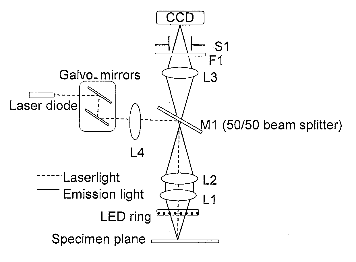
[0096]The intra-operative tumor margin assessment is performed by employing a fluorescence-based imaging system. One day...


PUM


Abstract

The invention provides systems and method for the removal of diseased cells during surgery.

Description

RELATED APPLICATIONS[0001]This application claims the priority benefit of provisional application U.S. Ser. No. 61 / 421,077 filed Dec. 8, 2011, the contents of which are herein incorporated by reference in its entirety.FIELD OF INVENTION[0002]The present invention relates generally to the ablation of abnormal cells guided by imaging of such cells during surgical procedures.BACKGROUND OF INVENTION[0003]A major challenge of oncology surgery is removing cancer cells from the tumor bed with certainty. Residual cancer, which refers to cancer cells left behind after the initial resection surgery, can lead to local recurrence, increased rates of metastasis, and poorer outcomes. Currently, there is a high rate of secondary surgeries because cancer cells are found at the margins of the resected mass during post-operative pathological analysis of the tumor. For example, 50% of breast conserving lumpectomies (Mullenix et al., Am. J. Surg., 187:643-646, 2004), 35% of limb-sparing sarcoma surgeri...

Claims


Application Information

Patent Timeline
Patent Type & Authority Applications(United States)
IPC IPC(8): A61B18/20A61B18/02A61B18/22A61B18/12A61B18/18
CPCA61B18/02A61B18/14A61B18/20A61B19/5202A61B19/5212A61B19/54A61B2018/00577A61B2018/2085A61B18/24A61B19/5244A61B2018/00029A61B34/20A61B90/30A61B90/361A61B90/39A61B2018/20351A61B2018/20359A61B2018/20361A61B18/12A61B2017/00057A61B2017/0011A61B2018/00351A61B2018/00434A61B2018/00565A61B2018/00642A61B2018/00886A61B2018/00904A61B2018/00982A61B2018/2283A61B2503/40A61B2562/028A61B2576/00A61B18/22
Inventor LEE, W. D.FERRER, JORGE M.STAMP, ANDREA
Owner LUMICELL
Why Eureka
Try Eureka