

Method for determining presence or absence of epithelial cancer-origin cell in biological sample, and molecular marker and kit therefor
a technology of epithelial cancer and biological sample, which is applied in the field of determination of the presence or absence of an epithelial cancerderived cell in a biological sample, and the use of a molecular marker and kit therefor, can solve problems such as patients' discomfor
- Summary
- Abstract
- Description
- Claims
- Application Information
AI Technical Summary
Benefits of technology
Problems solved by technology
Method used
Image

Examples
example 1
Comprehensive Methylation Analysis by Medip-Chip Method Using Cell Strains
[0097]Methylated DNA immunoprecipitation-microarray analysis (MeDIP-chip) was carried out to analyze the methylation status of breast cancer-derived cells.
[0098]Preparation Of Test Samples
(1) Methylated DNA Immunoprecipitation
[0099]Genomic DNA (4 μg) was extracted as biological samples from breast cancer-derived cell strains MCF7, MB-MDA231 and SKBR3 and a normal mammary epithelial cell strain HMEC and incubated overnight with the restriction enzyme MseI (NEB) at 37° C. to obtain fragments of 300 to 1000 bp. The biological samples after the reaction were denatured by heating them at 95° C. for 10 min to obtain a single-stranded genomic DNA.
[0100]The denatured biological samples were diluted with a dilution buffer included in Chromatin Immuoprecipitation assay kit (Upstate biotechnology) according to the instruction attached to the kit and added with Protein G Sepharose beads (GE Healthcare). The mixture was ro...
example 2
Analysis of Methylation of CpG Sites by Bisulfite Sequencing
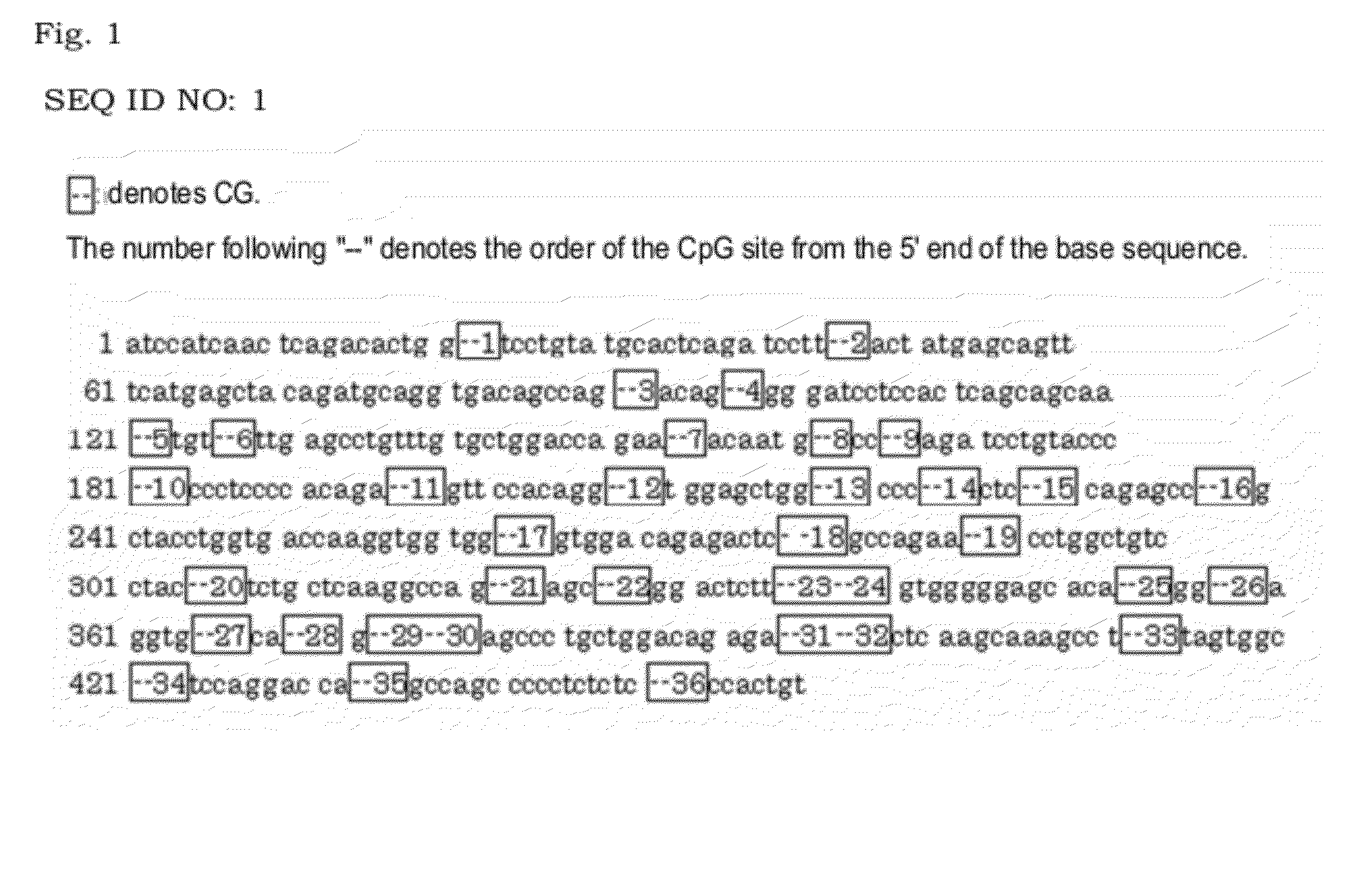
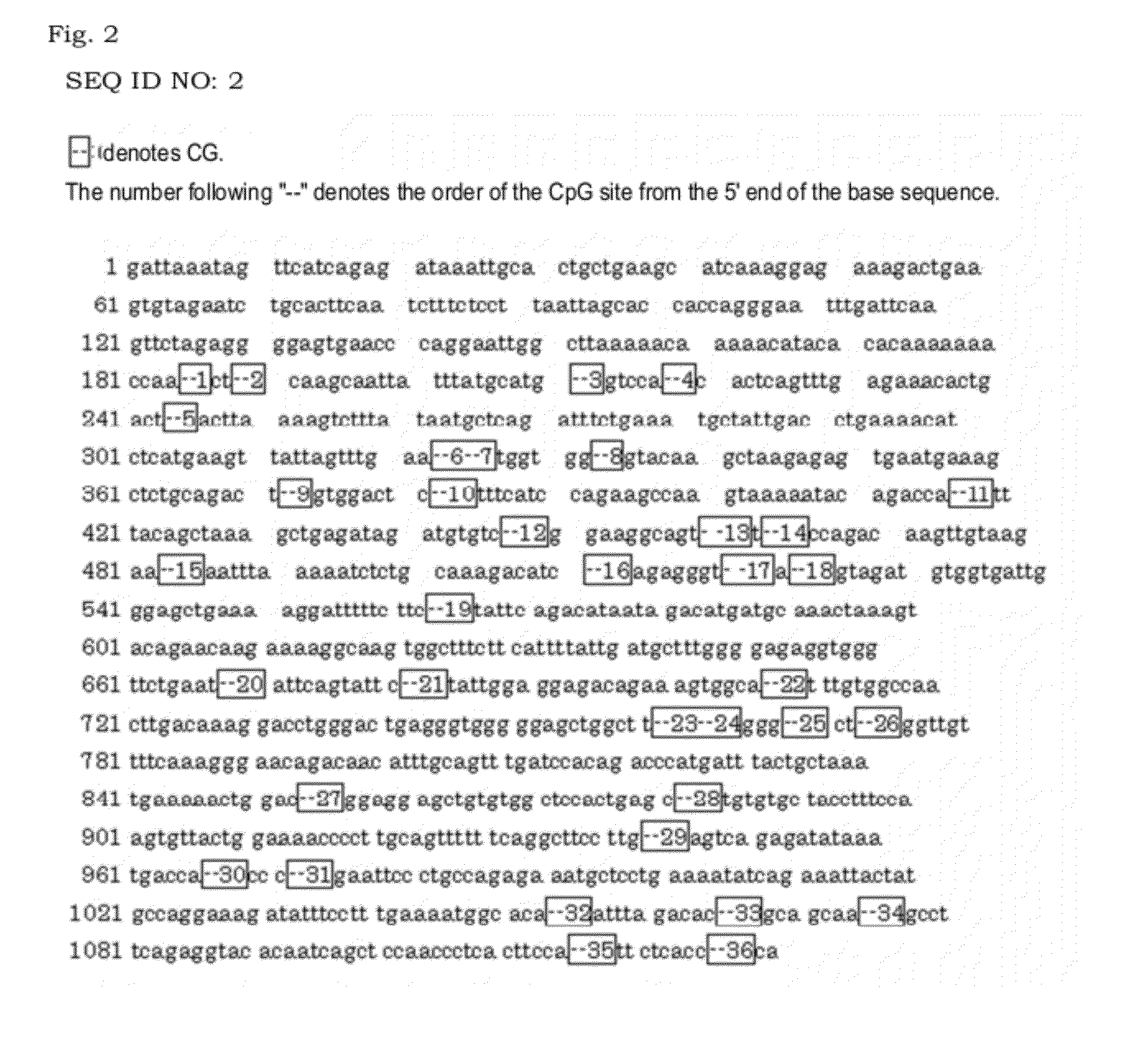
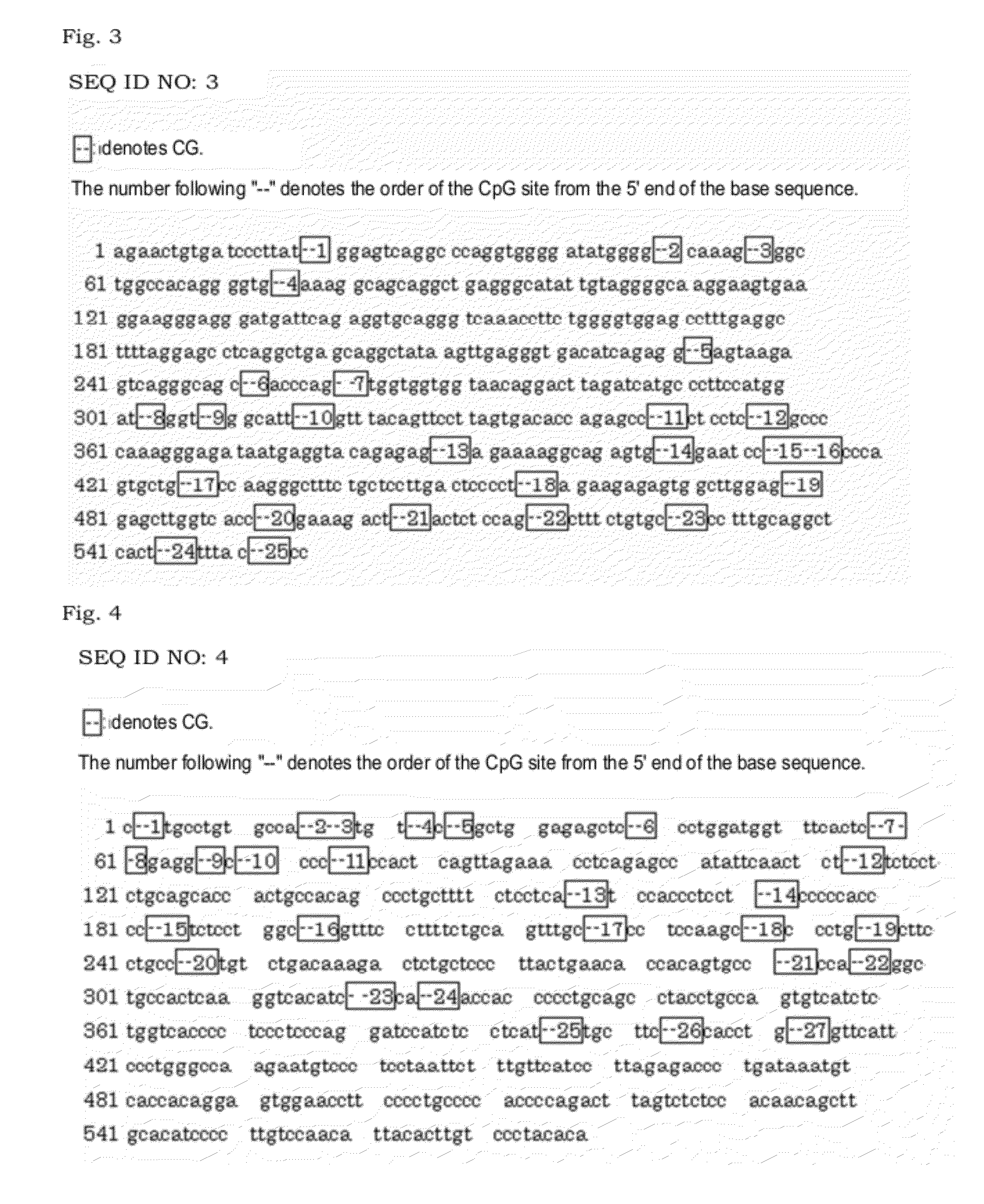
[0126]The methylation status of the CpG sites included in the base sequences of the regions identified in Example 1 was analyzed by bisulfite sequencing. The methylation frequency and the methylation ratio of each CpG site were determined based on the analysis results.
[0127]Bisulfite Treatment
[0128]In order to carry out bisulfite sequencing, analysis samples were prepared by treating DNA extracted from cell lines and tissue-derived genomic DNA with bisulfite.
[0129]Human normal mammary tissue-derived genomic DNA was obtained by mixing three different lots of human normal mammary tissue-derived genomic DNA (BioChain). Breast cancer tissue-derived genomic DNA was obtained by mixing three different lots of human breast cancer tissue-derived genomic DNA (BioChain).
[0130]Each genomic DNA (2 μg) was added with 300 μl of 0.3 M NaOH and the mixture was incubated at 37° C. for 10 min. For bisulfite treatment, 300 μl of a 10 M sodium ...
example 3
Detection of Breast Cancer-Derived Cells by Methylation Specific PCR
[0170]The methylation status of CpG sites in the base sequences SEQ ID NOs: 1, 3 and 4 in breast cancer genomic DNA was analyzed by methylation specific DNA.
[0171]Normal human mammary tissue-derived genomic DNA (BioChain) of three different lots and human breast cancer tissue-derived genomic DNA (BioChain) of three different lots were used as genomic DNAs.
[0172]The genomic DNA (2 μg) was added with 300 μl of 0.3 M NaOH and the mixture was incubated at 37° C. for 10 min. For bisulfite treatment, 300 μl of a 10 M sodium bisulfite solution was added and the mixture was incubated at 80° C. for 40 min. After the bisulfite treatment, DNA was purified from the solution using Qiaquick PCR purification kit (QIAGEN). Accordingly, the normal mammary tissue genomic samples A, B and C containing bisulfite-treated normal mammary tissue-derived genomic DNA and the breast cancer tissue samples D, E and F containing bisulfite-treate...
PUM
| Property | Measurement | Unit |
|---|---|---|
| Length | aaaaa | aaaaa |
| Length | aaaaa | aaaaa |
| Length | aaaaa | aaaaa |
Abstract
Description
Claims
Application Information