Medical image display processing method, device, and program

a medical image and display processing technology, applied in image enhancement, instruments, applications, etc., can solve the problem of placing a large and achieve the effect of reducing the burden on the physician during interpretation and easy and reliably extraction

Active Publication Date: 2012-08-02
DAI NIPPON PRINTING CO LTD
View PDF4 Cites 20 Cited by
  • Summary
  • Abstract
  • Description
  • Claims
  • Application Information

AI Technical Summary

Benefits of technology

[0032]According to the present invention, the region of cerebral parenchyma can be easily and reliably extracted from a functional image inputted by the ASL imaging method, and the resultant image can be displayed. Therefore, only the blood flow in the brain can be observed. Accordingly, a burden on a health professional such as a physician during interpretation of the image can be reduced, and useful diagnosis-assisting information can be provided to the health professional.

Problems solved by technology

However, in the PWIs and CBFs that are functional images inputted through the ASL imaging method containing cerebral surface regions that is located outside the cerebral parenchyma, not used for diagnosis of a particular brain disease, often contain high signal values corresponding to a blood flow as noise.
Therefore, a physician must correctly determine the region over which the cerebral parenchyma extends in each image, and that results in a problem in that a large burden is placed on the physician during interpretation of the images.

Method used


Image

  • Medical image display processing method, device, and program
  • Medical image display processing method, device, and program
  • Medical image display processing method, device, and program

Examples


first embodiment

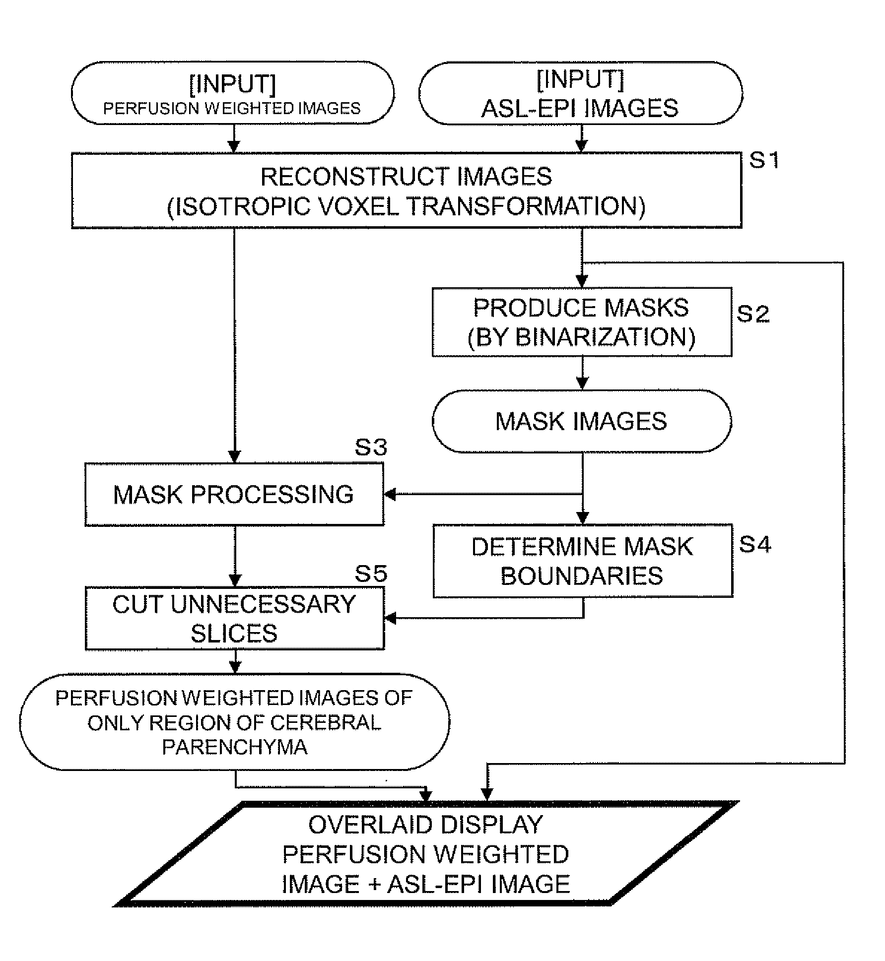
[0052]Next, basic processing procedures applied to an image display processing method of a first embodiment according to the present invention that is performed by the medical image display processing device will be described with reference to a flowchart shown in FIG. 3. In this flowchart, each ellipse represents an image or a processed product.

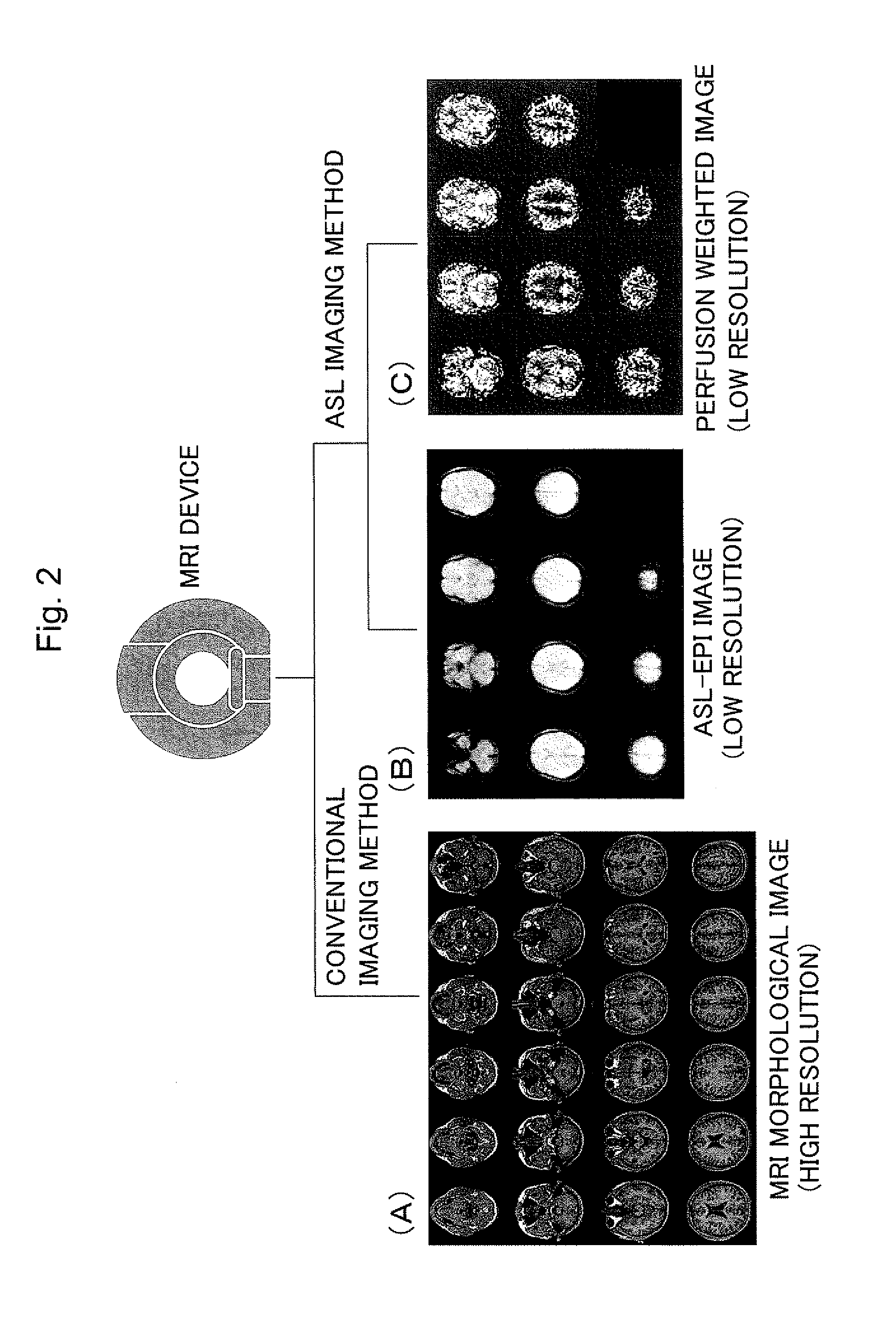
[0053]In the present embodiment, perfusion weighted images and ASL-EPI images are inputted as input images.

[0054]First, the two types of inputted images are subjected to image reconstruction (step 1). The image reconstruction is a process, in which the two types of images are re-sampled three-dimensionally and divided into voxels with equal size in all directions to make the units of the three-dimensional voxels, corresponding to pixels in two-dimensions, of the two types of images the same size.

[0055]Next, mask images are produced from the ASL-EPI images by mask production (step 2).

[0056]FIG. 4 shows a mask image (B) produced by binarizing ...

second embodiment

[0075]Next, an image display processing method of a second embodiment will be described with reference to a flowchart shown in FIG. 10.

[0076]In the present embodiment, as in the basic processing procedures, perfusion weighted images and ASL-EPI images are used as input images, and the same processing as in the above steps 1 to 4 is used to produce perfusion weighted images of only the region of the cerebral parenchyma (steps 21 to 23). However, the processing related to the determination of mask boundaries is omitted from the figure.

[0077]In the present embodiment, each of the perfusion weighted images in the subject brain coordinate system A that have been produced in step 23 is transformed into an image in the standard brain coordinate system. First, a spatial transformation field used to transform an ASL-EPI image into an image on the standard brain is estimated using spatial normalization in which the ASL-EPI image is fitted to a T1 weighted image serving as a template and store...

third embodiment

[0087]Next, an image display processing method of a third embodiment will be described with reference to a flowchart shown in FIG. 13.

[0088]In the present embodiment, perfusion weighted images and ASL-EPI images are used as input images, and the process until image reconstruction in step 31 is the same as that in the second embodiment. However, transformation fields to the standard brain are first formed from the reconstructed ASL-EPI images using spatial normalization in the same manner as in the above step 24 (step 32), and the transformation fields are applied to the perfusion weighted images before mask processing to transform them into images in the standard brain coordinate system (step 33).

[0089]Next, the perfusion weighted images transformed into the standard brain are subjected to mask processing using the known standard brain masks 38 stored in the database unit 30 (step 34).

[0090]Although not illustrated, perfusion weighted images of only the region of the cerebral parenc...


PUM


Abstract

When brain images are inputted through MRI (Magnetic Resonance Imaging) and subjected to image processing to assist the diagnosis of brain diseases, functional and morphological images in an ASL coordinate system are inputted from the head of a subject by an ASL (Arterial Spin Labeling) imaging method using an MRI device. The inputted functional images are subjected to mask processing to extract only the region of cerebral parenchyma, and functional images of only the extracted region of the cerebral parenchyma are thereby produced and displayed to be overlaid on the morphological images. In this manner, a functional image such as a perfusion weighted image of only the region of the cerebral parenchyma can be extracted and displayed overlaid on a morphological image.

Description

TECHNICAL FIELD[0001]The present invention relates to medical image display processing methods, devices, and programs and particularly to a medical image display processing method, device, and program suitably used when brain images inputted through MRI (Magnetic Resonance Imaging) are subjected to image processing to assist the diagnosis of brain diseases.BACKGROUND ART[0002]With the advent of an aging society, the number of patients with dementia is increasing year by year. There are various types of dementia, and it is important to determine the type of dementia by diagnosis so that the disease is treated appropriately according to the determined type.[0003]To respond to such a demand, an ASL imaging method (hereinafter may be simply referred to as an ASL method) based on ASL (Arterial Spin Labeling) is recently being used as a novel perfusion weighted imaging technique using MRI. PWT (Perfusion Weighted Imaging) (also referred to as perfusion imaging) is a technique for imaging ...

Claims


Application Information

Patent Timeline
Patent Type & Authority Applications(United States)
IPC IPC(8): G06K9/46
CPCA61B5/0263A61B5/055A61B5/4064G01R33/4838G01R33/5608G06T2207/30104G06T7/0014G06T7/0081G06T2207/10088G06T2207/20221G06T2207/30016G01R33/56366G06T7/11
Inventor MATSUBA, SEIJIONO, TETSUTAROGOTO, TOMOAKIKASAHARA, KUNIHIKOMATSUDA, HIROSHI
Owner DAI NIPPON PRINTING CO LTD
Why Eureka
Try Eureka