
Tissue removal device and method of use

Active Publication Date: 2012-08-16
MERIT MEDICAL SYST INC
View PDF11 Cites 16 Cited by
  • Summary
  • Abstract
  • Description
  • Claims
  • Application Information

AI Technical Summary

Benefits of technology

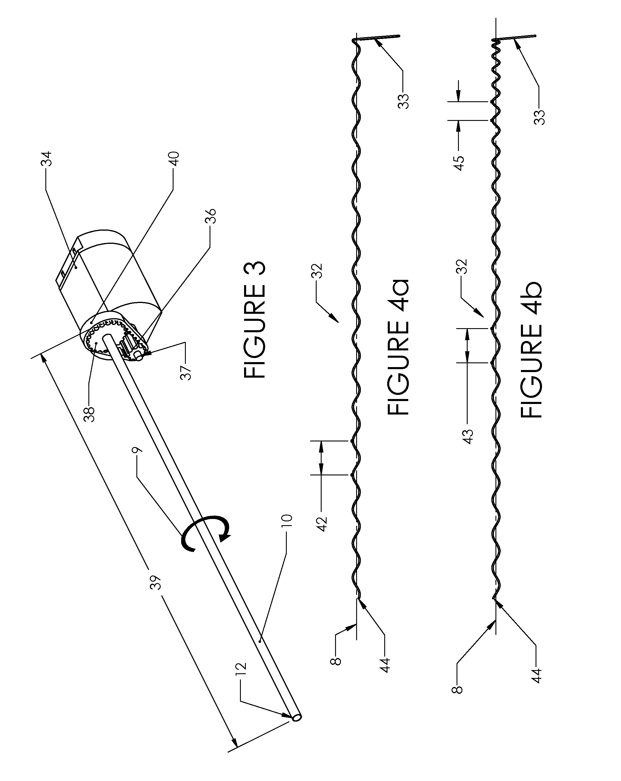
[0013]The tissue samples may be rotationally stationary, or nearly rotationally stationary, relative to the internal element. A spinning outer element may be composed of at least a transport tube. The transport tube can have a sharp distal edge and a spiral feature on the internal face. Nested within the transport tube, the internal element can be a stationary wire with a rectangular cross-section which can pierce the tissue. As the transport tube spins and is advanced into the tissue, the transport tube may core a section of tissue. The internal element can prevent rotation of the sample relative to the internal element without restricting axial motion of the tissue sample. As the spiral feature on the outer element spins relative to the tissue sample, the spiral feature can force the sample proximally into a collection chamber.
[0014]The tissue transport systems described above may be used in a side-cutting tool. For instance, a stationary tube may have a closed distal end functioning as a trocar. Proximal to the closed end may be a window cut into the wall of the tube that could allow passage of tissue samples into the inner lumen. A second transport tube may be placed concentrically inside the stationary tube, such that the radial gap between them is less than about 0.1 mm (0.005 in), but large enough such that the second transport tube and the stationary tube can move freely relative to each other. The internal transport tube may be actuated forward or backward to control movement of tissue through the window. A vacuum may be applied to draw tissue through the window. The actuation of the internal transport tube to close the window may sever any tissue from the mass of tissue that has passed through the window. A tissue transport system, as described herein, could then transport the samples proximally, where the internal transport tube may be spun relative to a third internal element.

Problems solved by technology

This procedure, which may be repeated several times, is time-consuming and traumatic for the patient.
The mechanisms of SIMS devices are generally more complex and expensive to manufacture than SISS tools.
In addition, they are often quite large devices, as a tissue capturing element is often moved fully distally to obtain the sample, then fully proximally to store the tissue sample.
Other known devices use tissue augers to transport the tissue, but these have not yet been employed in a system that can provide large contiguous samples.

Method used


Image

  • Tissue removal device and method of use
  • Tissue removal device and method of use
  • Tissue removal device and method of use

Examples


Embodiment Construction

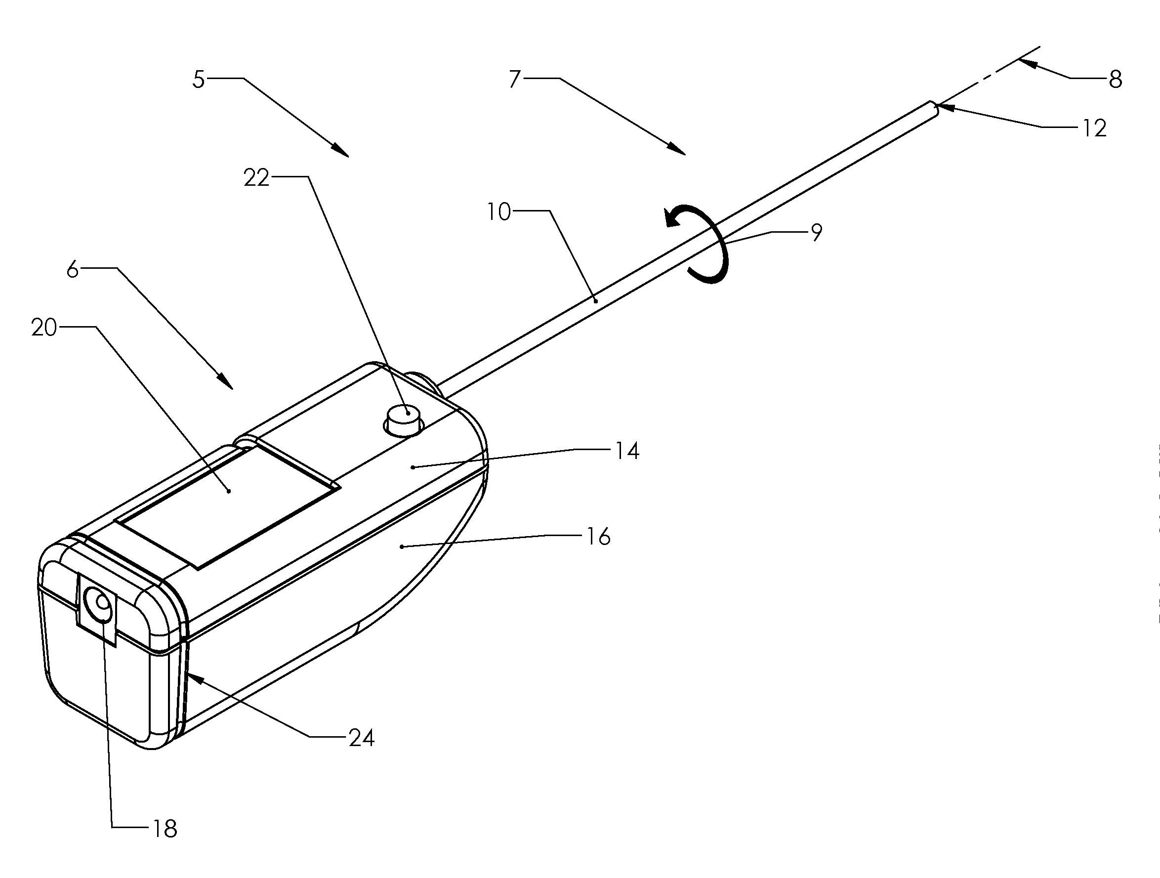
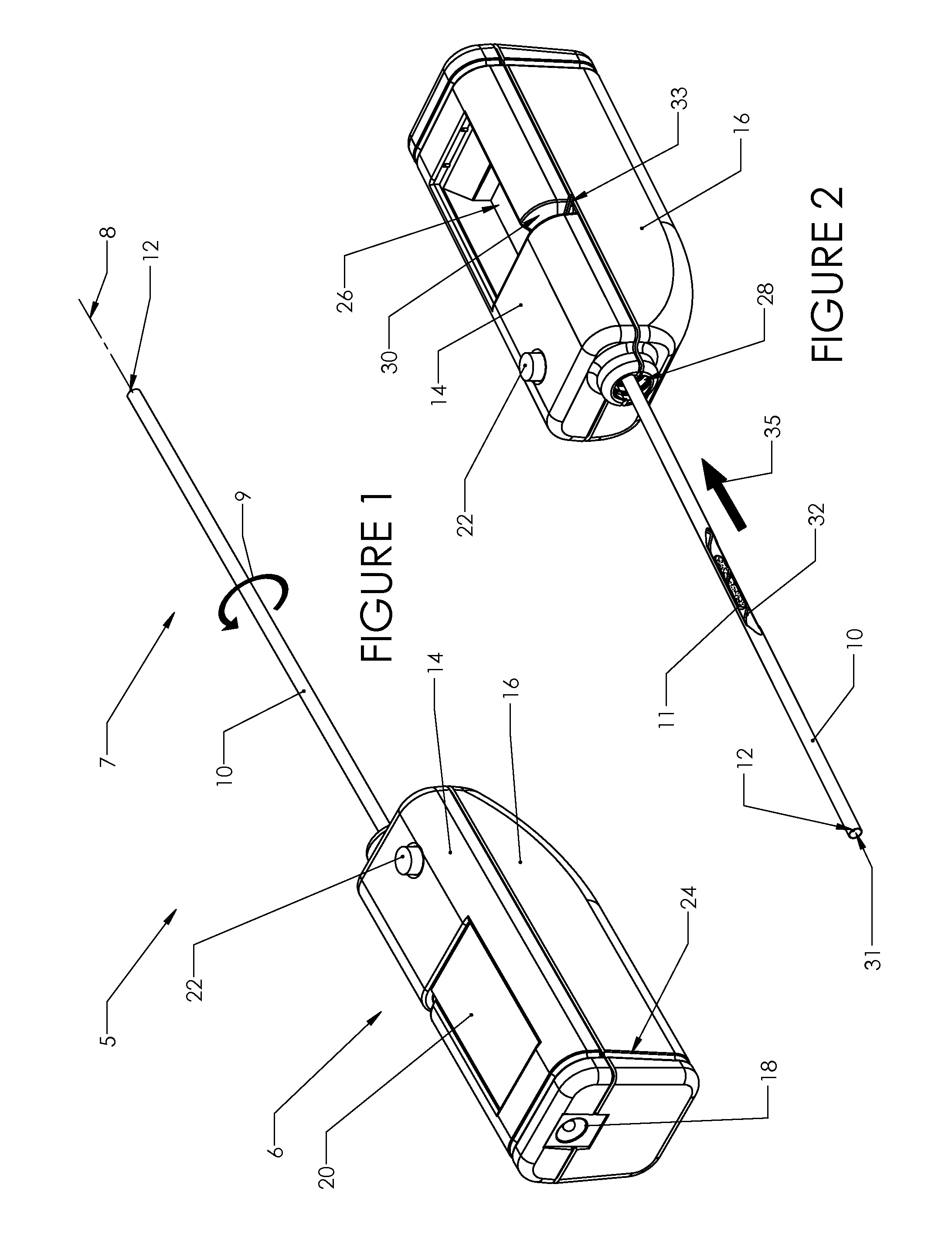
[0047]FIG. 1 illustrates a tool 5. The tool 5 may be sterilized. The tool 5 may have a handle 6 and a tissue transport system 7. The handle 6 can have a handle top portion 14 and a handle bottom portion 16, or a handle left portion and a handle right portion. The handle top portion 14 and handle bottom portion 16 may be joined together to form an ergonomic handle which the operator may hold. The handle top portion 14 and handle bottom portion 16 may be injection molded. The tissue transport system 7 can have a tissue-engaging first external outer element and a tissue-engaging second internal inner element. The tissue-engaging first element (e.g., a tissue-engaging outer element) can be radially outside of the tissue-engaging second element (e.g., a tissue-engaging inner element). The tissue-engaging first element can be or have a transport tube 10. The tissue-engaging inner element can be or have a coiled helical element 32, spiral element 74, flat stationary element 78, curved stat...


PUM


Abstract

A biopsy device for acquiring more than one tissue sample is disclosed. The biopsy device can have at least two tissue engaging elements, where at least one element contains a helical feature. A control mechanism can be used to spin an outer element relative to an internal element, resulting in transport of multiple tissue samples from the mass of tissue to an accessible collection chamber. The outer element may have a sharp distal end which may sever the samples from the mass of tissue. Samples may be stored sequentially in the collection chamber and be removed by the operator.

Description

CROSS-REFERENCE TO RELATED APPLICATIONS[0001]This application is a continuation-in-part of PCT International Application No. PCT / US 2011 / 061089 filed Nov. 16, 2011, which claims the benefit of U.S. Provisional Application No. 61 / 415,850 filed Nov. 21, 2010, which are both incorporated by reference herein in their entireties.BACKGROUND OF THE INVENTION[0002]1. Field of the Invention[0003]This invention relates to medical instrumentation. More particularly, a tool used for acquiring tissue and a method for using the same are disclosed.[0004]2. Description of the Prior Art[0005]A number of medical procedures require the removal of tissue samples from a patient. These operations can range from the removal of suspicious tissue, as in the biopsy of a cancerous lesion, to cell harvesting, as in a bone marrow donation. A number of different biopsy tools are used for retrieving these tissue samples from patients, falling into two broad categories: Single-Insertion, Single-Sample (SISS) tools...

Claims


Application Information

Patent Timeline
IPC IPC(8): A61B10/00
CPCA61B10/0266A61B10/0283A61B2017/00685A61B2010/0208A61B17/32002A61B17/320783
Inventor PELIKS, ROBERT BILGOR
Owner MERIT MEDICAL SYST INC
Learn More