Eureka AIR delivers breakthrough ideas for toughest innovation challenges, trusted by R&D personnel around the world.

Ultrasound diagnostic apparatus and ultrasound image producing method

a diagnostic apparatus and ultrasound technology, applied in the field of ultrasonic diagnostic equipment and ultrasound image producing method, can solve the problems of difficult to give high-accuracy diagnosis based solely on a b mode image and a hepatorenal contrast, and achieve the effect of high-accuracy diagnosis of a subject's liver diseas

Inactive Publication Date: 2012-10-11
FUJIFILM CORP
View PDF1 Cites 18 Cited by
  • Summary
  • Abstract
  • Description
  • Claims
  • Application Information

AI Technical Summary

Benefits of technology

The present invention provides an ultrasound diagnostic apparatus and method that can accurately diagnose liver disease by measuring the sound speed on the liver and kidneys using a B mode image and a sound speed map. This is useful in cases where the hepatorenal contrast is increased by chronic hepatitis or a fatty liver. The apparatus includes a transducer array, transmission and reception circuits, an image producer, a monitor, a region of interest setting unit, a controller, and a hepatorenal contrast calculator. The method involves producing a B mode image, setting a region of interest containing the liver and kidneys, transmitting and receiving ultrasonic beams to obtain reception data, producing a sound speed map based on the reception data, calculating the average luminance in the liver and kidneys, and calculating the hepatorenal contrast based on the average luminances. The technical effect of this invention is to provide a more accurate diagnosis of liver disease.

Problems solved by technology

However, the hepatorenal contrast is known to be increased by, for example, chronic hepatitis as well as a fatty liver, making it difficult in some cases to give a high accuracy diagnosis based solely on a B mode image and a hepatorenal contrast.

Method used


Image

  • Ultrasound diagnostic apparatus and ultrasound image producing method
  • Ultrasound diagnostic apparatus and ultrasound image producing method
  • Ultrasound diagnostic apparatus and ultrasound image producing method

Examples


embodiment 1

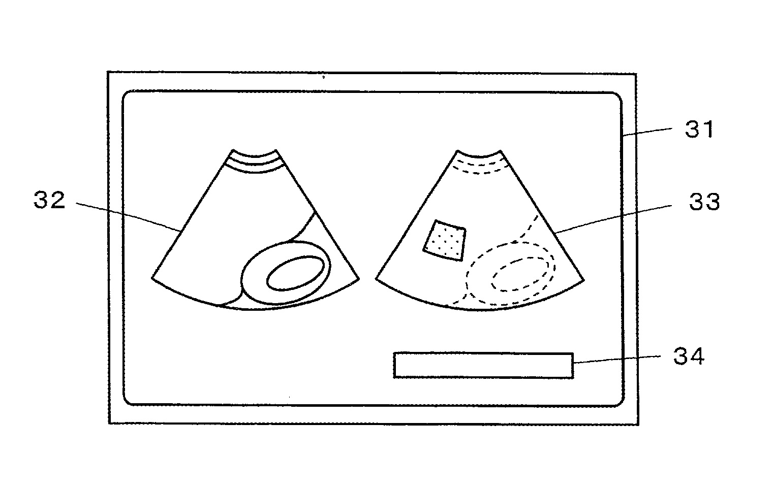
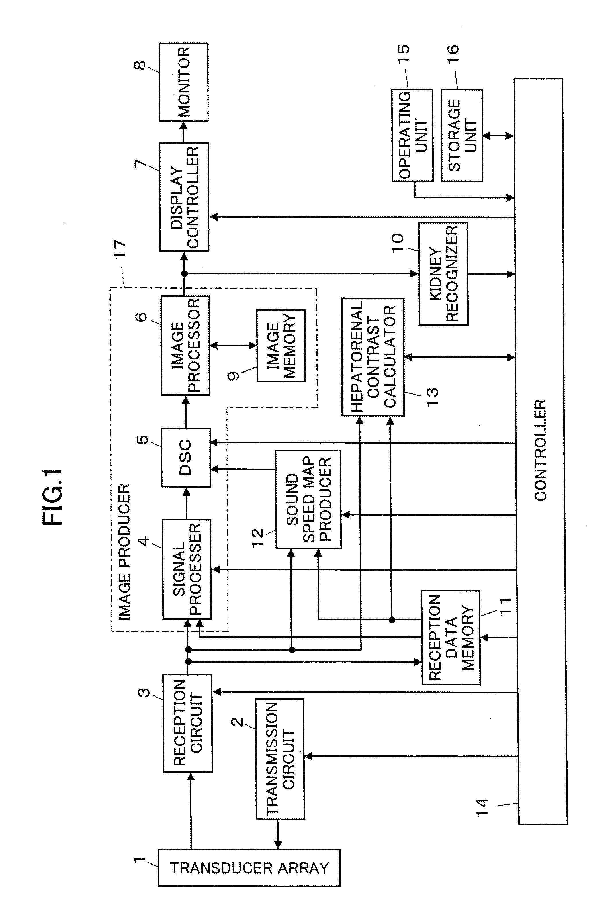
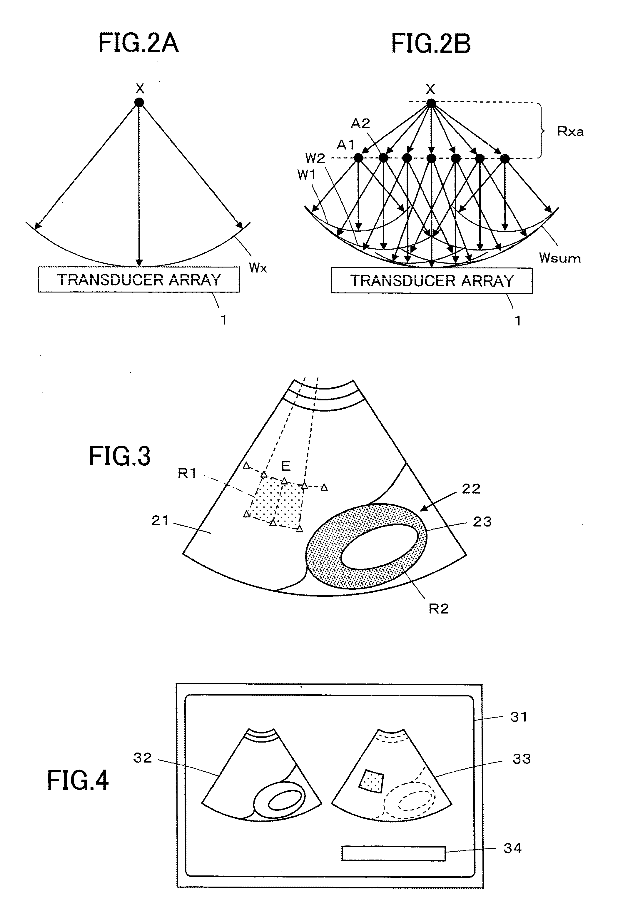
[0035]FIG. 1 illustrates a configuration of an ultrasound diagnostic apparatus according to Embodiment 1 of the invention. The ultrasound diagnostic apparatus of the invention comprises a transducer array 1, which is connected to a transmission circuit 2 and a reception circuit 3. A signal processor 4, a DSC (Digital Scan Converter) 5, an image processor 6, a display controller 7, and a monitor 8 are connected to the reception circuit 3. in sequences. An image memory 9 and a kidney recognizer 10 are connected to the image processor 6.

[0036]A reception data memory 11, a sound speed map producer 12 and a hepatorenal contrast calculator 13 are connected to the reception circuit 3. A controller 14 is connected to the transmission circuit 2, the reception circuit 3, the signal processor 4, the DSC 5, the display controller 7, the kidney recognizer 10, the reception data memory 11, the sound speed map producer 12 and the hepatorenal contrast calculator 13. An operating unit 15 and a stora...


PUM


Abstract

An ultrasound diagnostic apparatus includes a region of interest setting unit for setting a region of interest containing at least part of a liver and at least part of a kidney in a B mode image, a controller for transmitting and receiving ultrasonic beams to obtain reception data for measuring a sound speed, a sound speed map producer for producing a sound speed map of a liver region based on the reception data for measuring a sound speed, and a hepatorenal contrast calculator for calculating an average luminance in the liver region and an average luminance in a kidney region of the region of interest and calculating a hepatorenal contrast based on the calculated average luminances.

Description

BACKGROUND OF THE INVENTION[0001]The present invention relates to an ultrasound diagnostic apparatus and an ultrasound image producing method and particularly to an ultrasound diagnostic apparatus that produces a B mode image by transmitting and receiving ultrasonic waves through a transducer array of an ultrasound probe.[0002]Conventionally, ultrasound diagnostic apparatus using ultrasound images are employed in medicine. In general, this type of ultrasound diagnostic apparatus comprises an ultrasound probe having a built-in transducer array and an apparatus body connected to the ultrasound probe. The ultrasound probe transmits an ultrasonic beam toward the inside of a subject's body, receives ultrasonic echoes from the subject, and the apparatus body electrically processes the reception signals to produce an ultrasound image.[0003]In diagnosis of fatty liver or like disease, use is often made of the ratio of average liver luminance to kidney average luminance or a so-called hepato...

Claims


Application Information

Patent Timeline
Patent Type & Authority Applications(United States)
IPC IPC(8): A61B8/14
CPCA61B8/14A61B8/5223A61B8/469G16H50/30
Inventor TASHIRO, RIKA
Owner FUJIFILM CORP
Why Eureka
Eureka Blog
Learn More