Preparation device and preparation method for exosome liquid biopsy sample and method for analyzing exosome liquid biopsy sample prepared thereby

a technology for exosome liquid biopsy and preparation device, which is applied in the direction of biochemistry apparatus and processes, instruments, ict adaptation, etc., can solve the problems of only providing tissue biopsy, invasive surgical tissue sampling that poses potential risks, and difficult to fully understand the composition of the target tumor, so as to minimize the interference of non-specific exosomes, minimize the interference of specific exosome signals, and minimize the effect of noise during analysis

Pending Publication Date: 2022-04-28
SOL BIO CORP
View PDF0 Cites 2 Cited by
  • Summary
  • Abstract
  • Description
  • Claims
  • Application Information

AI Technical Summary

Benefits of technology

[0019]According to the present invention, a subpopulation of exosomes having exosome-specific markers (primary separation markers) can be separated from a population containing exosomes relatively less associated with a target disease and the specific exosomes highly associated with the target disease can be enriched and recovered for subsequent isolation. A sub-subpopulation of exosomes having exosome-specific markers (secondary separation markers) in the primary separation solution can be separated from a population and the exosomes containing biomarkers involved in the progression of the target disease can be enriched to provide a liquid biopsy sample.
[0020]In addition, a subpopulation, a sub-subpopulation or a subset thereof containing exosomes associated with a specific disease can be separated and recovered from a population sample containing exosomes released from cells in the body to prepare a liquid biopsy sample. The liquid biopsy sample can be analyzed by biomarker profiling to discover biomarkers (e.g., proteins and nucleic acids) specific to the target disease.
[0021]Furthermore, since the liquid biopsy sample separated and concentrated from the exosome subpopulation, sub-subpopulation or subset thereof is used as an analyte, non-specific exosomes are removed so that noise can be minimized during analysis and specific exosome signals can be maximized. The use of such exosomes as biomarkers for a specific disease such as cancer minimizes the interference of non-specific exosomes resulting from the heterogeneity of exosomes, which is a current problem, enabling accurate recognition of information on the physiological properties and lineage of parent cells from the liquid biopsy sample.
[0022]Moreover, the present invention can be used to diagnose a specific disease such as a cancer disease, degenerative disease, heart disease or infectious disease with significantly high accuracy and is thus effective in early clinical diagnosis of the disease. The present invention enables acquisition of information on the progression or decline of a target disease depending on a therapeutic agent for the disease. Therefore, the present invention can also be applied to companion diagnostics for personalized treatment.

Problems solved by technology

In contrast, tissue biopsy usually involves expensive, invasive surgical tissue sampling that poses potential risks.
In contrast, tissue biopsy only provides a snapshot of a single disease lesion when necessary and fails to monitor the progression and treatment of diseases every moment.
In contrast, tissue biopsy collects diverse tumors, making it difficult to fully understand the composition of a target tumor.
In contrast, the limited size of samples for tissue biopsy makes it impossible to divide and use them for several diagnostic purposes.
In contrast, the restricted accuracy of tissue biopsy limits the clinical usefulness of test results.
Although circulating tumor cells are excellent targets in terms of enrichment, selectivity, and stability of isolated nucleic acids, their extremely low blood level makes it difficult to use them for early diagnosis of cancer diseases.
Cell-free DNA is difficult to enrich and select despite its high blood level and their stability after isolation is low.
Moreover, due to the presence of a large amount of non-specific DNA that is not associated with target diseases, overcoming the problem of noise during analysis is becoming a major issue.
However, since the size and density of exosome particles and the compositions of protein and nucleic acid components present in exosome particles are extremely heterogeneous, only disease-associated exosomes need to be isolated for liquid biopsy.
However, since exosomes derived from all cells exist in a mixed state in body fluids, there is a limitation in accurately diagnosing specific diseases such as cancer from a bulk population of exosome samples separated from body fluids.
However, ultracentrifugation has disadvantages in that the use of an expensive system is required and it takes a long time for isolation.
However, the existing exosome isolation techniques are limited in selectively detecting trace amounts of exosomes derived from specific cells such as cancer cells because all exosomes present in body fluids are provided in the form of single populations.
In the case where a bulk population containing all exosomes present in a body fluid is used as a liquid biopsy sample for the diagnosis of a specific disease such as cancer, high diagnostic accuracy is very difficult to achieve because sample matrix effects (e.g., non-specific binding of non-specific exosomes) cause negative results in analytical specificity and sensitivity.
However, the use of the multiple biomarkers has limited synergistic effects because most exosomal markers are non-specifically associated with the disease and their specificity for the disease is fundamentally low.

Method used


Image

  • Preparation device and preparation method for exosome liquid biopsy sample and method for analyzing exosome liquid biopsy sample prepared thereby
  • Preparation device and preparation method for exosome liquid biopsy sample and method for analyzing exosome liquid biopsy sample prepared thereby
  • Preparation device and preparation method for exosome liquid biopsy sample and method for analyzing exosome liquid biopsy sample prepared thereby

Examples


first embodiment

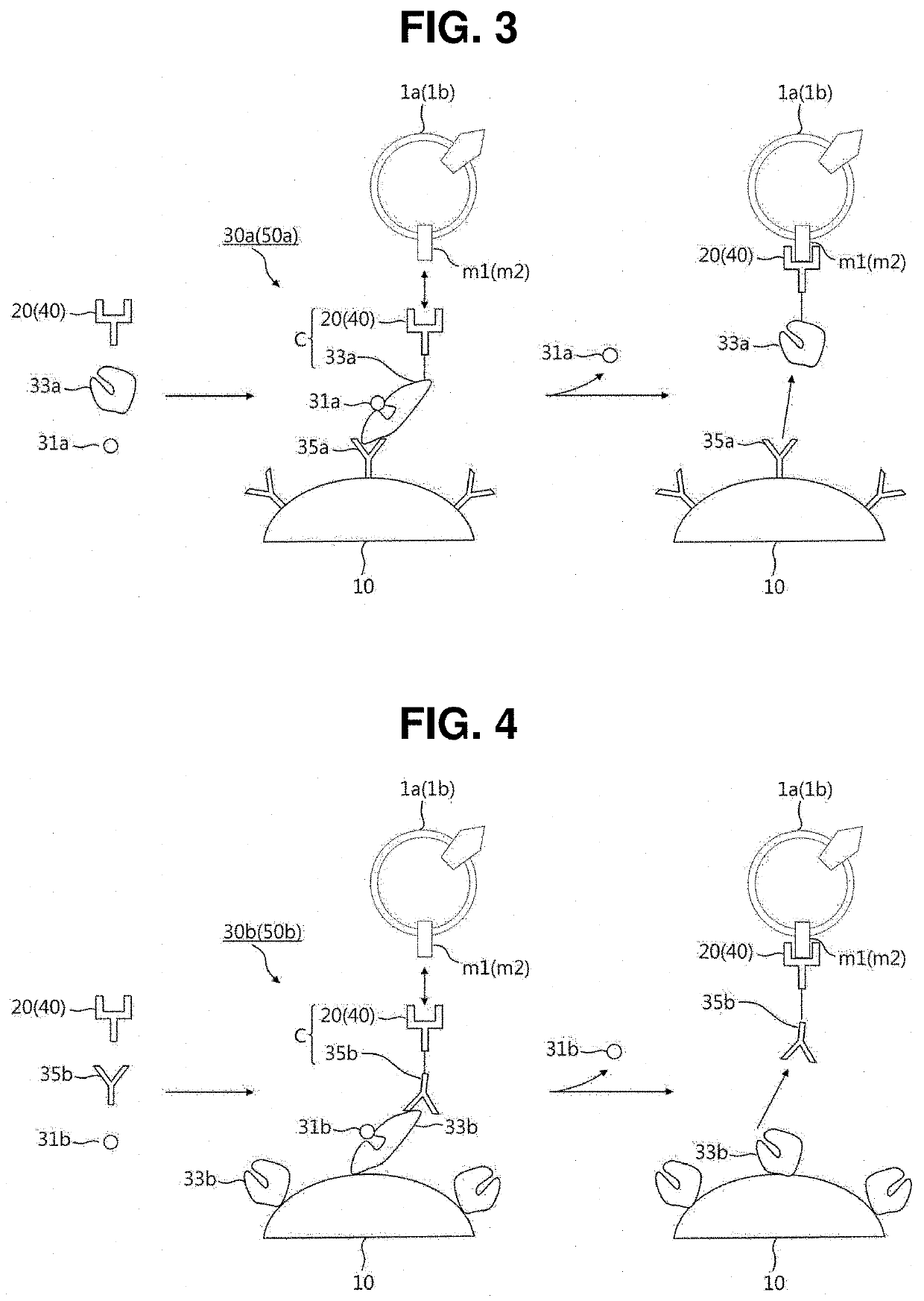
[0078]As illustrated in FIG. 3, each of the reversible linkers 30a and 50a according to the present invention includes a ligand 31a, a binder 33a, and a recognition material 35a.

[0079]The ligand 31a is a material capable of detachably binding to the binder 33a and may be selected from the group consisting of sugar molecules, ions, substrates, antigens, peptides, vitamins, growth factors, hormones, and combinations thereof.

[0080]The binder 33a is a material whose conformation is changed when bound with the ligand 31a. The binder 33a may be selected from the group consisting of sugar-binding proteins, ion-binding proteins, enzymes, antibodies, avidins, aptamers, cell receptors, nanostructures, and combinations thereof. Accordingly, the binder 33a and the ligand 31a can form a binder-ligand pair such as a sugar-binding protein-sugar molecule pair, an ion-binding protein-ion pair, a biotin-avidin pair, an enzyme-substrate pair, an antigen-antibody pair, an aptamer-ligand pair, a cell r...

second embodiment

[0083]Referring to FIG. 4, each of the reversible linkers 30b and 50b according to the present invention includes a ligand 31b, a binder 33b, and a recognition material 35b.

[0084]The ligand 31b, the binder 33b, and the recognition material 35b of the reversible linker 30b or 50b according to the second embodiment correspond to the ligand 31a, the binder 33a, and the recognition material 35a of the reversible linker 30a or 50a according to the first embodiment, respectively, and only the differences will be mainly described below.

[0085]In the reversible linker 30a or 50a according to the first embodiment, the binder 33a is conjugated with the capture material 20 or 40 to form the polymer C and the recognition material 35a is immobilized on the surface of the immobilization member 10. In contrast, in the reversible linker 30b or 50b according to the second embodiment, the binder 33b is immobilized on the surface of the immobilization member 10 and the recognition material 35b is conj...

third embodiment

[0087]As illustrated in FIG. 5, each of the reversible linkers 30c and 50c according to the present invention includes a ligand 31c and a binder 33c.

[0088]The ligand 31c of the reversible linker 30c or 50c according to the third embodiment is conjugated with a first capture material 20 and / or a second capture material 40 to form a capture material-ligand conjugate C.

[0089]The binder 33c of the reversible linker 30c or 50c is immobilized on the surface of the immobilization member 10 and binds with the capture material-ligand conjugate C specifically bound to an exosome 1a or 1b, with the result that the exosome 1a or 1b is captured by the immobilization member 10. The binder 33c bound with the ligand 31c binds with another ligand 32 competing with the ligand 31c. As a result, the ligand 31c is detached and the exosome 1a or 1b can be separated and recovered from the immobilization member 10. Here, the ligand 31c may be selected from the group consisting of polyhistidine-tags (His-t...


PUM

PropertyMeasurementUnit
sizeaaaaaaaaaa
diameteraaaaaaaaaa
volumeaaaaaaaaaa

Abstract

The present invention relates to a technique in which a target exosome subpopulation, a sub-subpopulation, or a lower population in human fluid, which is associated with a specific disease, is isolated and recovered in its intact form at high yield to prepare a liquid biopsy sample and analyzed.

Description

TECHNICAL FIELD[0001]The present invention relates to a technology in which a liquid biopsy sample is prepared by separating a population sample into a subpopulation, a sub-subpopulation or a subset thereof containing exosomes associated with a specific disease such as a cancer disease and is analyzed.BACKGROUND ART[0002]Recently, the concept of “liquid biopsy” has been introduced to minimize pain during screening for specific diseases such as cancer and degenerative diseases and to achieve early diagnosis of the diseases. Liquid biopsy refers to a non-invasive method for extracting body fluids, including blood, saliva, and urine, in a relatively simple manner and analyzing specific disease-associated cells or components (e.g., peptides, proteins, nucleic acids, organelles, and specific substances) released therefrom in the body fluids. Liquid biopsy helps to ascertain the occurrence of specific diseases at an early stage. For this reason, liquid biopsy has attracted attention as an...

Claims


Application Information

Patent Timeline
Patent Type & Authority Applications(United States)
IPC IPC(8): C12Q1/6886G16H70/60
CPCC12Q1/6886G16H70/60C12N5/06G01N33/569G01N33/574G01N33/68G16B40/10Y02A90/10G01N33/57484G01N33/5082G01N33/54326G01N33/543
Inventor PAEK, SUNG-HOCHOI, DA-YEON
Owner SOL BIO CORP
Why Eureka
Try Eureka