

Differential gene expression for detecting and/or differentiating lung disease
a technology of differentiating gene expression and lung disease, applied in the field of lung disease, can solve the problems of no effective medical treatment, high morbidity and mortality, and fibrotic lung disease with progressive onset and progression
- Summary
- Abstract
- Description
- Claims
- Application Information
AI Technical Summary
Problems solved by technology
Method used
Image

Examples
example 1
Lung Sample Collection and Preparation
[0080]Nine (9) normal lung tissue samples from 8 individuals and 10 ILD lung tissue samples were collected from three different sources, as shown in Table 1 below.
TABLE 1Patient Sample Table indicating type of tissue, diagnosis, pathology report,CT scan, and outcome.Outcome asPatholgyof MarchSubject IDType of tissueDiagnosisreportCT scan20111007-033explantIPFIPFILDdiedAug. 20, 2008 dueto bacterialpneumonia1007-033pneumonectomyIPFIPFILDdiedAug. 20, 2008 dueto bacterialpneumonia1008-060explantIPFIPFsubpleuraldied a coupleinterstitialof weeksfibrosispost-transplantmost likelyfrompulmonaryembolism1008-062explantIPFIPFpulmonarydied 15fibrosis,months post-emphysema,transplantbronchiectasisdue tochronicrejection1009-092biopsyIPFUIPperipheral andworked upbasilarfor transplantpredominantlist but is toointerstitialwell to bechanges andlistedhoneycombingcompatible withpulmonaryfibrosis.1009-097biopsyNSIPcellular andinterstitialclinicallyfibrosingfibrosisst...
example 2
Reverse Transcription (RT)-PCR
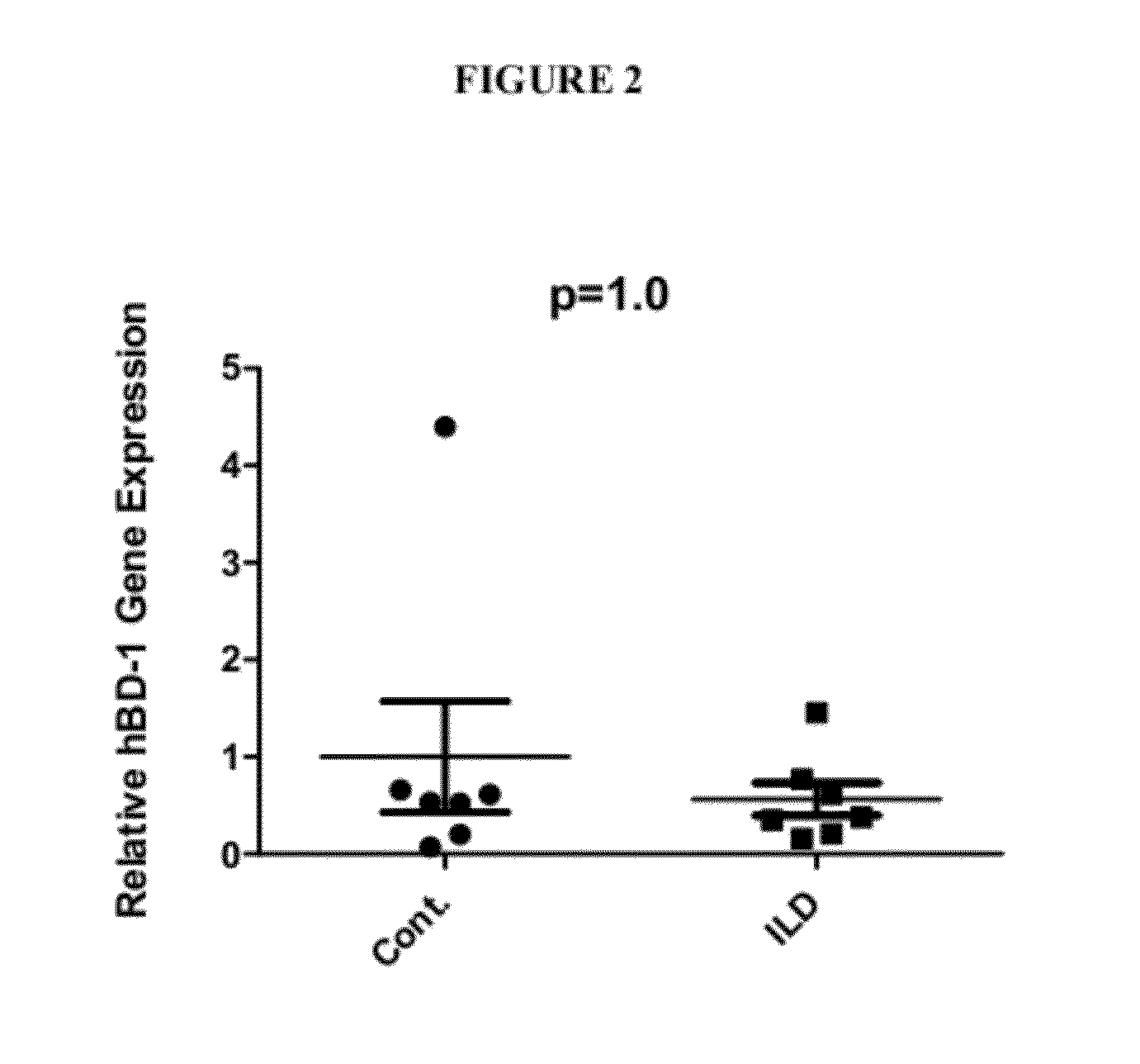
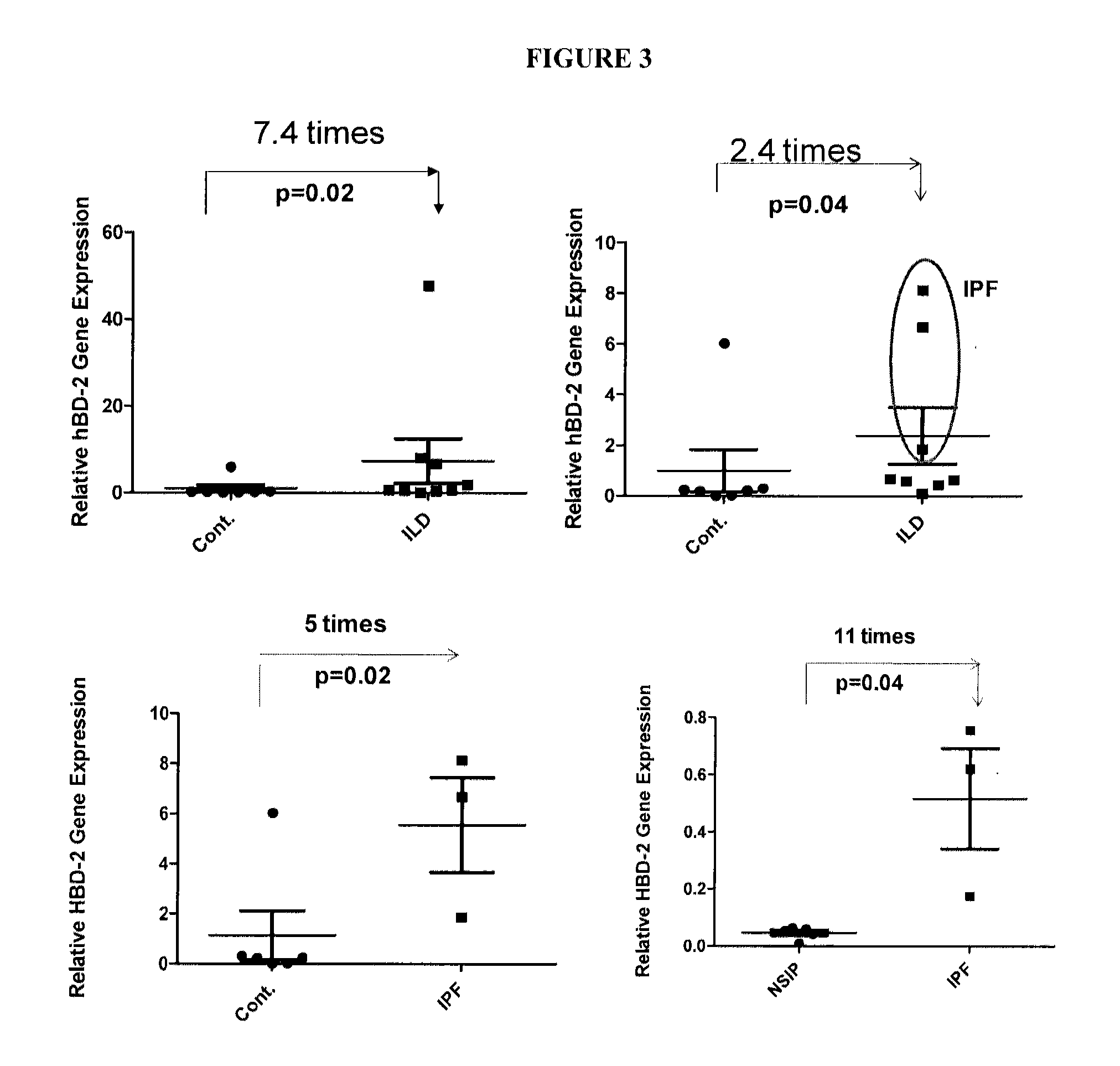
[0082]Real-time RT-PCR was performed to analyze antimicrobial peptide gene expression.
[0083]RT-PCR analysis was performed in a MyiQ Single Color Real-Time PCR Detection System (BioRad Laboratories) according to the manufacturer's instructions. Briefly, 2 μg of total RNA were reverse-transcribed (SuperScript™ III Reverse Transcriptase, Invitrogen). Template cDNA corresponding to 50 ng of RNA was added to a 20 μl reaction: 0.2 μM each primer and 1X iQ™ SYBR® Green Supermix (BioRad Laboratories). Samples were incubated in a 96-well PCR plate in the MyiQ Single Color Real-Time PCR Detection System. Initial denaturing: 95° C. for 3 min; 40 cycles consisting of 95° C. for 15 s, 56° C. (for hBD-1; other peptides, see Table 2) for 15 s and 72° C. for 20 s. SYBR Green fluorescence was detected at 72° C. at the end of each cycle. Melting curve profiles were produced (cooling the sample to 60° C. for 1 min and then heating slowly at 0.5° C. / s up to 95° C. with con...
example 3
Analysis of RT-PCR Results
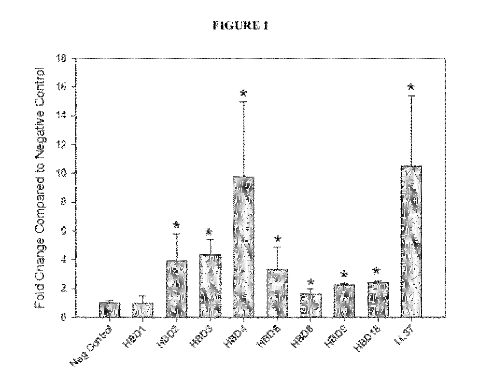
[0085]Previously, global gene expression patterns in the IPF-AEx lung were analyzed and compared with that of stable IPF and control lung. Gene expression of α-defensins (DEFA3 and DEFA4) were found to be significantly increased in IPF-AEx lungs compared with stale IPF in the microarray data, which was confirmed by qRT-PCR. Their levels in the plasma of patients with IPF-AEx were considerably higher in patients with IPF-AEx compared with control subjects or patients with stable IPF as well [50], indicating antimicrobial peptides may play a role in the pathogenesis of IPF or IPF-AEx.
[0086]Microarray or DNA chip technology is a high through-put technology. Gene expression profiling is the most common application of this technology. Hybridization between complementary nucleic acids is the basis of the technology. While the microarray is a powerful tool that can give a lot of information, analyzing a large scale of samples and interpretation of microarray can b...
PUM
| Property | Measurement | Unit |
|---|---|---|
| median survival time | aaaaa | aaaaa |
| molecular weight | aaaaa | aaaaa |
| real time qRT- | aaaaa | aaaaa |
Abstract
Description
Claims
Application Information