Positron Emission Tomography Detector Based on Monolithic Scintillator Crystal

a tomography detector and monolithic technology, applied in tomography, x/gamma/cosmic radiation measurement, instruments, etc., can solve the problems of reducing the accuracy of positioning at areas with high crystal thickness, affecting the simultaneous event of scintillator plates, and affecting the accuracy of positioning. , to achieve the effect of reducing the edge effect and significantly deteriorating the resolution

Active Publication Date: 2013-01-10
SIEMENS MEDICAL SOLUTIONS USA INC +1
View PDF7 Cites 33 Cited by
  • Summary
  • Abstract
  • Description
  • Claims
  • Application Information

AI Technical Summary

Benefits of technology

[0012]Further provided is a plurality of configurations with the solid-state photosensor pixel arrays on up to four vertical surfaces of the scintillator block, or five total surfaces. This aids in reducing the edge effects that usually deteriorate the resolution significantly when only one fac

Problems solved by technology

The drawback of this approach is that the positioning becomes less accurate at areas of high crystal thickness, especially near the edges of the scintillator plate.
In the case of Nal

Method used


Image

  • Positron Emission Tomography Detector Based on Monolithic Scintillator Crystal
  • Positron Emission Tomography Detector Based on Monolithic Scintillator Crystal
  • Positron Emission Tomography Detector Based on Monolithic Scintillator Crystal

Examples


Embodiment Construction

[0018]As required, disclosures herein provide detailed embodiments of the present invention; however, the disclosed embodiments are merely exemplary of the invention that may be embodied in various and alternative forms. Therefore, there is no intent that specific structural and functional details should be limiting, but rather the intention is that they provide a basis for the claims and as a representative basis for teaching one skilled in the art to variously employ the present invention.

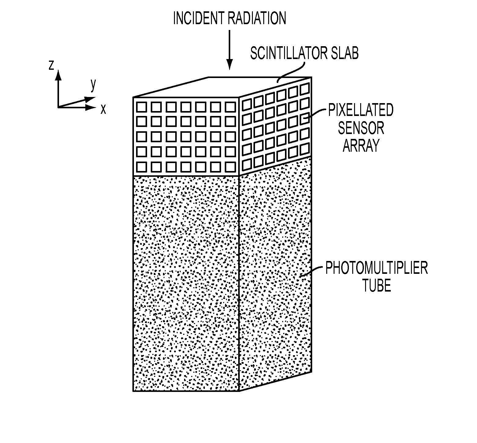
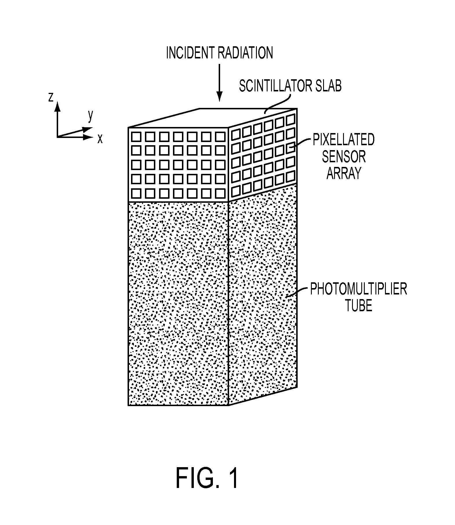
[0019]FIG. 1 shows a PET detector block in accordance with one embodiment of the invention. The detector includes a monolithic scintillator crystal block or scintillator slab, a photomultiplier tube (PMT), and one or more arrays of pixelated solid-state photosensors. The scintillator face opposite the incident radiation is coupled to the PMT, either directly or via an additional light guide that is not shown in the drawing. The PMT can have a square, rectangular or rounded entrance window. The si...


PUM


Abstract

A high-resolution nuclear imaging detector for use in systems such as positron emission tomography includes a monolithic scintillator crystal block in combination with a single photomultiplier tube read-out channel for timing and total energy signals, and one or more solid-state photosensor pixels arrays on one or more vertical surfaces of the scintillator block to determine event position information.

Description

CROSS-REFERENCE TO RELATED APPLICATIONS[0001]This application claims the benefit of U.S. Provisional Patent Application 61 / 504,820, filed on Jul. 6, 2011, the entire disclosure of which is hereby incorporated by reference.[0002]TECHNICAL FIELD[0003]The current invention relates to the field of nuclear medical imaging systems. Particularly, the invention relates to providing a high-resolution Positron Emission Tomography (PET) detector by using a monolithic scintillator crystal block.BACKGROUND OF THE INVENTION[0004]Medical imaging is one of the most useful diagnostic tools available in modern medicine. Medical imaging allows medical personnel to non-intrusively look into a living body in order to detect and assess many types of injuries, diseases, conditions, etc. Medical imaging allows doctors and technicians to more easily and correctly make a diagnosis, decide on a treatment, prescribe medication, perform surgery or other treatments, etc.[0005]There are medical imaging processes ...

Claims


Application Information

Patent Timeline
IPC IPC(8): G01T1/202G01T1/164
CPCG01T1/1642A61B6/037G01T1/1644G01T1/2985G01T1/20187
Inventor SCHMAND, MATTHIAS J.HENSELER, DEBORA
Owner SIEMENS MEDICAL SOLUTIONS USA INC
Why Eureka
Try Eureka