Preterm labor monitor

a preterm labor and monitor technology, applied in the field of measuring and monitoring uterine cervical activity indicative, can solve the problems of 70% of neonatal morbidity and mortality, no way of accurately detecting preterm labor, and continued plague of preterm labor diagnosis in the obstetric community

Active Publication Date: 2013-02-28
ROSE HUANG +6
View PDF13 Cites 16 Cited by
  • Summary
  • Abstract
  • Description
  • Claims
  • Application Information

AI Technical Summary

Problems solved by technology

Despite recent technological medical breakthroughs, the issue of diagnosing preterm labor has continued to plague the obstetric community.
Preterm births lead to 70% percent of neonatal morbidity and mortality, and cost the United States over $26.2 billion in 2005 alone.
Currently there is no way of accurately detecting preterm labor, which often leads to preterm birth.
Current detection methods, such as the commonly used tocodynamometer, lack the ability to detect critical cervical changes and uterine activity and / or contractions at early gestational ages.
This prevents timely diagnosis and treatment of preterm labor.
A method and device that detects preterm labor early in its course in patients is currently lacking.
Preterm delivery does not allow the fetus enough time to develop within the womb, resulting in severe short and long-term health issues for the neonate.
The results of preterm delivery directly correlate to vast increases in medical costs.
All of the current methods employed to screen for preterm labor are ineffective, insufficient, or inaccurate.
The tocodynamometer, used to detect contractions from the abdominal surface, is often unable to detect contractions and cervical changes at an early gestational age.
Transvaginal ultrasound, while able to detect cervical changes even at very early gestational ages, cannot usually detect the contractions that are often present before these changes are evident.
Symptomatic monitoring is insufficient because patients are most often not evaluated until the time for meaningful intervention has passed.
Even with increased surveillance, the failings of current methods have led to an annual preterm birth rate of twelve percent in the United States.
Failure to detect preterm labor early in its course means that by the time cervical changes have truly manifested, delivery can only be delayed by a few days.
Additional difficulty arises in striking a balance between avoiding unnecessary intervention and making timely diagnosis and treatment.

Method used


Image

  • Preterm labor monitor
  • Preterm labor monitor
  • Preterm labor monitor

Examples


example 1

Use of Medical Device with a Patient

[0072]According to one embodiment, the medical device 100, 200, 302 may be adapted to be applied directly to the cervix by the clinician. For example, when a symptomatic or highrisk patient visits her physician for a weekly or bi-weekly routine check-up, she may undergo a series of tests, including monitoring via a tocodynamometer, a digital examination and / or a transvaginal ultrasound to assess cervical length. This evaluation can last from two hours to 24 hours while the woman is probed and monitored. The disposable cervical elastic ring portion of the medical device may be placed on the cervix of the patient after the initial digital examination, and remain for the full duration of the observation period. This may allow the physician to closely monitor the patient's dilatation, effacement, and contractions without constantly being in attendance. The readings may be provided by a separate monitor and saved to a hard-drive. At each evaluation for...

example 2

Direct Application to the Cervix

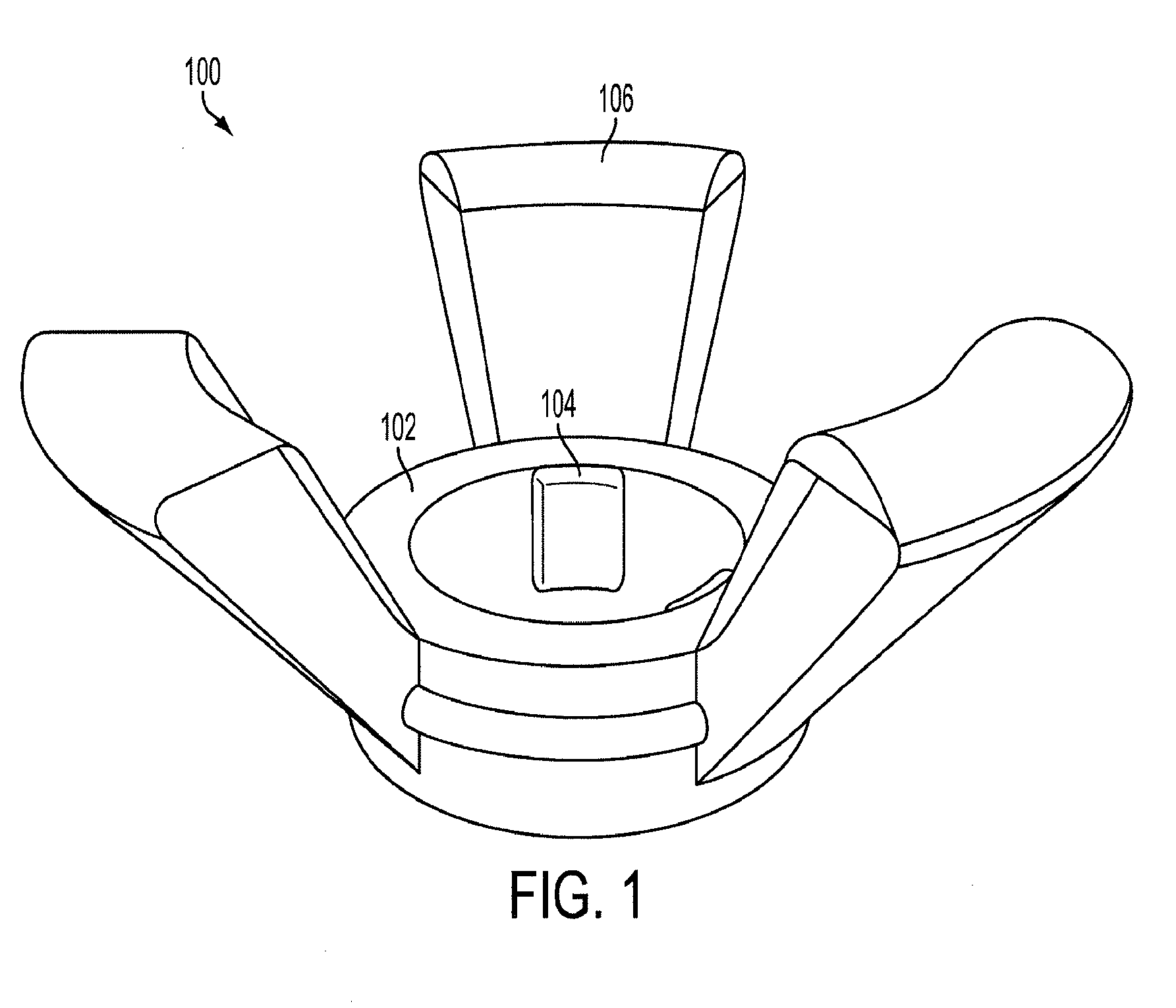
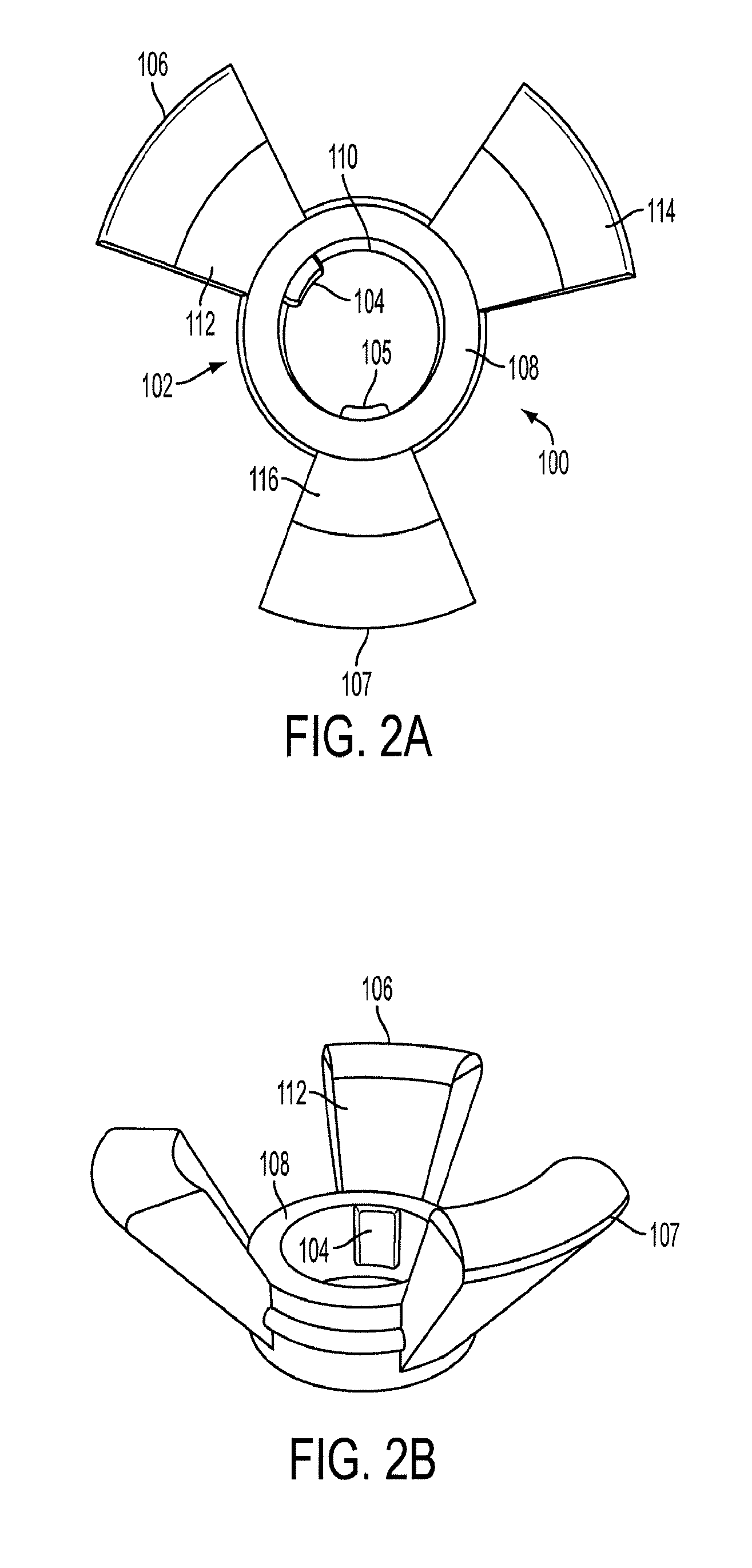
[0074]According to an embodiment, the medical device 100, 200, 302 may be comprised of flexible, biocompatible and sterilizable components that conform to a normal pregnant cervix. The outputs that address these inputs may be realized in the design of the cervical ring through the specified characteristics of materials, fixation points and overall form factor.

[0075]According to one embodiment, the elastic ring 108 may have an initial inner diameter of approximately 20 mm and a stretched inner diameter of approximately 40 mm. The flexible band may have a thickness of approximately 4 mm and a depth of approximately 10 mm. Each wing portion may have a width of approximately 10 mm and a depth of approximately 7 mm.

[0076]According to another embodiment, the device may be made of biocompatible medical grade polymer, for example, but not limited to, a medical grade silicone elastomer. The optimal Young's modulus, sometimes referred to as the elastic modulus ...

example 3

Direct Detection of Cervical / Uterine Activity

[0077]According to one embodiment, the medical device 100, 200, 302 may be comprised of uterine contractility sensors (measuring cervical and vaginal electrical potential), effacement sensors (measuring tissue thickness) and dilatation sensors (measuring diameter). The outputs that address these inputs are realized in the selection of the cervical ring sensors through the specified characteristics and placement of the selected electrodes, LED emitter and detector pair and embedded stretch gauge.

[0078]According to another embodiment, unipolar electrodes may be used to detect both cervical and uterine contractions and / or electric potentials. Such electrodes may be made from, for example, 316L stainless steel and / or sintered silver chloride (Ag—AgCl). The electrodes may have an approximately 8 mm diameter. According to one embodiment, the electrodes may comprise EMG electrodes having a measurement range of approximately 50 to 3000 Hertz. Acc...


PUM


Abstract

A system, method and apparatus for monitoring uterine and/or cervical activity indicative of labor in a patient. The system includes a medical device and a data processor in communication with the medical device. The medical device includes a structural component, a first electrode attached to the structural component, and a second electrode attached to the structural component. The structural component is structured to be in contact with a cervical surface and a vaginal surface of the patient, such that said first electrode is in electrical contact with said cervical surface and said second electrode is in electrical contact with said vaginal surface. The first electrode is adapted to receive an electrical activity of the cervical surface and the second electrode is adapted to receive an electrical activity of the uterus through the vaginal surface. The data processor is adapted to process the electrical activity of the electrodes to detect contractions on a surface of the patient indicative of labor.

Description

CROSS-REFERENCE OF RELATED APPLICATION[0001]This application claims priority to U.S. Provisional Application No. 61 / 305,616 filed Feb. 18, 2010, the entire contents of which are hereby incorporated by reference.BACKGROUND[0002]1. Field of Invention[0003]The field of the currently claimed embodiments of this invention relates to systems and methods for measuring and monitoring uterine cervical activity indicative of labor.[0004]2. Discussion of Related Art[0005]Despite recent technological medical breakthroughs, the issue of diagnosing preterm labor has continued to plague the obstetric community. In order to fully understand the gravity of this need, it is important to recognize the regrettable outcomes and heavy costs related to preterm birth. Preterm births lead to 70% percent of neonatal morbidity and mortality, and cost the United States over $26.2 billion in 2005 alone. Currently there is no way of accurately detecting preterm labor, which often leads to preterm birth. Current ...

Claims


Application Information

Patent Timeline
Patent Type & Authority Applications(United States)
IPC IPC(8): A61B5/04A61B6/00A61B5/296
CPCA61B5/0084A61B5/04882A61B5/0492A61B5/4356A61B5/435A61B5/6882A61B5/04A61B5/1076A61B5/391A61B5/296A61B5/24
Inventor AINA-MUMUNEY, ABIMBOLASAGARAM, DEEPIKACOURVILLE, CHRISTOPHER BRANDONHWANG, KARINHUANG, YI ROSEBLAKEMORE, KARIN J.SUNWOO, SUNG JINACHARYA, SOUMYADIPTA
Owner ROSE HUANG
Why Eureka
Try Eureka