Method for diagnosing melanocytic proliferations

a melanocytic and proliferation technology, applied in the field of melanocytic proliferation diagnosis, can solve problems such as characteristic staining sac pattern, and achieve the effect of diagnosing melanocytic proliferation

Active Publication Date: 2013-03-14
CORNELL UNIVERSITY
View PDF3 Cites 6 Cited by
  • Summary
  • Abstract
  • Description
  • Claims
  • Application Information

AI Technical Summary

Benefits of technology

The invention is a method for diagnosing melanocytic proliferations by staining them with an antibody against soluble adenylyl cyclase (sAC) and interpreting the staining pattern. The method has been found to have a distinctive staining pattern that can be used to confirm or disaffirm a diagnosis made through conventional histologic examination. The invention also provides a kit for interpreting melanocytic proliferations.

Problems solved by technology

Application of this method yields characteristic staining sAC patterns, which are complex, but appear discriminatory and distinctive according to the nature of the melanocytic proliferation.

Method used


Image

  • Method for diagnosing melanocytic proliferations
  • Method for diagnosing melanocytic proliferations
  • Method for diagnosing melanocytic proliferations

Examples


example 1

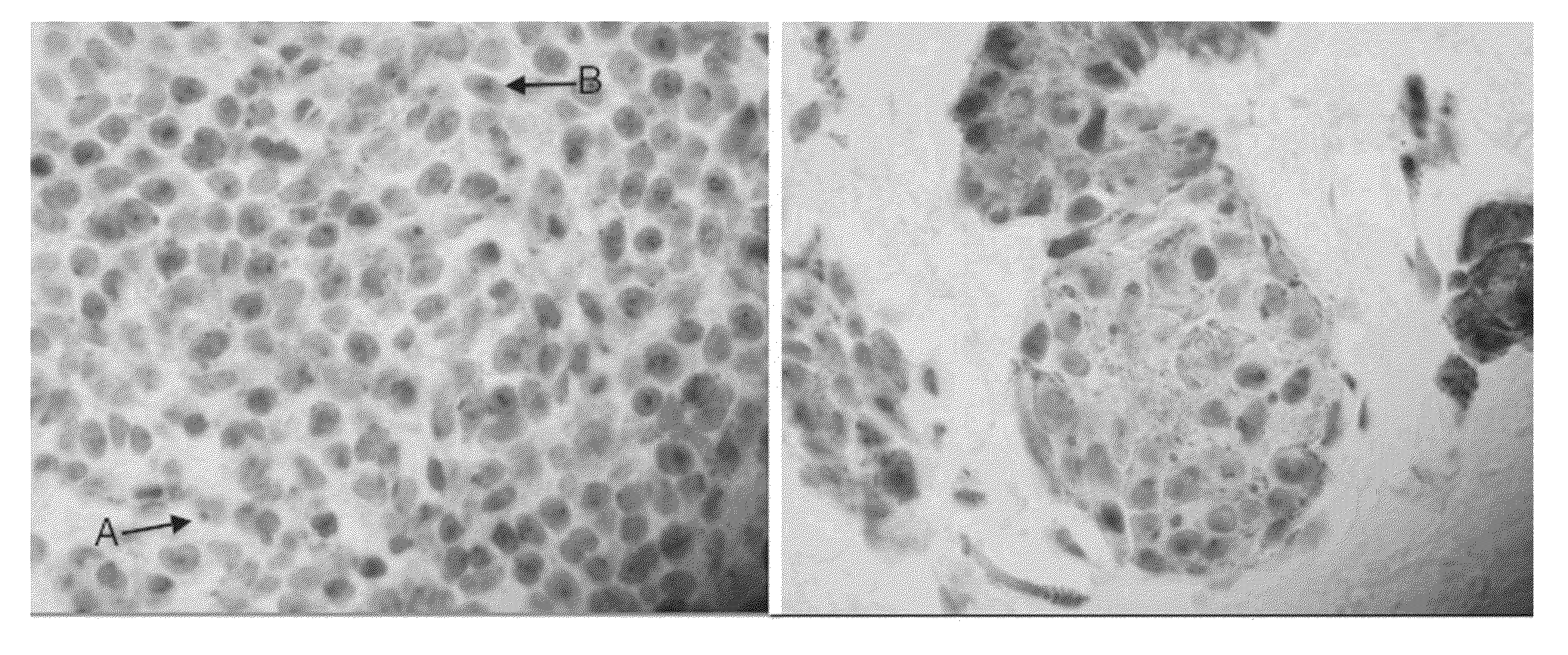
[0110]This example demonstrates the staining of a tissue sample with R21 antibody against sAC to provide an sAC staining pattern. All steps were performed using the Leica Microsystems BOND-MAX™ autostainer (Bannockburn, Ill.).

[0111]Dewaxing: Formalin-fixed, paraffin embedded samples of lesional melanocytes were baked at 60° C. for 30 minutes. Slides were then treated with a Leica Microsystems BOND DEWAX SOLUTION™ (AR9222) for 3 minutes at 72° C., followed by washes in BOND DEWAX SOLUTION™ first at 72° C. and then at ambient temperature. Finally, slides were washed three times with ethyl alcohol 200-proof (Pharmco-Aaper, Brookfield, Conn., 111000200) and three times with Leica Microsystems BOND WASH SOLUTION™ (AR9590).

[0112]Pre-treatment and blocking: Following the dewaxing procedure, the sections were pre-treated by two washes in Leica Microsystems BOND EPITOPE RETRIEVAL SOLUTION 1™ (AR9961), followed by BOND EPITOPE RETRIEVAL SOLUTION 1™ pre-treatment for 30 minutes at 100° C., and...

example 2

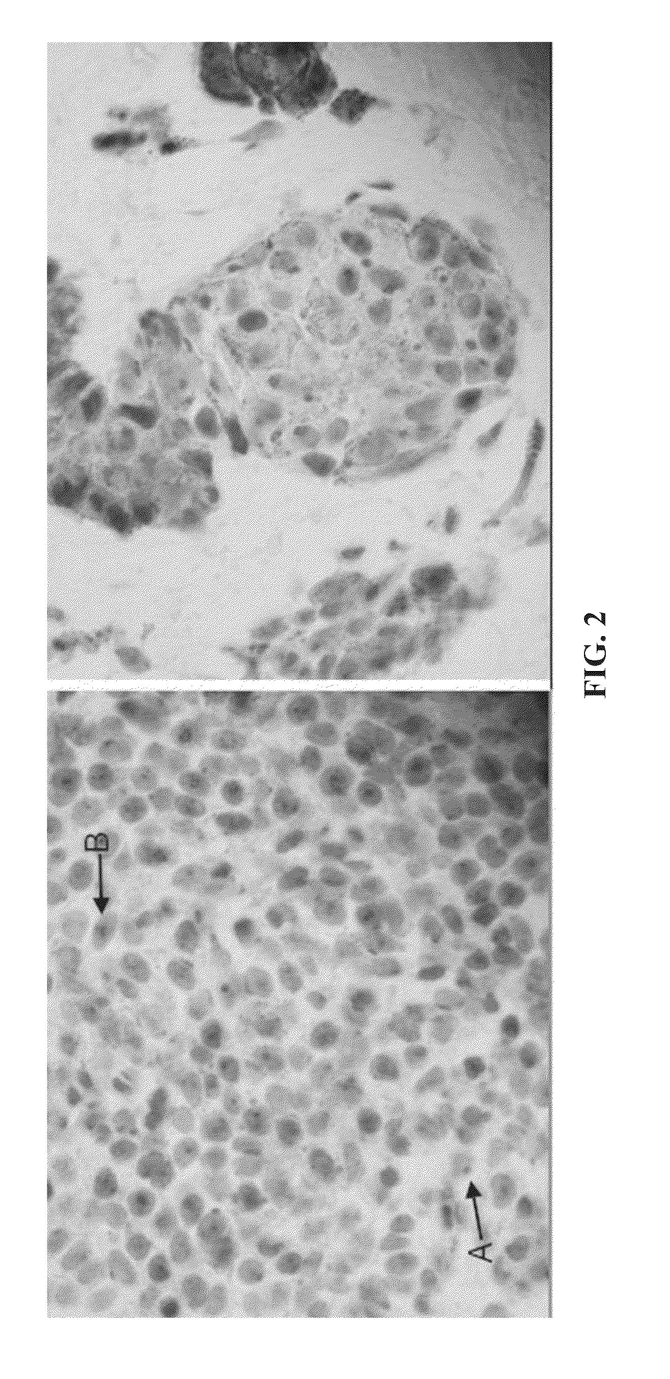
[0116]This example demonstrates that the analysis of a sAC staining pattern of a sample of lesional melanocytes from a subject can be used to provide a diagnosis of melanocytic proliferations for the subject.

[0117]Samples of lesional melanocytes were obtained from 140 different subjects and subjected to the procedures described in Example 1 to obtain or discern sAC staining patterns. The 140 sAC staining patterns were analyzed and grouped into categories pertaining to different diagnoses of melanocytic proliferations. The results of this analysis provided the following sAC staining pattern characteristics for each of the diagnoses of melanocytic proliferations.

(1) Benign nevus and capsular nevus of lymph nodes[0118](a) Dot-like Golgi staining in >50% of lesional melanocytes[0119](b) Weak nucleolar and incomplete granular nuclear staining in 50% of lesional melanocytes, on average[0120](c) Pan nuclear staining in <10% of lesional melanocytes

(2) Atypical nevus of special sites[0121](a...

example 3

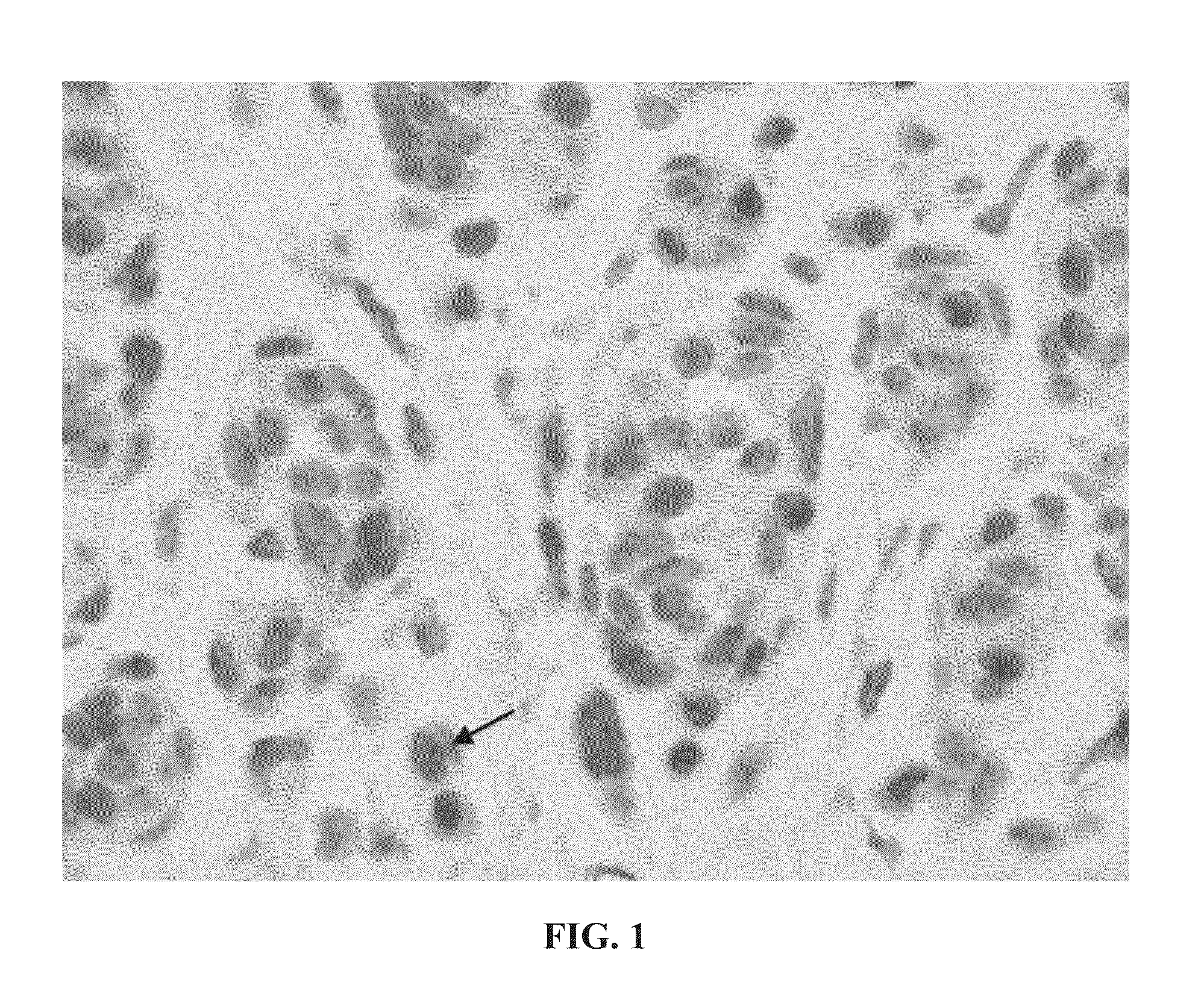
[0163]This example compares the dot-like Golgi and pan-nuclear staining patterns in dysplastic nevi to the dot-like Golgi and pan-nuclear staining patterns in melanoma.

[0164]Loss of dot-like Golgi staining with gain of pan-nuclear staining appears to be indicative of melanoma. In the following table (Table 1), loss of dot-like Golgi staining (DG) is assumed when 25% total melanocytes have this staining pattern (range for nevi 0-25%; range for melanomas 50-90%). Melanomas were compared to a combined group of all moderately and severely dyspalstic nevi, with “all melanomas” representing superficial spreading, acral lentiginous, nodular, and lentigo maligna melanomas, and “SSM” representing superficial spreading melanomas.

[0165]To determine if particular staining patterns were present or absent in a given type of lesion more often than simple chance, a binomial proportion test was performed (50% assumption being simple chance) with a two-sided student t-test. To determine if there was ...


PUM

PropertyMeasurementUnit
sizeaaaaaaaaaa
temperatureaaaaaaaaaa

Abstract

The invention provides a method for diagnosing a melanocytic proliferation in a subject comprising staining a sample of lesional melanocytes with an antibody against soluble adenylyl cyclase (sAC) and interpreting the sAC staining pattern, which is associated with a diagnosis of a melanocytic proliferation. The sAC staining pattern, which is complex, is discriminatory and distinctive according to the nature of the melanocytic proliferation. The sAC staining pattern comprises one or more of dot-like Golgi staining, broad granular Golgi staining, diffuse cytoplasmic staining, nucleolar staining, incomplete granular nuclear staining, and pan-nuclear staining. The method of the invention is particularly useful in confirming or disaffirming a diagnosis reached through conventional histologic examination of a sample. Additionally, the invention provides a kit for use in interpreting melanocytic proliferations.

Description

CROSS-REFERENCE TO RELATED APPLICATIONS[0001]This application claims the benefit of U.S. Provisional Patent Application No. 61 / 326,174, filed Apr. 20, 2010, the entire content of which is incorporated herein by reference.INCORPORATION-BY-REFERENCE OF MATERIAL SUBMITTED ELECTRONICALLY[0002]Incorporated by reference in its entirety herein is a computer-readable nucleotide / amino acid sequence listing submitted concurrently herewith and identified as follows: One 21,813 Byte ASCII (Text) file named “707638_ST25.TXT,” created on Apr. 6, 2011.BACKGROUND OF THE INVENTION[0003]Melanocytes, i.e., pigment-producing cells in the skin, eye, and mucosal surfaces, give rise to a spectrum of proliferative lesions ranging from benign nevi to overtly malignant melanoma. Melanoma is a disease with high metastatic potential even at very early stages of development. Melanoma accounts for less than 5% of skin cancer cases, but causes a large majority of skin cancer deaths (Cancer Facts &Figures, America...

Claims


Application Information

Patent Timeline
Patent Type & Authority Applications(United States)
IPC IPC(8): G01N33/574G01N33/577
CPCC12Q1/527G01N33/5026G01N33/5044G01N2800/56G01N33/5743G01N33/6893G01N2800/20G01N33/5076
Inventor MAGRO, CYNTHIAZIPPIN, JONATHANLEVIN, LONNY R.BUCK, JOCHEN
Owner CORNELL UNIVERSITY
Why Eureka
Try Eureka