
Medical tomosynthesis system

a tomosynthesis and synthesis technology, applied in the field of medical tomosynthesis system, can solve the problem that the accuracy of the x-position (horizontal) may propagate into an uncertain z-position

Inactive Publication Date: 2013-04-11
KONINKLIJKE PHILIPS ELECTRONICS NV
View PDF4 Cites 3 Cited by
  • Summary
  • Abstract
  • Description
  • Claims
  • Application Information

AI Technical Summary

Benefits of technology

The invention replaces two regular exposures with multiple left and right exposures to improve the three-dimensional visualization of a target for biopsy. By using a dose-saving technique and a three-dimensional geometrical model of the biopsy unit, the invention saves X-ray dose and provides a wider field of use. The invention also simplifies the workflow because it does not have to consider the shadowing caused by the biopsy needle and holder. Overall, the invention provides a more accurate and efficient tool for biopsy procedures.

Problems solved by technology

However, the accuracy of the x-position (horizontal) may propagate into an uncertain z-position.

Method used


Image

  • Medical tomosynthesis system
  • Medical tomosynthesis system
  • Medical tomosynthesis system

Examples


Embodiment Construction

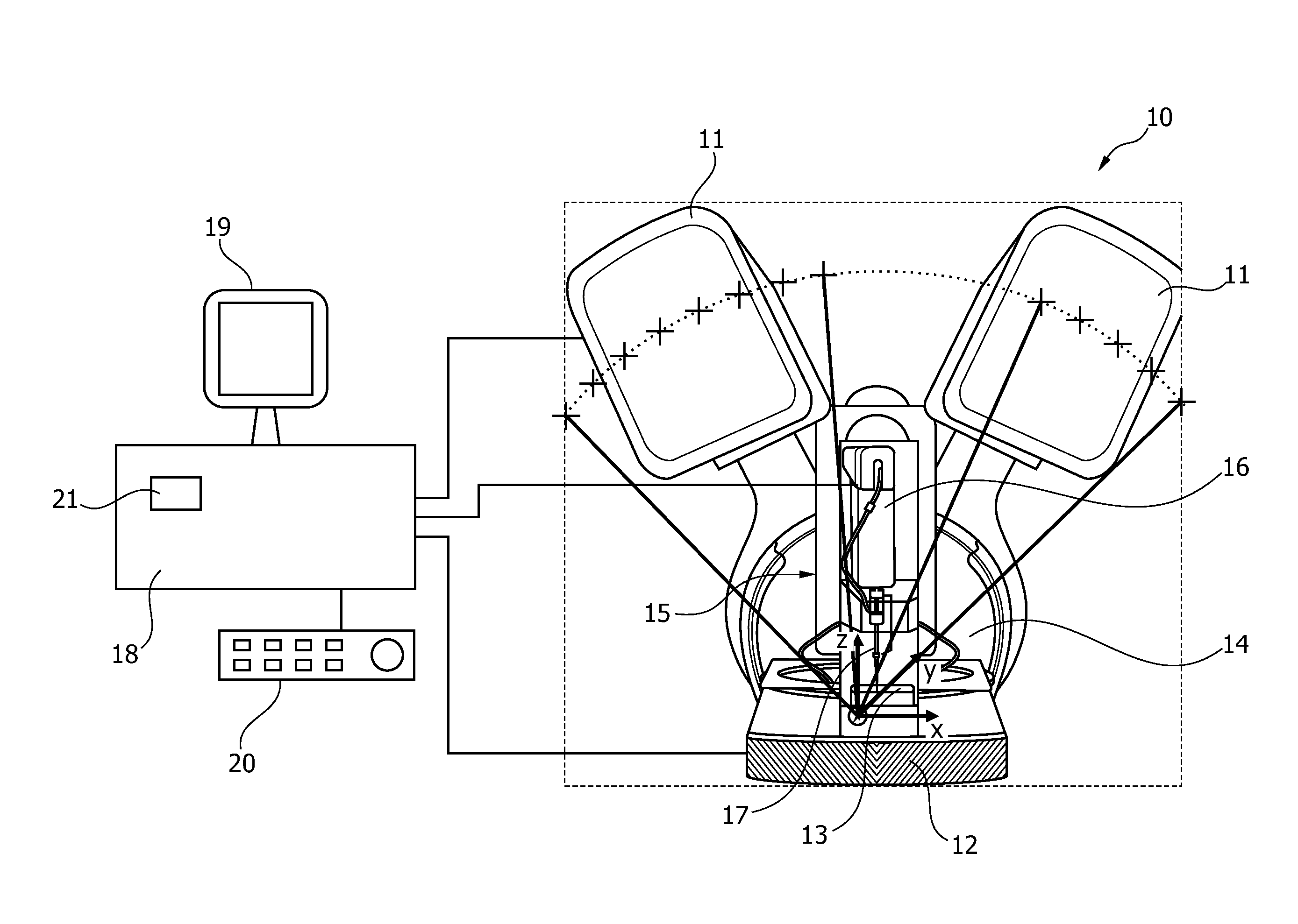
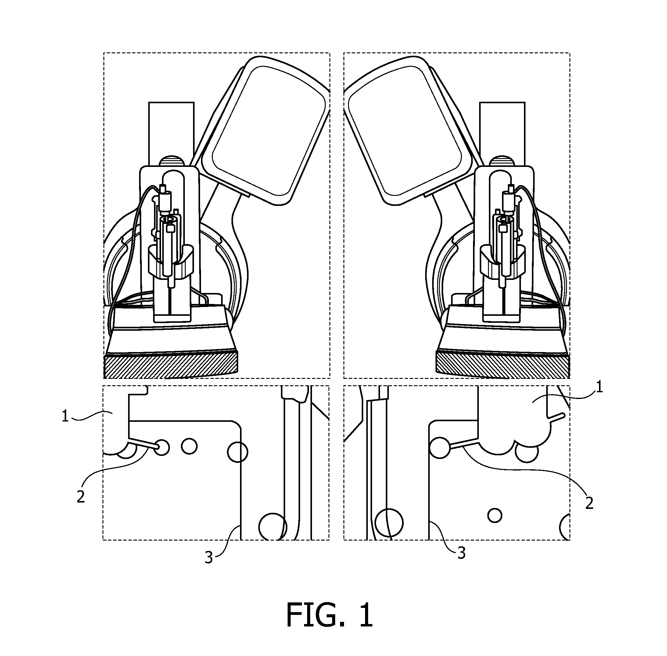
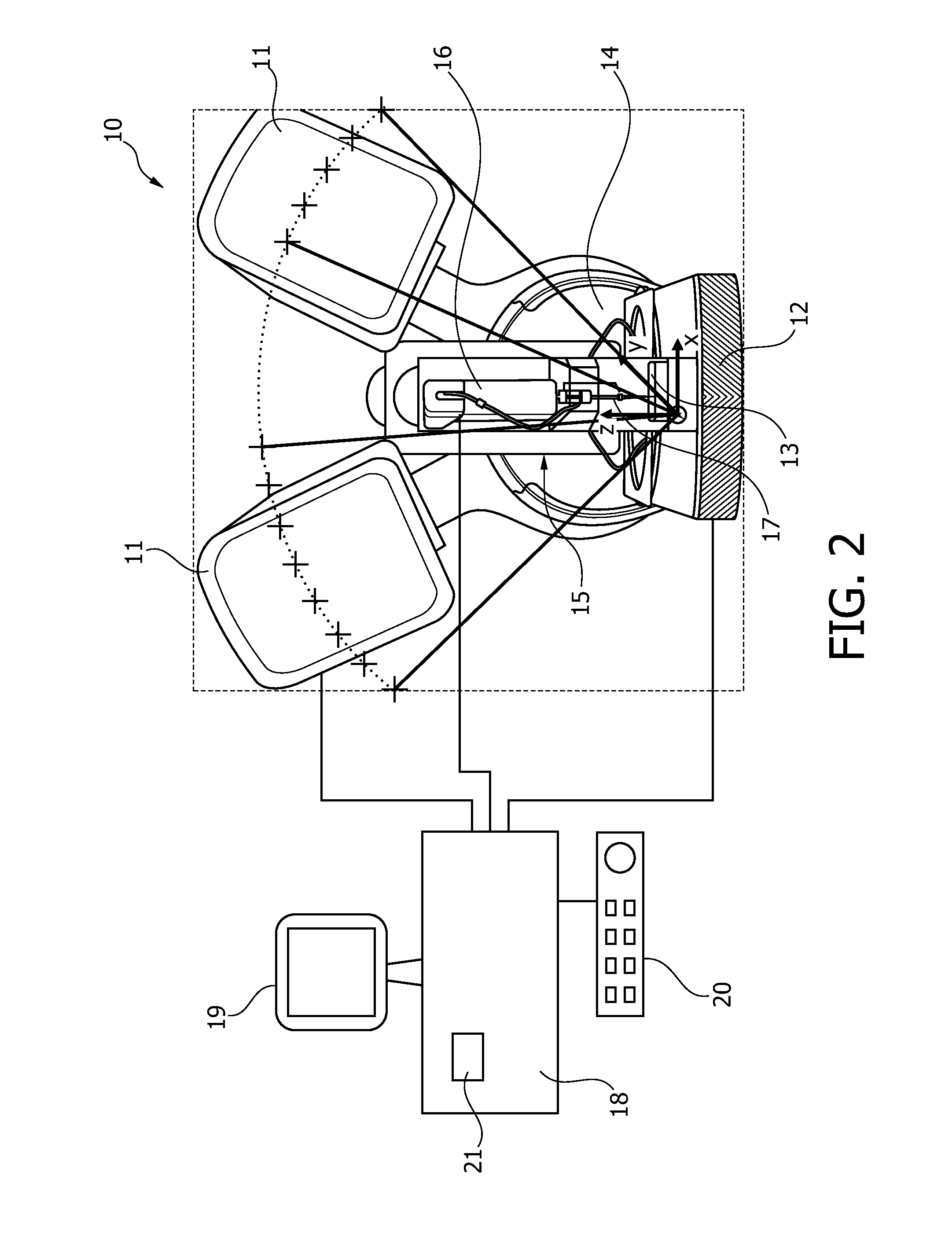
[0020]The present invention is directed to a medical tomosynthesis system which can be used for diagnosing breast cancer and for guiding an interventional tool or device, such as a biopsy needle, to a lesion for taking a tissue sample. Tomosynthesis is a medical imaging method which acquires several x-ray images at different angles from a subject volume, such as a human breast. From this set of projection images a three-dimensional (3D) data set is derived. This 3D data set is the basis for reconstructing two-dimensional (2D) slices or slabs at desired positions and with a desired orientation, wherein the slices or slabs are usually in parallel to the xy-plane, xz-plane or yz-plane. One slice represents a very thin volume element in form of a plane being 0.5 to 1 mm thick. One slab represents an average of some adjacent slices and corresponds to a volume element of some mm or cm of the subject volume. Only the first imaging for determining the 3D model of the biopsy unit is done as ...


PUM


Abstract

The invention relates to a medical tomosynthesis system (10) having an interventional device (15) and an image acquisition device (11, 12) for acquiring images of a subject volume in a plurality of angular positions around the subject volume. In the system, a three-dimensional geometrical model of the interventional unit is used to identify the projection angles of the image acquisition device that actually can be used. Preferably, this three-dimensional model is achieved by reconstructing it from projection images acquired with the X-ray image acquisition device. The invention also relates to a method for acquiring images with such a system.

Description

FIELD OF THE INVENTION[0001]The present invention relates to a medical tomosynthesis system, a method for acquiring medical images and a computer program for executing such a method.BACKGROUND OF THE INVENTION[0002]Regular stereoscopic guidance is done using two predefined angular positions of an X-ray tube for X-ray images, wherein one angular position is left and the other one is right of the centre position. The two coordinates of a specific lesion in the two images can be used to identify all three coordinates of the lesion in the object of examination. To identify the z-position of a lesion, a cross hair cursor has to be set in the two stereo images. The z-position can be calculated from these values. However, the accuracy of the x-position (horizontal) may propagate into an uncertain z-position. A tissue sample can be taken at this position using a biopsy needle.[0003]In tomosynthesis imaging several x-ray images are acquired in different angles from a subject volume, such as ...

Claims


Application Information

Patent Timeline
Patent Type & Authority Applications(United States)
IPC IPC(8): A61B6/02A61B10/02A61B6/00A61B6/12
CPCA61B6/022A61B6/025A61B6/0414A61B6/12A61B10/0233A61B6/502A61B6/582A61B2019/507A61B6/5205A61B6/4494A61B2034/107
Inventor MAACK, HANNS-INGO
Owner KONINKLIJKE PHILIPS ELECTRONICS NV
Learn More