
Apparatus and methods for cooling positron emission tomography scanner detector crystals

a technology of positron emission tomography and detector crystals, which is applied in tomography, x/gamma/cosmic radiation measurement, instruments, etc., can solve the problems of insufficient control of localized temperature zones in the scanner gantry, and achieve the effect of increasing the sensitivity of the detector and stabilizing the gantry

Inactive Publication Date: 2013-05-16
SIEMENS MEDICAL SOLUTIONS USA INC
View PDF2 Cites 24 Cited by
  • Summary
  • Abstract
  • Description
  • Claims
  • Application Information

AI Technical Summary

Benefits of technology

The technical effect of this patent is to maintain a stable temperature for the detector scintillation crystals in a PET scanner during operation, which increases sensitivity and reduces the amount of cooling energy needed. This is achieved without reducing the sensitivity of the scanner.

Problems solved by technology

However, localized temperature zones within the scanner gantry are not controlled with sufficient precision to assure constant operational temperature needed to maximize scintillation crystal detector sensitivity.

Method used


Image

  • Apparatus and methods for cooling positron emission tomography scanner detector crystals
  • Apparatus and methods for cooling positron emission tomography scanner detector crystals
  • Apparatus and methods for cooling positron emission tomography scanner detector crystals

Examples


Embodiment Construction

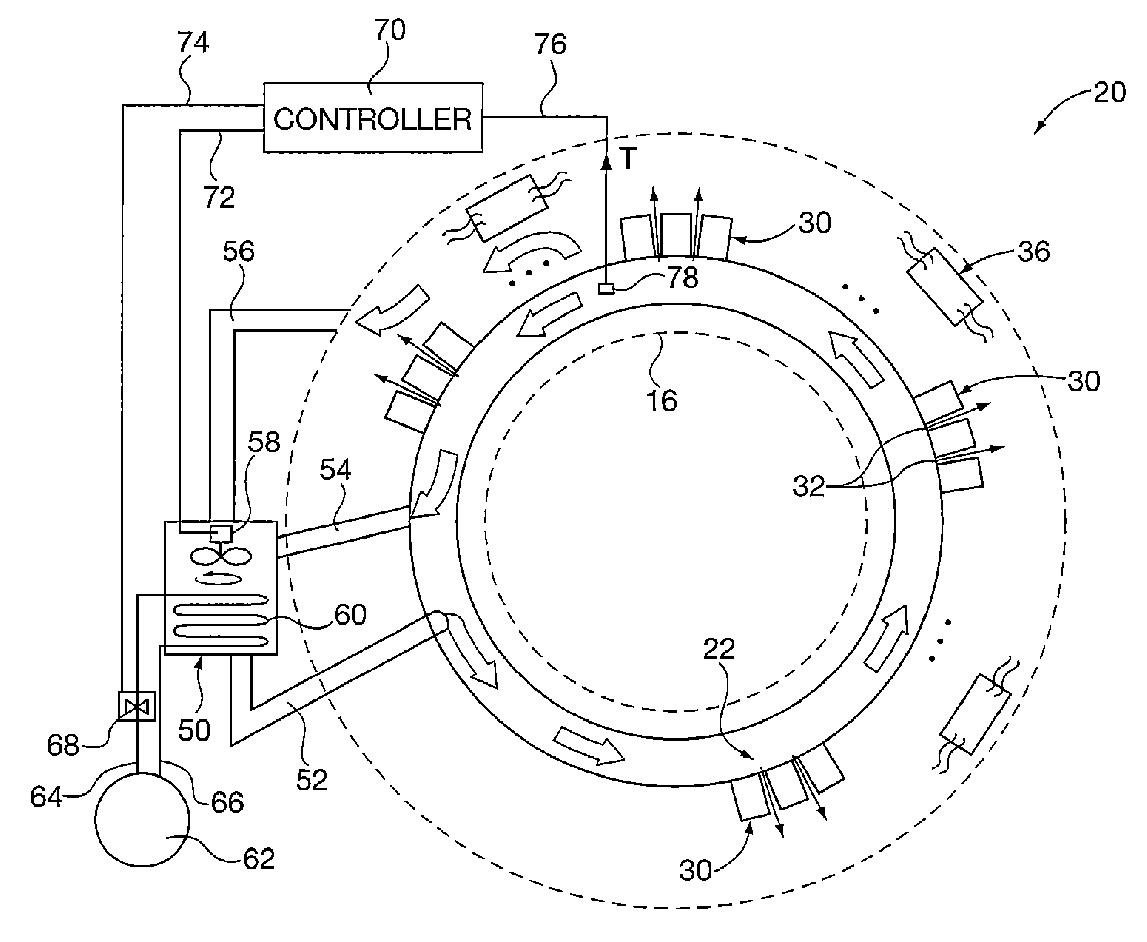
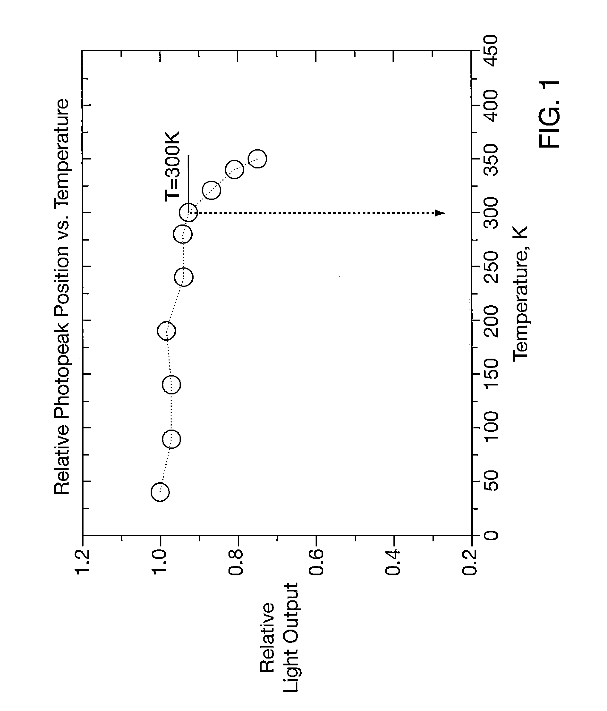
[0023]After considering the following description, those skilled in the art will clearly realize that the teachings of the present invention can be readily utilized in positron emission tomography (PET) scanner apparatus to cool detector crystals. Further, cooling the detector crystals is achieved without adding materials between the detector crystal face and the patient—thus not sacrificing scanner sensitivity. In the case of LSO material detector crystals, maintaining an operating temperature below 81° F. (27° C.) enhances their light output, and hence detector sensitivity.

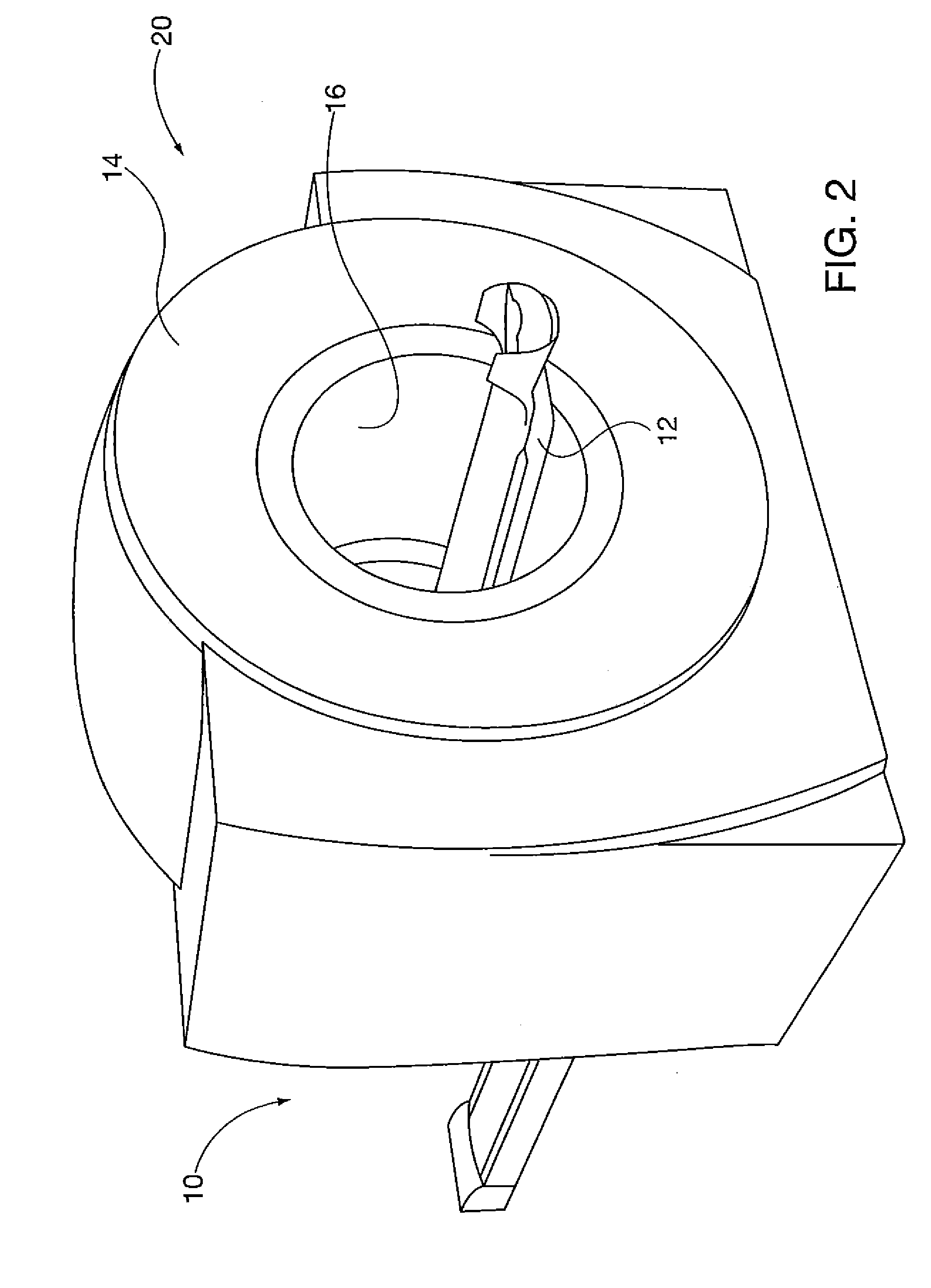
[0024]FIGS. 2-7 show embodiments of the detector crystal cooling apparatus and methods of the present invention. FIG. 2 shows a PET scanner 10 having a patient bed 12 that translates relative to a scanning field. General structure and operation of the scanning components used to form an image representative of the scanned area of interest in the patient is known to those skilled in the art, and for brevity is no...


PUM


Abstract

Detector crystals in a positron emission tomography (PET) apparatus gantry are cooled by directing cooling gas flow into a cooling duct bounded by the crystals and a cover defining the patient scanning field within the gantry. The cooling gas cools the crystals. Cooling gas may also be directed radially outwardly from the cooling duct into spatial gaps defined between detector enclosures that include the crystals, further isolating heat generated by other components within gantry from the detector crystals. Cooling gas is provided by a cooling system that may be incorporated within the gantry, external the gantry or a combination of both. Cooling gas can be provided by directing air within the gantry in contact with internal gantry cooling tubes and routing cooled air directly into the cooling duct with a powered fan.

Description

BACKGROUND OF THE DISCLOSURE[0001]1. Field of the Invention[0002]The invention relates to positron emission tomography (PET) scanners used to image areas of interest in patients, and particularly apparatus and methods for cooling PET scanner detector scintillation crystals, so that they are maintained at a stabilized temperature selected for desired detector performance.[0003]2. Description of the Prior Art[0004]PET scanners image areas of interest in patients who have ingested radioactive imaging solutions. The scanner utilizes an array of scintillation crystals in a generally annular scanner gantry to detect radioactive particle emissions from the patient; and then correlates those emissions with patient tissue structure. One known scintillation crystal material is lutetinium oxy-orthosilicate (LSO). In PET scanner operation, a radioactive particle emitted from the patient striking a detector crystal within a detector element causes a light emission. That light emission is in turn...

Claims


Application Information

Patent Timeline
Patent Type & Authority Applications(United States)
IPC IPC(8): G01T1/164
CPCG01T1/1648A61B6/037A61B6/4488
Inventor MARTIN, MATTHEW ERIC
Owner SIEMENS MEDICAL SOLUTIONS USA INC
Learn More