Methods of inhibiting photoreceptor apoptosis
a photoreceptor and apoptosis technology, applied in the direction of drug compositions, peptide/protein ingredients, immunological disorders, etc., can solve the problem of lack of molecular specificity of therapies
- Summary
- Abstract
- Description
- Claims
- Application Information
AI Technical Summary
Benefits of technology
Problems solved by technology
Method used
Image
Examples
example 1
Compositions and Methods
Experimental Model of Retinal Detachment
[0062]All experiments performed during the development of embodiments of the present invention were performed in accordance with the ARVO Statement for the Use of Animals in Ophthalmic and Vision Research and the guidelines established by the University Committee on Use and Care of Animals of the University of Michigan. Detachments were created in adult male Brown-Norway rats (300-400 g) (Charles River Laboratories, Wilmington, Mass.), wildtype C57BL mice (age 3-6 weeks) (Jackson Laboratory, Bar Harbor, Me.), and IL-6− / − mice on a C57BL background (age 3-6 weeks) (Jackson Laboratory, Bar Harbor, Me.)(Zacks et al. Arch Ophthalmol. 2007; 125(10):1389-1395; herein incorporated by reference in its entirety). Rodents were anesthetized with a 50:50 mix of ketamine (100 mg / mL) and xylazine (20 mg / mL), and pupils were dilated with topical phenylephrine (2.5%) and tropicamide (1%). A 20-gauge microvitreoretinal blade (Walcott Sc...
example 2
Interleukin-6 as a Photoreceptor Neuroprotectant in an Experimental Model of Retinal Detachment
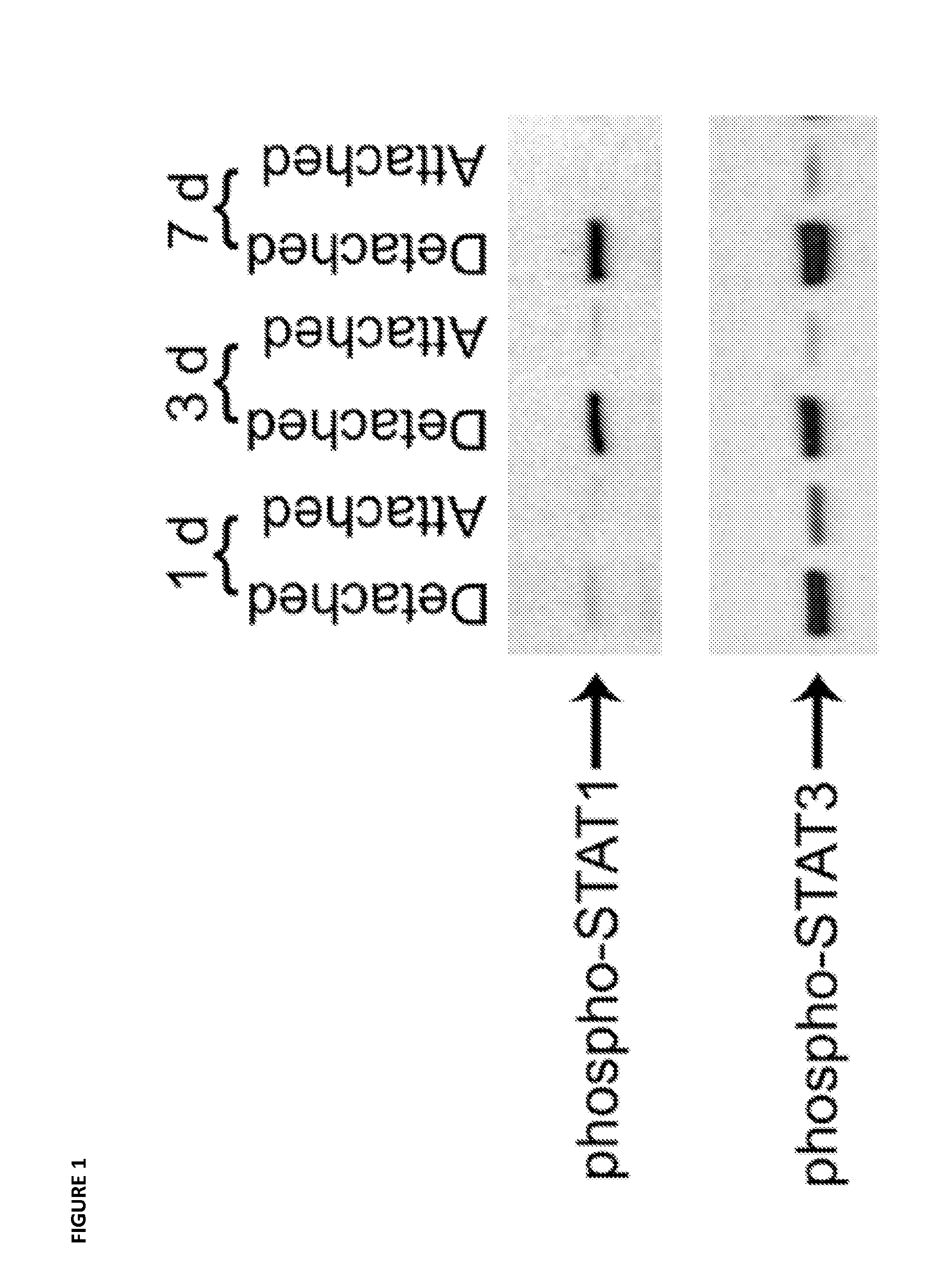
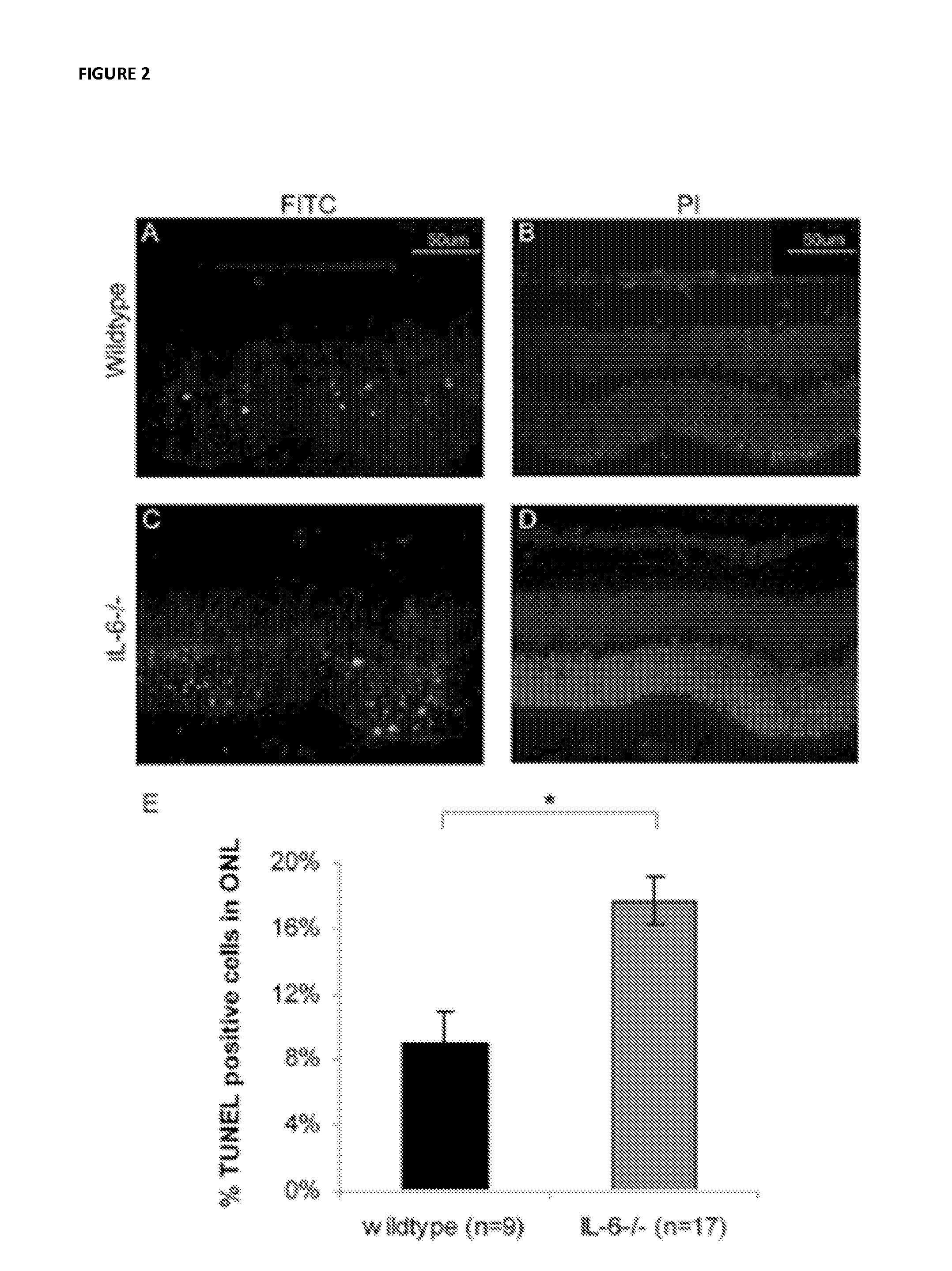
[0067]The immediately downstream transducers of IL-6 receptor signaling are Signal Transducers and Activators of Transcription (STATs) (Heinrich et al. Biochem J. 2003; 374(Pt 1):1-20, Samardzija et al. FASEB J. 2006; 20(13):2411-2413; herein incorporated by reference in their entireties). In the context of retinal-RPE separation, STAT1 and STAT3 transcript and protein levels are increased (Zacks et al. Invest Ophthalmol Vis Sci. 2006; 47(4):1691-1695, herein incorporated by reference in its entirety). Increased phosphorylation (i.e., activation) of STAT1 and STAT3 in detached retinas compared to attached retinas. Injection of the IL-6 neutralizing antibody into the subretinal space at the time of detachment resulted in approximately a 50% reduction in the level of phosphorylated STAT3. There was not any reduction in the level of phosphorylated STAT1. These data suggest that after retinal-...
example 3
Compositions and Methods Relating to X-Link Inhibitor of Apoptosis
Animals
[0072]Adult male, Brown Norway rats, at least 6 weeks of age, were purchased from Harlan (Indianapolis, Ind.) and Charles River Laboratories (Wilmington, Mass.). Animals were maintained under standard laboratory conditions and all procedures conformed to both the ARVO Statement for the Use of Animals in Ophthalmic and Vision Research and the guidelines of the University of Ottawa Animal Care and Veterinary Service. Rats were divided into two groups, with one group receiving XIAP gene therapy and the other receiving the green fluorescent protein (GFP) control.
Construction of the Recombinant Adeno-Associated Virus (rAAV) Vectors
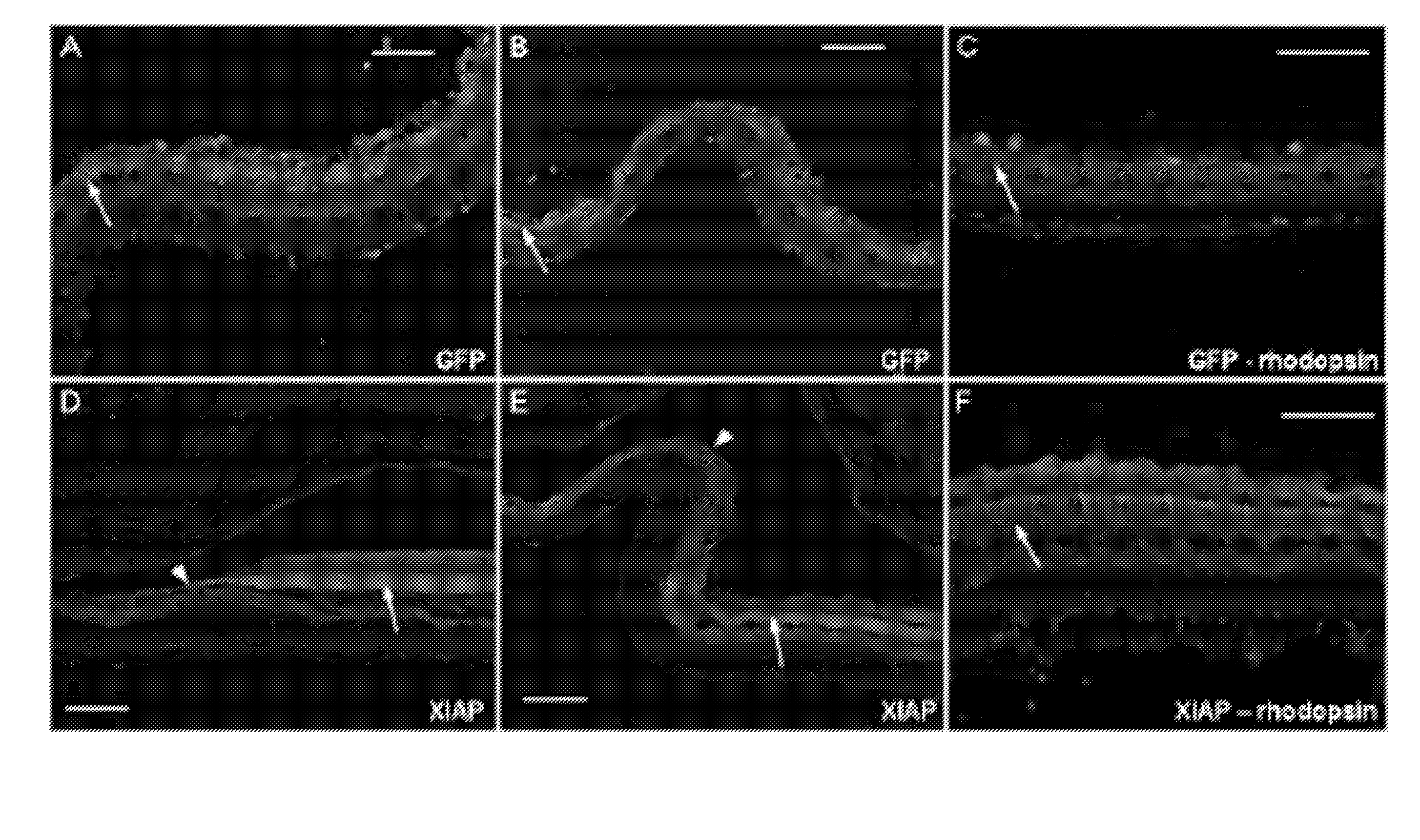
[0073]A cDNA construct encoding the full-length, open-reading frame of human XIAP with an Nterminal hemagglutinin (HA) tag was inserted into a pTR vector under the control of the chicken β-actin promoter. A GFP construct was similarly generated for use as a surgical and viral control. X-li...
PUM
 Abstract
Description
Claims
Application Information
 - R&D
- Intellectual Property
- Life Sciences
- Materials
- Tech Scout
- Unparalleled Data Quality
- Higher Quality Content
- 60% Fewer Hallucinations
Browse by: Latest US Patents, China's latest patents, Technical Efficacy Thesaurus, Application Domain, Technology Topic, Popular Technical Reports.
© 2025 PatSnap. All rights reserved.Legal|Privacy policy|Modern Slavery Act Transparency Statement|Sitemap|About US| Contact US: help@patsnap.com


