Circulating tumor cells (CTC's): early assessment of time to progression, survival and response to therapy in metastatic cancer patients

a tumor cell and tumor prognosis technology, applied in the field of cancer prognosis and survival in metastatic cancer patients, can solve the problems of difficult detection and elimination, inability to treat all patients successfully, and inability to improve the survival rate of cancer patients over the past two decades, so as to achieve less side effects, improve survival rate, and improve the effect of quality of li

Inactive Publication Date: 2007-02-15
JANSSEN DIAGNOSTICS LLC
View PDF4 Cites 79 Cited by
  • Summary
  • Abstract
  • Description
  • Claims
  • Application Information

AI Technical Summary

Benefits of technology

[0023] The present invention is a method and means for cancer prognosis, incorporating diagnostic tools, such as the Cell Spotter® System, in assessing time to disease progression, survival, and response to therapy based upon the absolute number, change, or combinations of both of circulating epithelial cells in patients with metastatic cancer. The system immunomagnetically concentrates epithelial cells, fluorescently labels the cells, identifies and quantifies CTC's for positive enumeration. The statistical analysis of the cell count predicts survival.
[0024] More specifically, the present invention provides the apparatus, methods, and kits for assessing patient survival, the time to disease progression, and response to therapy in MBC. Prediction of survival is based upon a threshold comparison of the number of circulating tumor cells in blood with time to death and disease progression. Statistical analysis of long term follow-up studies of patients diagnosed with cancer established a threshold for the number of CTC found in blood and prediction of survival. An absence of CTC's is defined as fewer than 5 morphologically intact CTC's. The presence or absence of CTC's to predict survival is useful in making treatment choices. For example, the absence of CTC's in a woman previously untreated for metastatic breast cancer could be used to select hormonal therapy vs chemotherapy with less side effects and higher quality of life. In contrast, the presence of CTC's could be used to select chemotherapy which has higher side effects but may prolong survival more effectively in a high risk population. Thus, the invention has a prognostic role in the detection of CTC's in women with metastatic breast cancer.

Problems solved by technology

Despite efforts to improve treatment and management of cancer patients, survival in cancer patients has not improved over the past two decades for many cancer types.
Unfortunately, the metastatic colonies are difficult to detect and eliminate and it is often impossible to treat all of them successfully.
These and other debris antigens are thought to be derived from destruction of both circulating and non-circulating tumor cells, and thus their presence may not always reflect metastatic potential, especially if the cells rupture while in an apoptotic state.
Assessing enzyme activity in this type of analysis can involve time-consuming laboratory procedures such as gel electrophoresis and Western blot analysis.
However in both cases, the base levels during remission, or even in healthy normals, are relatively high and may overlap with concentrations found in patients, thus requiring multiple testing and monitoring to establish patient-dependent baseline and cut-off levels.
However, PSA or the related PSMA testing leaves much to be desired.
For example, elevated levels of PSA weakly correlate with disease stage and appear not to be a reliable indicator of the metastatic potential of the tumor.
This is being done through the laborious procedure of isolating all of the mRNA from the blood sample and performing reverse transcriptase PCR.
Additionally, false positives are often observed using this technique.
There is an added drawback, which is that there is a finite and practical limit to the sensitivity of this technique based on the sample size.
Again using tyrosinase mRNA as a soluble tumor marker is subject to the previously cited limitations of soluble tumor antigens as indicators of metastatic potential and patient survival.
The aforementioned and other studies, while seemingly prognostic under the experimental conditions, do not provide for consistent data with a long follow-up period or at a satisfactory specificity.
Accordingly, these efforts have proven to be somewhat futile as the appearance of mRNA for antigens in blood have not been generally predictive for most cancers and are often detected when there is little hope for the patient.
These results suggest that tumor cells were shed into the bloodstream (possibly during surgical procedures or from micro metastases already existing at the time of the operation), and resulted in poor patient outcomes in patients with colorectal cancer.
As mentioned, these detection ranges are based on unreliable conversions of amplified product to the number of tumor cells.
Further, PCR-based assays are limited by possible sample contamination, along with an inability to quantify tumor cells.
Most importantly, methods based on PCR, flowcytometry, cytoplasmic enzymes and circulating tumor antigens cannot provide essential morphological information confirming the structural integrity underlying metastatic potential of the presumed CTC and thus constitute functionally less reliable surrogate assays than the highly sensitive imaging methods embodied, in part, in this invention.
Unfortunately, the same spreading of malignant cells continues to be missed by conventional tumor staging procedures.
But these invasive techniques are deemed undesirable or unacceptable for routine or multiple clinical assays compared to detection of disseminated epithelial tumor cells in blood.
Currently available prognostic protocols have not demonstrated a consistently reliable means for correlating CTC's to predict progression free- or overall survival in patients with cancers such as metastatic breast cancer (MBC).

Method used


Image

  • Circulating tumor cells (CTC's): early assessment of time to progression, survival and response to therapy in metastatic cancer patients
  • Circulating tumor cells (CTC's): early assessment of time to progression, survival and response to therapy in metastatic cancer patients
  • Circulating tumor cells (CTC's): early assessment of time to progression, survival and response to therapy in metastatic cancer patients

Examples


example 1

Enumeration of Circulating Cytokeratin Positive Cells Using CellPrep™

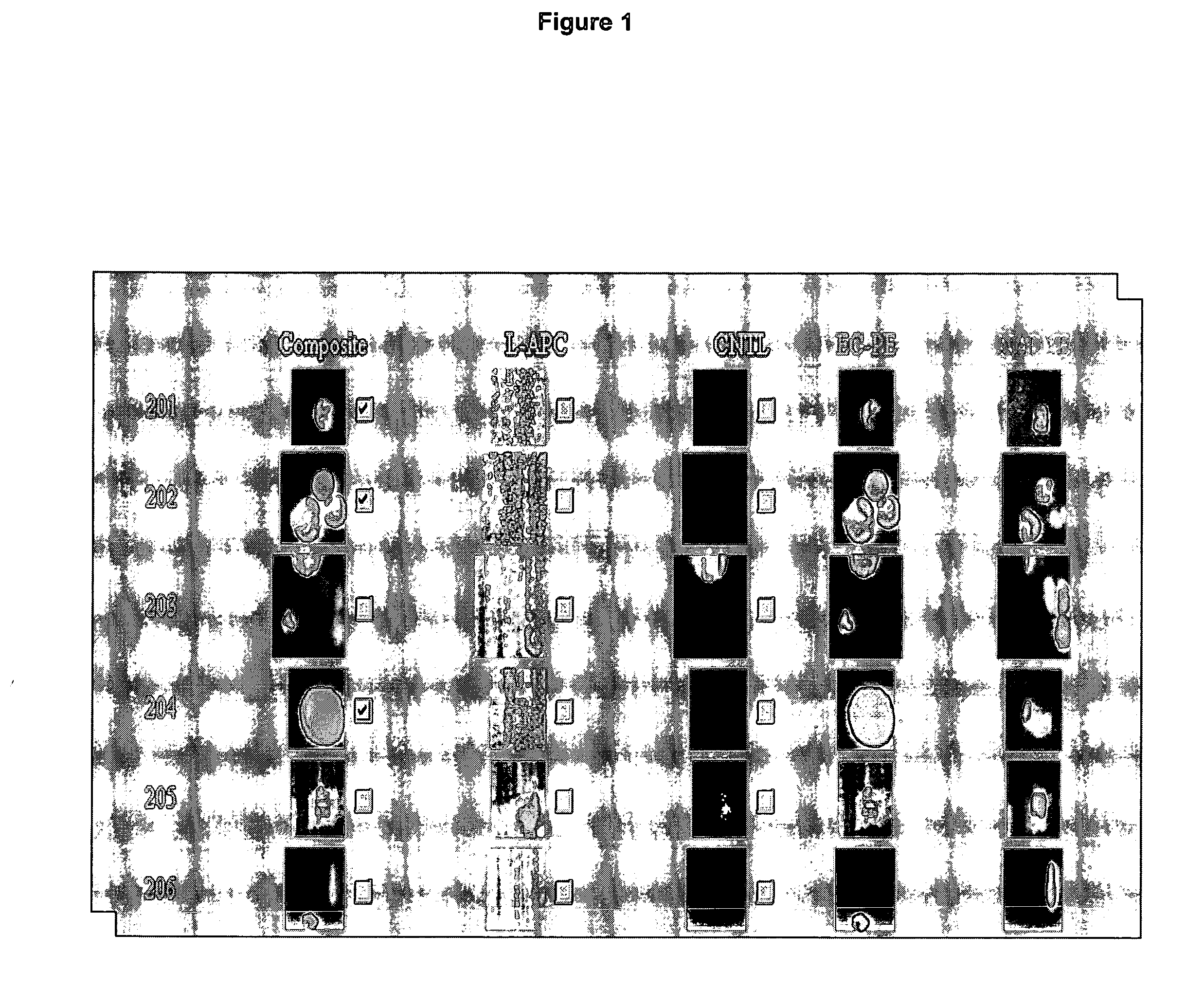
[0044] Cytokeratin positive cells are isolated by the CellPrep™ System using a 7.5 ml sample of whole blood. Epithelial cell-specific immunomagnetic fluid is added and incubated for 20 minutes. After magnetic separation for 20 minutes, the cells bound to the immunomagnetic-linked antibodies are magnetically held at the wall of the tube. Unbound sample is then aspirated and an isotonic solution is added to resuspend the sample. A nucleic acid dye, monoclonal antibodies to cytokeratin (a marker of epithelial cells) and CD 45 (a broad-spectrum leukocyte marker) are incubated with the sample for 15 minutes. After magnetic separation, the unbound fraction is again aspirated and the bound and labeled cells are resuspended in 0.2 ml of an isotonic solution. The sample is suspended in a cell presentation chamber and placed in a magnetic device whose field orients the magnetically labeled cells for fluorescence microscopic...

example 2

Assessment of the Tumor Load: Comparison between Radiographic Image Analysis and the Absolute Number of CTC's.

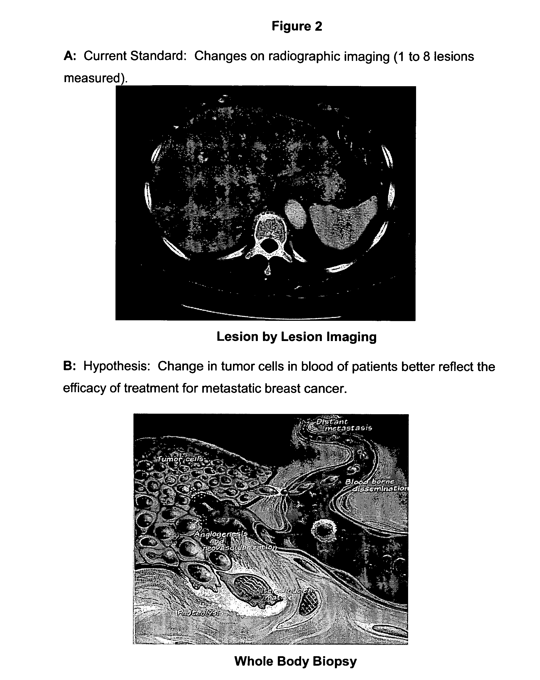
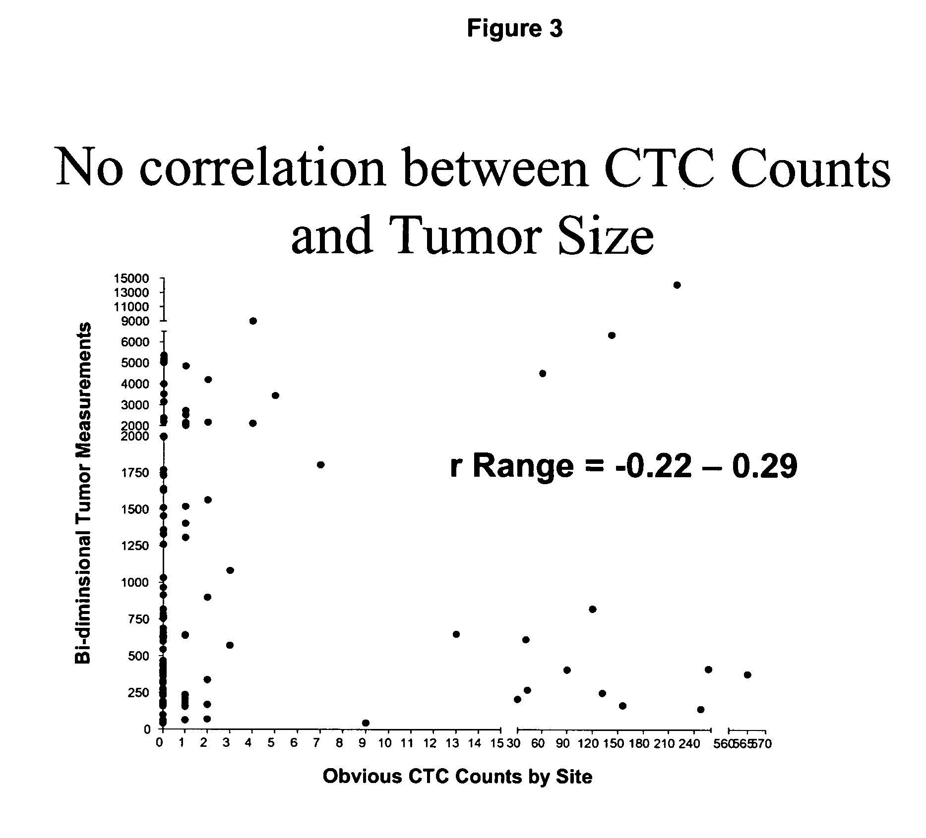
[0045] Radiographic measurements of metastatic lesions are currently used to assess tumor load in cancer patients with metastatic disease. In general, the largest lesions are measured and summed to obtain a tumor load. An example of a bidimensional measurement of a liver metastasis in a breast cancer patient is illustrated in FIG. 2A. A model depicting the necessity for measuring tumor load in the blood stream is illustrated in FIG. 2B as a measurement of the actual active tumor load, and thus a better measure of the overall activity of the disease. To determine whether or not the absolute number of CTC's correlated with the dimension of the tumor measured by imaging a prospective study in patients with MBC was performed.

[0046] The Cell Spotter® System was used to enumerate CTC's in 7.5 ml of blood from 69 patients with measurable MBC. Tumor load was assessed by bi-dimens...

example 3

Assessment of the Tumor Load: Comparison Between Changes in the Radiographic Image and Changes in the Absolute Number of CTC's.

[0049] Radiographic imaging is the current standard to assess whether a particular disease is responding, stabilizing, or progressing to treatment. The interval between radiographic measurements must be at least 3 months in order to give meaningful results. Consequently, a test that could predict response to therapy earlier during the treatment cycle would improve the management of patients treated for metastatic diseases, potentially increase quality of life and possibly improve survival. In this study, patients starting a new line of treatment for MBC were assessed to determine whether a change in the number of CTC's correlated with a change in patient status as measured by imaging.

[0050] The Cell Spotter® System was used to enumerate CTC's in 7.5 ml of blood in MBC patients about to start a new therapy, and at various time points during the treatment c...


PUM

PropertyMeasurementUnit
timeaaaaaaaaaa
magnetic fieldaaaaaaaaaa
densityaaaaaaaaaa

Abstract

A cancer test having prognostic utility in predicting time to disease progression, overall survival, and response to therapy in patients with MBC based upon the presence and number of CTC's. The Cell Spotter® System is used to enumerate CTC's in blood. The system immunomagnetically concentrates epithelial cells, fluorescently labels the cells and identifies and quantifies CTC's. The absolute number of CTC's detected in the peripheral blood tumor load is, in part, a factor in prediction of survival, time to progression, and response to therapy. The mean time to survival of patients depended upon a threshold number of 5 CTC's per 7.5 ml of blood. Detection of CTC's in metastatic cancer represents a novel prognostic factor in patients with metastatic cancers, suggests a biological role for the presence of tumor cells in the blood, and indicates that the detection of CTC's could be considered an appropriate surrogate marker for prospective therapeutic clinical trials.

Description

CROSS-REFERENCE TO RELATED APPLICATIONS [0001] This is a non-provisional application which claims priority to U.S. Provisional Applications 60 / 450,519, filed Feb. 27, 2003, and 60 / 524,759, filed Nov. 25, 2003. Each of the aforementioned applications is incorporated in full by reference herein.BACKGROUND [0002] 1. Field of the Invention [0003] The invention relates generally to cancer prognosis and survival in metastatic cancer patients, based on the presence of morphologically intact circulating cancer cells (CTC) in blood. More specifically, diagnostic methods, reagents and apparatus are described that correlate the presence of circulating cancer cells in 7.5 ml of blood of metastatic breast cancer patients with time to disease progression and survivability. Circulating tumor cells are determined by highly sensitive methodologies capable of isolating and imaging 1 or 2 cancer cells in approximately 5 to 50 ml of peripheral blood, the level of the tumor cell number and an increase i...

Claims


Application Information

Patent Timeline
Patent Type & Authority Applications(United States)
IPC IPC(8): C12Q1/68G01N33/574G06F19/00
CPCG01N33/574G01N33/54326
Inventor ALLARD, JEFFREY W.CRISTOFANILLI, MASSIMOTERSTAPPEN, LEON W.M.M.
Owner JANSSEN DIAGNOSTICS LLC
Why Eureka
Try Eureka