Detection of Damage to DNA

Inactive Publication Date: 2013-07-11
THE UNIV OF NORTH CAROLINA AT CHAPEL HILL
View PDF0 Cites 14 Cited by
  • Summary
  • Abstract
  • Description
  • Claims
  • Application Information

AI Technical Summary

Benefits of technology

The invention provides a faster and easier way to evaluate DNA damage, which can be done with fewer cells and DNA. This method can detect multiple types of damage and can be done with a single DNA fiber preparation. The results are similar to those achieved with the current gold standard method.

Problems solved by technology

When cells cannot efficiently eliminate ROS, they suffer the consequences of oxidative stress, including increased ROS-induced damage of DNA.

Method used


Image

  • Detection of Damage to DNA
  • Detection of Damage to DNA
  • Detection of Damage to DNA

Examples


example 1

Materials and Methods

Cell Lines and Culture Condition

[0148]Avian DT40 cells (38, 39) were grown at 39.5° C., the normal Avian body temperature, in a humidified atmosphere supplemented with 5% CO2 as a suspension in RPMI-1640 (Invitrogen) supplemented with 10% fetal bovine serum (Sigma), 1% chicken serum (Sigma and Invitrogen), and containing 100 μg / ml penicillin and 100 μg / ml streptomycin (Invitrogen).

Detection of Endogenous AP Sites by Slot-Blot Analysis

[0149]DNA was isolated from DT40 cells in normal culture conditions or DT40 cells experiencing oxidative stress and processed for slot-blot analysis as described previously (2).

DNA Labeling and Fiber Spreading

[0150]The detection of areas undergoing replication in isolated DNA fibers was originally performed by Bensimon (40, 41) and later modified by Jackson and Pombo (42) to generate DNA fibers directly from lysed cells instead of using purified DNA. The DNA fiber extension methodology used in this paper is a modified version of the...

example 2

Results and Discussion

Average Number of AP Sites in DNA Fiber Spreads

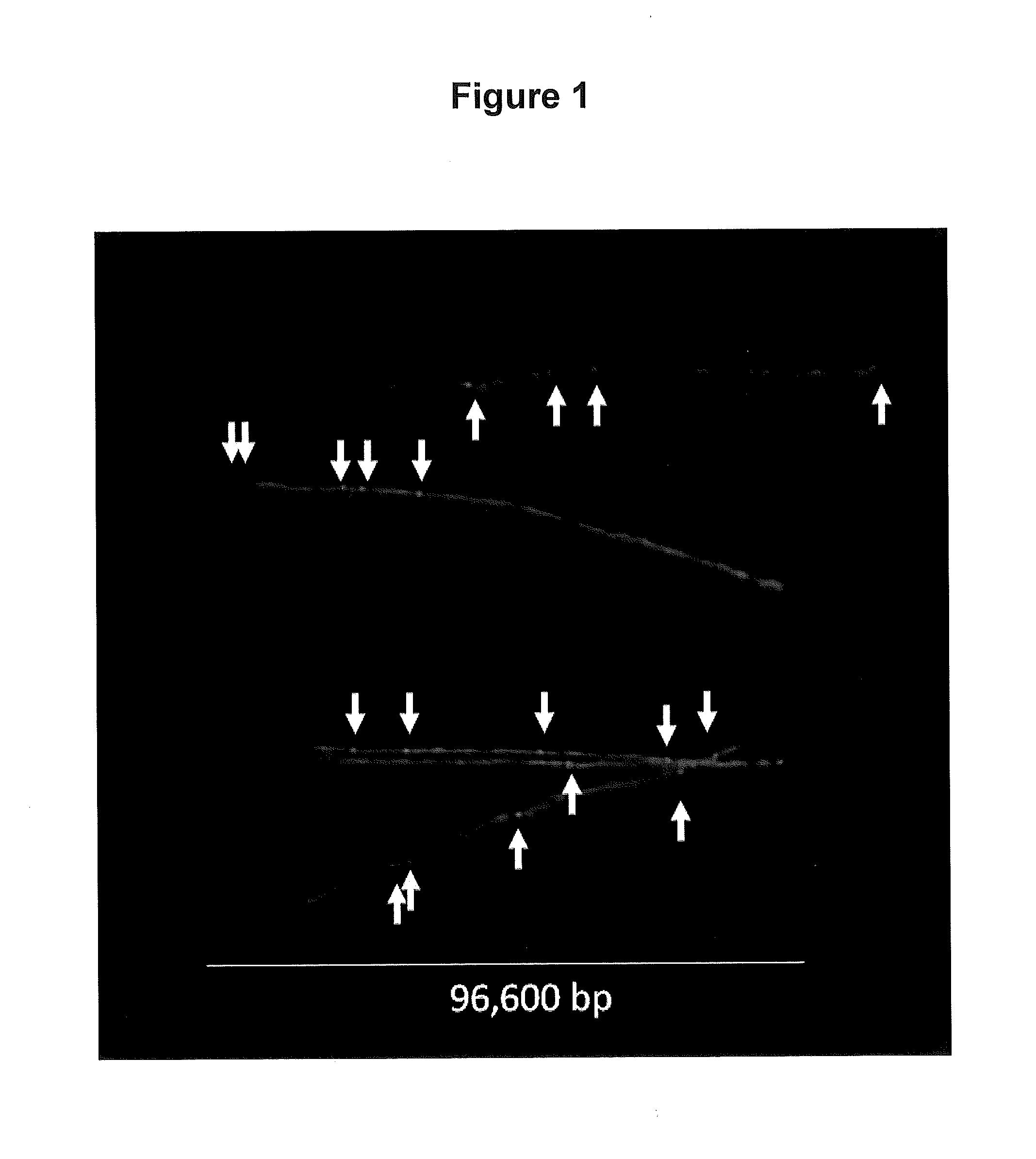
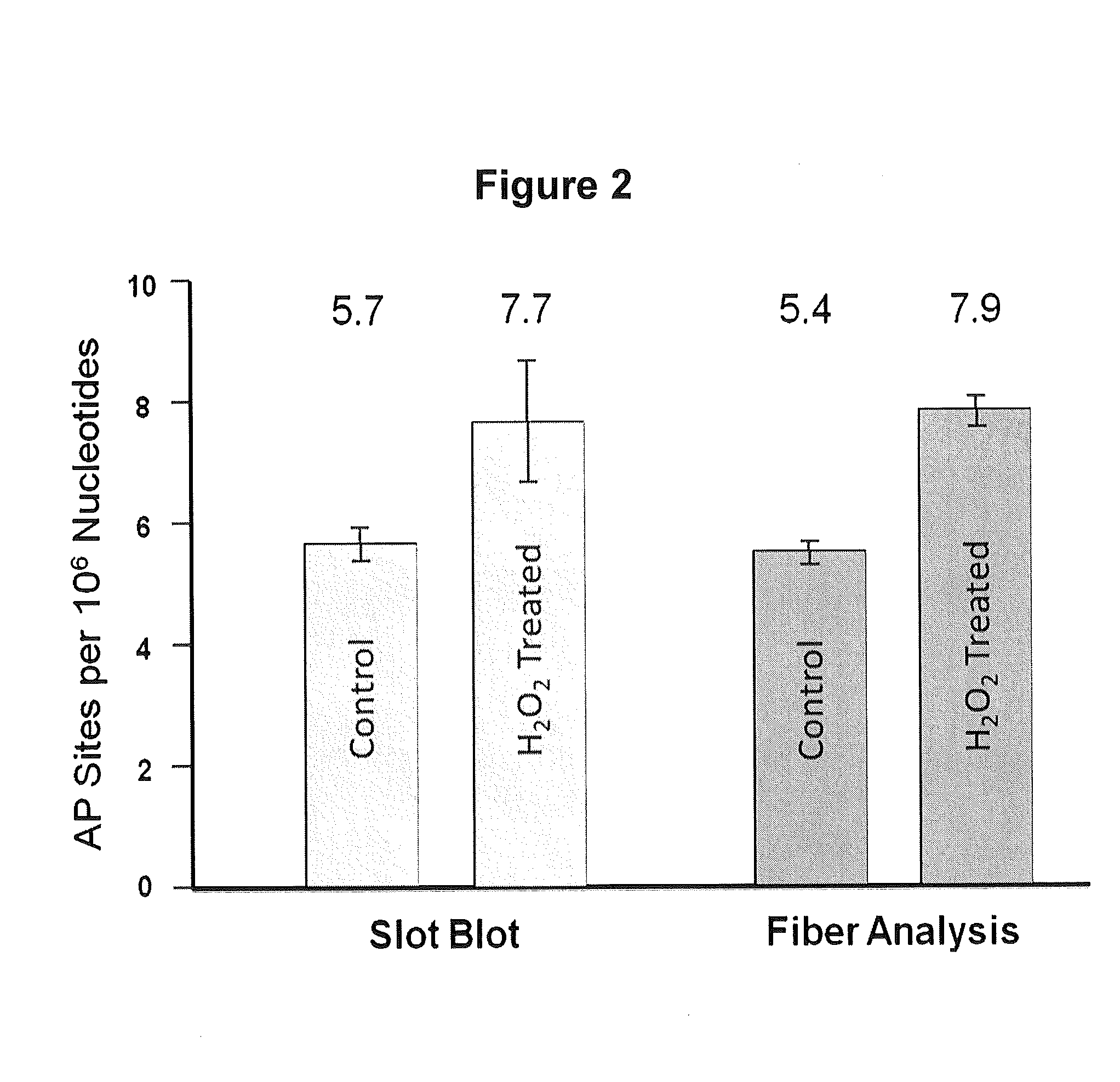
[0165]To quantify the number of AP sites per 106 nt in the YOYO-labeled DNA fiber spreads, we determined the total amount of DNA (expressed in nucleotides, nt) in a given image by dividing the total green fluorescence intensity of the image by the average intensity per nt for that image, as outlined in Material and Methods (Example 1). We then determined the total number of AP sites located in well-defined DNA fibers in the image by counting the number of AP sites that co-localized to those fibers (see Material and Methods Section; Example 1) FIG. 1 presents a composite image of a number of isolated DNA fibers that were observed in our samples. Using this approach, we analyzed over 109 nt of DNA and determined that our sample of DNA from cells growing in normal culture conditions contained a basal value of 5.4 AP sites per 106 nt, while slot blot analysis gave a value of 5.7 AP sites per 106 nt (FIG. 2). The number...

example 3

Detection of Multiple Types of DNA Damage in DNA Fibers

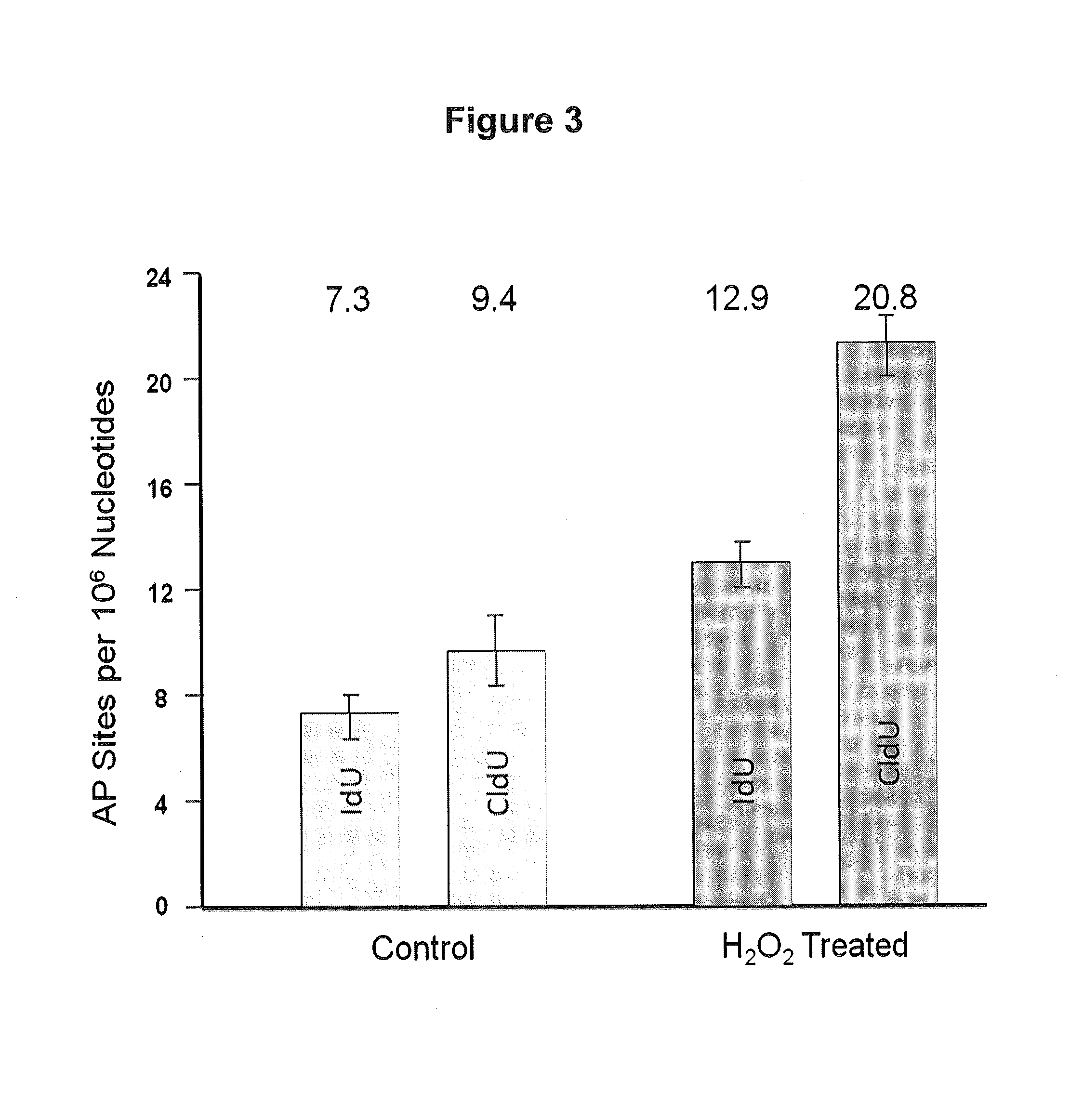
[0172]DNA fibers can be used to assess two or more types of DNA damage. Logarithmically growing human normal human fibroblasts were pulsed with iododeoxyuridine (IdU), exposed to 50 μM hydrogen peroxide, and then pulsed with chlorodeoxyuridine (CIdU). Approximately 400 cells were applied to a glass slide and treated with lysis buffer as described in Example 1 after which the slides were placed at an angle (about 30 degrees from horizontal) causing the DNA fibers to flow out (combing). The slides were then fixed. Immunostaining with fluorescent antibodies (AlexaFlour 594 for IdU and Alexaflour 488 for CIdU) was then performed to detect the presence of the halogenated uracil with red staining indicating regions with incorporated IdU and green staining regions with incorporated CIdU. DNA damage was detected by first using a chemical reagent that tags the damage with biotin and then that tag is immunostained with fluorescent antibod...


PUM

PropertyMeasurementUnit
Angleaaaaaaaaaa
Electric potential / voltageaaaaaaaaaa
Frequencyaaaaaaaaaa

Abstract

The present invention relates to methods of assessing damage to cellular DNA including the type, frequency and / or distribution of the DNA damage in the genome. The invention further provides methods of evaluating DNA damage in a cell caused by an agent and / or event as well as methods of determining a subject's prior exposure to an agent and / or event that is known or suspected to cause DNA damage. Further provided are methods of determining whether a subject is at an increased risk for a disease or disorder as a result of cellular DNA damage.

Description

RELATED APPLICATION INFORMATION[0001]This application is a continuation-in-part application of International Application Serial No. PCT / US2011 / 037858; Filed: May 25, 2011, which claims the benefit of U.S. Provisional Application No. 61 / 348,168; filed May 25, 2010, the disclosures of which are incorporated by reference herein in their entireties.STATEMENT OF FEDERAL SUPPORT[0002]This invention was made with government support under Grant Nos. CA084493, CA125337, ES010126. ES005948, ES015856 and ES007017 awarded by The National Institutes of Health. The government has certain rights in the invention.FIELD OF THE INVENTION[0003]The present invention relates to methods of detecting damage to DNA. In particular, the present invention relates to methods of detecting the frequency, type and / or distribution of DNA damage in a cell, of evaluating DNA damage in a cell caused by an agent and / or event as well as methods of determining a subject's prior exposure to an agent and / or event that is ...

Claims


Application Information

Patent Timeline
IPC IPC(8): C12Q1/68G01N33/58
CPCC12Q1/68C12Q2600/156G01N33/582G01N33/58C12Q2565/601
Inventor CHASTAIN, PAULKAUFMAN, DAVIDCOHEN, STEPHANIE M.
Owner THE UNIV OF NORTH CAROLINA AT CHAPEL HILL
Why Eureka
Try Eureka