
Compositions And Methods For Neovascularization

a technology of neovascularization and compositions, applied in the field of tissue engineering, advanced wound care and implantology, to achieve the effect of enhancing the angiogenic

Active Publication Date: 2013-08-01
MASSACHUSETTS INST OF TECH +1
View PDF0 Cites 6 Cited by
  • Summary
  • Abstract
  • Description
  • Claims
  • Application Information

AI Technical Summary

Benefits of technology

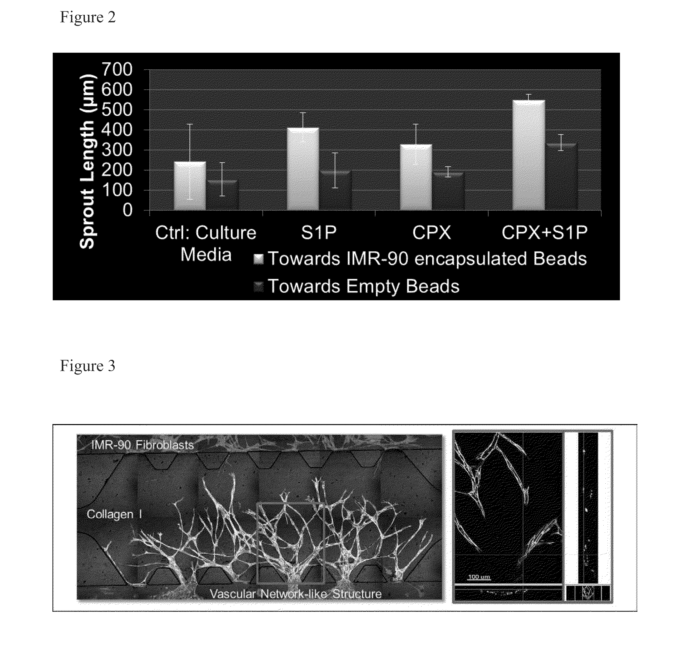
The patent describes a way to study how certain substances affect the growth of blood vessels using a special technique called microfluidic. The researchers found that when certain substances are used alone, they cause endothelial cells to sprout and grow towards mesenchymal cells (a type of connective tissue). However, when both a substance that induces a protein called HIF-1α and a substance called lysophospholipid are used together, the angiogenic effects are significantly increased. This may have important implications for developing new treatments for diseases that affect blood vessel growth, such as cancer and diabetes.

Problems solved by technology

Angiogenesis represents a major challenge in regenerative medicine and tissue engineering.

Method used


Image

  • Compositions And Methods For Neovascularization
  • Compositions And Methods For Neovascularization
  • Compositions And Methods For Neovascularization

Examples


example 1

Induction of Angiogenesis in Microfluidic Devices Using Prolyl Hydroxylase Inhibitors and Sphingosine-1 Phosphate

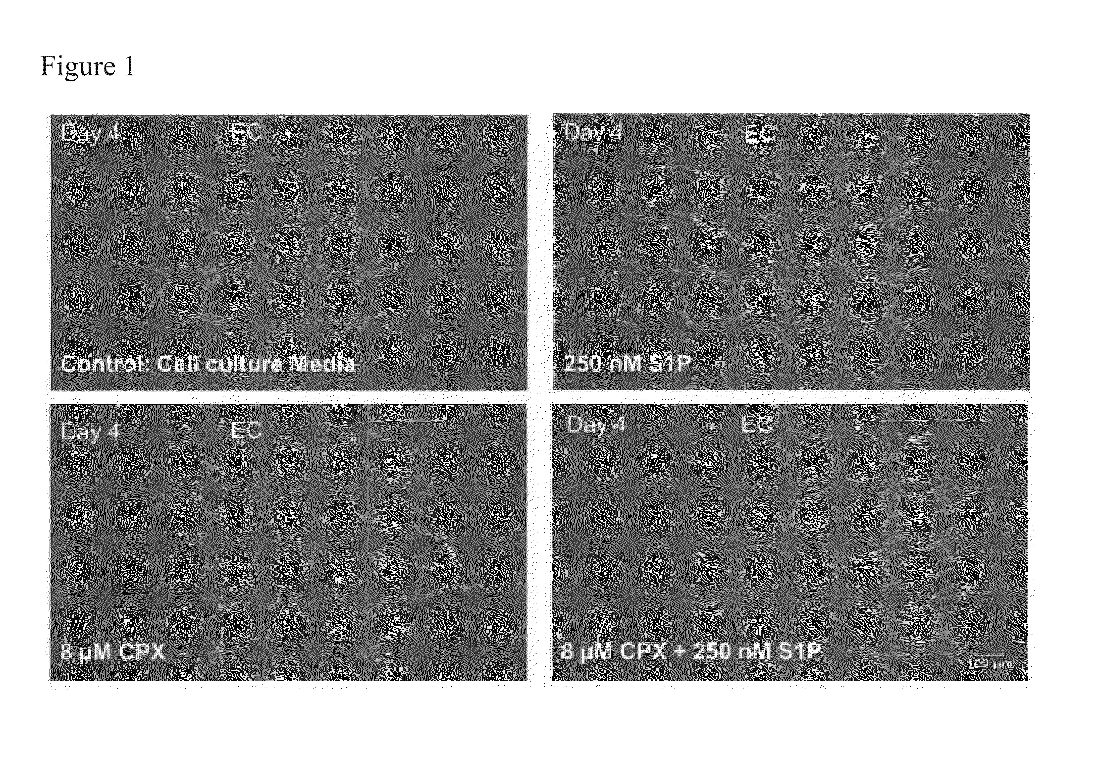
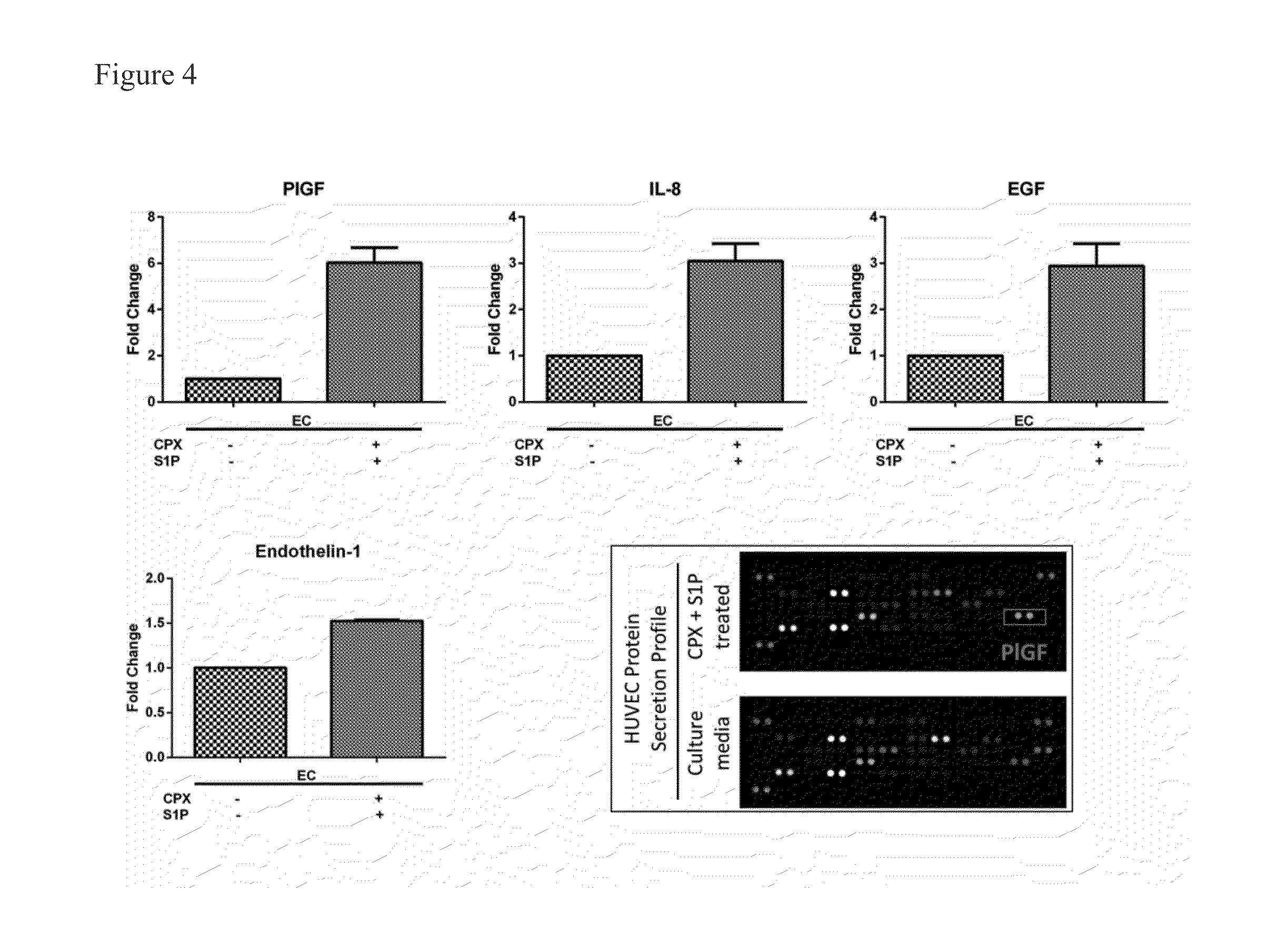
[0085]Angiogenesis represents a major challenge in regenerative medicine and tissue engineering. The synergistic effects of PHis and S1P were studied in an in vitro microfluidic platform where endothelial cells were co-cultured with fibroblast cells and subjected to the stimulation of PHis and S1P. Shown herein is that the interactions between endothelial cells and mesenchymal cells or fibroblasts maintain immature sprouts and turn them into functional vessels with lumens and these effects are most prominent with PHis and S1P. Thus, PHis worked synergistically with S1P.

[0086]The pro-angiogenic effects of PHis and S1P were studied on human umbilical venous endothelial cells (HUVEC) whereby fibroblasts in a neighbouring channel served as angiogenic secretory cells when stimulated by PHis and S1P. As shown herein, synergistic effects of PHis and S1P developed functional vasc...

example 2

CPX Induces Secretion of Complementary Angiogenic Proteins from Both Fibroblasts and Endothelial Cells

[0094]As the experimental set-up favoured soluble factors as messengers between the two cell types the secretion of angiogenic factors by fibroblasts and endothelial cells in the presence of CPX and S1P, respectively, was assessed.

[0095]Proteome profiler data indicated the increased secretion of several proteins including PlGF (6 fold), IL-8 (3 fold), EGF (2.9 fold) and endothelin-1 (1.5 fold) by CPX+S1P by endothelial cells. Highly comparable increases were found in the presence of CPX alone, and none were observed with S1P only.

[0096]In fibroblasts, CPX+S1P expression of HGF (1.4 fold), IGFBP-2 (1.8 fold), uPA (1.6 fold) and VEGF (11 fold). Highly comparable increases were found in the presence of CPX alone, and none were observed with S1P only. These data were validated for PlGF and VEGF through ELISA. It was found that CPX+S1P can increase the PlGF secretion rate in endothelia f...

example 3

Endothelial Cells CM Increases MCP-1 Secretion by IMR90

[0097]To study the influence of endothelial cells on fibroblasts IMR90 cells were cultured in EC conditioned medium with or without the addition of CPX+S1P, and quantified factors secreted by fibroblasts by ELISA. Fibroblasts showed a basal secretion of MCP-1. Endothelial cell CM induced the secretion of MCP-1 by fibroblasts by 3-fold in the absence of any CPX+S1P induction, however, the combination of both pharmacological agents increased MCP-1 secretion by a further 20%. As monosubstances, neither S1P nor CPX were able to increase basal MCP-1 secretion. See FIG. 5.


PUM

PropertyMeasurementUnit
concentrationsaaaaaaaaaa
diameteraaaaaaaaaa

Abstract

The invention is directed to a method of inducing angiogenesis at a site in an individual in need thereof comprising administering locally to the site an effective amount of one or more agents that induce hypoxia induced factor 1α (HIF-1α). In another aspect, the invention is directed to a method of inducing angiogenesis at a site in an individual in need thereof comprising administering locally to the site an effective amount of one or more agents that induce hypoxia induced factor 1α (HIF-1α) and one or more lysophospholipids. In addition, the invention is directed to methods of generating prevascularized tissue, methods of generating a vascular network in a device and compositions thereof.

Description

RELATED APPLICATION(S)[0001]This application claims the benefit of U.S. Provisional Application No. 61 / 587,713, filed on Jan. 18, 2012. The entire teachings of the above application are incorporated herein by reference.BACKGROUND OF THE INVENTION[0002]Angiogenesis represents a major challenge in regenerative medicine and tissue engineering.[0003]Thus, a need exists for compositions and methods for inducing angiogenesis.SUMMARY OF THE INVENTION[0004]The invention relates to the fields which include tissue engineering, advanced wound care and implantology e.g., where the induction of blood vessel growth in, into and / or around a biomaterial with less fibrotic capsule formation is highly desirable. Described herein are compositions and methods for use in induction (e.g., local induction; pharmacological (local) induction) of angiogenesis which involves the use of an (one or more) agent that induces hypoxia-induced factor 1α (HIF-1α), or a combination of an (one or more) agent that induc...

Claims


Application Information

Patent Timeline
Patent Type & Authority Applications(United States)
IPC IPC(8): A61K31/4412C12N5/07
CPCA61K31/4412C12N5/06A61K31/688C12N2501/998C12N5/069A61K2300/00
Inventor RAGHUNATH, MICHAELLIM, SEI HIENKAMM, ROGER DALE
Owner MASSACHUSETTS INST OF TECH
Learn More