

Apparatus for Systematic Single Cell Tracking of Distinctive Cellular Events
a single cell and event technology, applied in the field of single cell quantitative identification of rare distinctive cellular events, can solve the problems of inability to readily allow in vitro genotoxicity and mutagenicity tests, inability to and inability to reliably detect major and frequent events. , to achieve the effect of saving administration
- Summary
- Abstract
- Description
- Claims
- Application Information
AI Technical Summary
Benefits of technology
Problems solved by technology
Method used
Image

Examples
example microscope
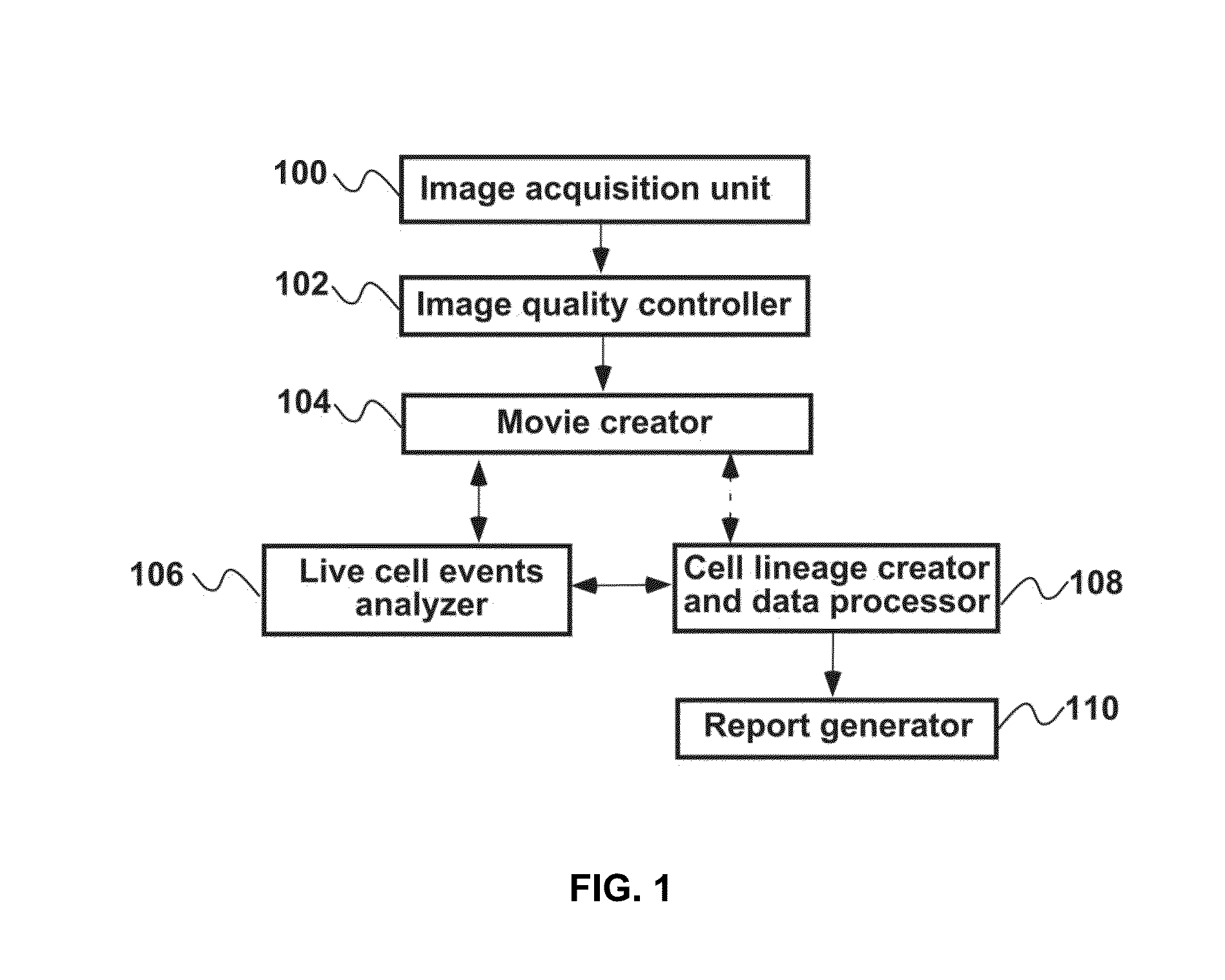
[0099 System
[0100]Different microscope systems can be designed for use in the present system. The following description is for an example microscope system.
[0101]The cells should be kept in condition for optimal cell viability and uncorrupted cell division. The microscope system should facilitate tracking of large numbers of individual cells. In some embodiments, different subsets of cells are imaged in parallel under the same time frame and conditions. Image quality and sampling frequency should be of sufficient quality to allow identification of detailed cell features required for automated tracking of cell division events and cell viability.
[0102]Differential Interference Contrast (DIC) with near infrared (NIR) light (>700 nm) provides the best prophylactic lighting conditions and required resolution for continuous cell imaging. DIC imaging provides sufficient detail to track cell behavior while providing high contrast data for computer analysis. The microscope system uses Back t...
example experiment
[0116
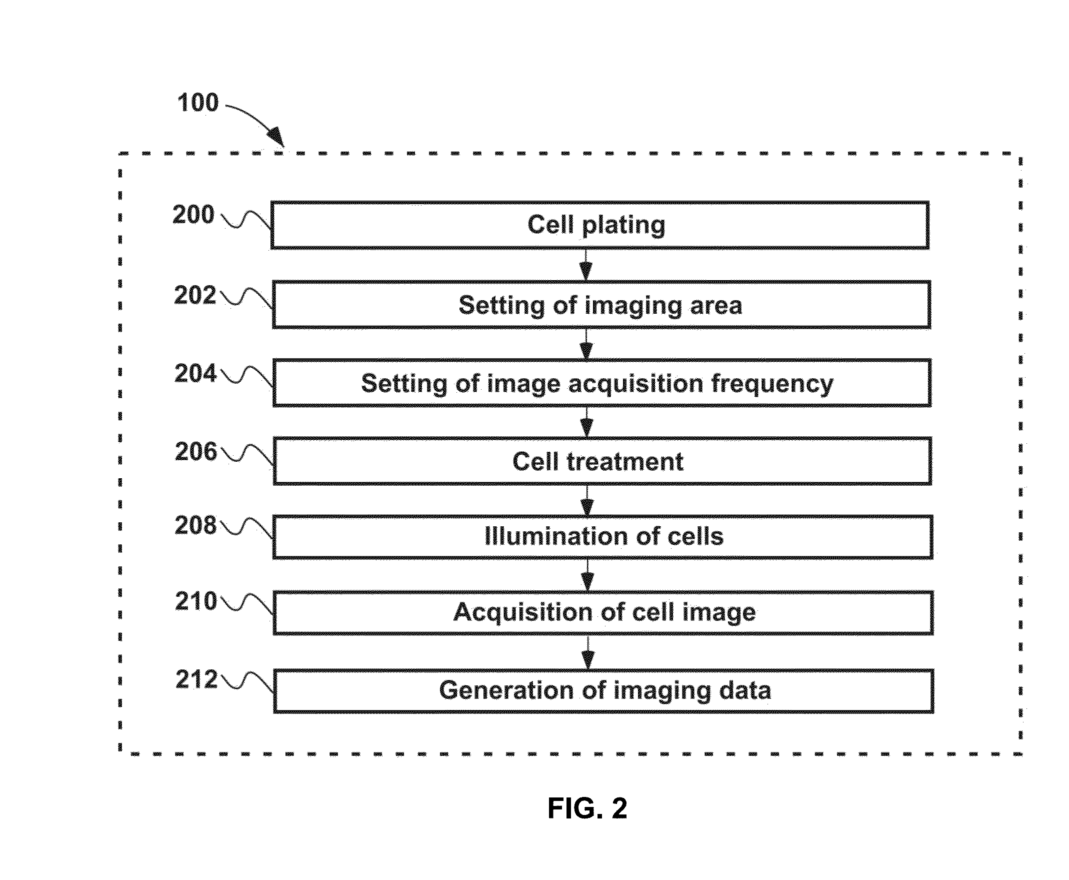
[0117]HeLa cells were purchased from ATCC. HeLa cells (1×10⁴ cells in 50 μl per well) were carefully plated in the center of a coverglass Lab-Tek 8 wells chamber for optimal optical observation. After cells were stably attached, 500 μl of culture medium was added. HeLa cells were used after 24 hrs of plating. Cultures were maintained until cells occupied over 90% of the surface of each chamber.
[0118]By employing a 8 well-chambered coverglass, cells treated with various doses of substances can be monitored simultaneously, allowing real-time cell biological assays to be performed on the microscope stage with their appropriate control (FIG. 8).
[0119]The HeLa cells showed optimal mobility since the cells moved around an area of about 10 to 50 μm diameter. Furthermore, their reasonable mobility decreased the chance of pilling up of HeLa cells, allowing precise individual cell tracking.
[0120]A Quorum WaveFX Spinning Disc Confocal System (Quorum Technologies Inc., Canada) with a Leica...
PUM
| Property | Measurement | Unit |
|---|---|---|
| time | aaaaa | aaaaa |
| time | aaaaa | aaaaa |
| time | aaaaa | aaaaa |
Abstract
Description
Claims
Application Information