
Ophthalmologic apparatus

a technology of ophthalmologic equipment and ophthalmology, which is applied in the field of ophthalmologic equipment, can solve the problems of eye pressure rapidly increasing, optic nerve damage, and difficulty for an examiner to accurately perform diagnosis, and achieve the effect of accurate diagnosis

Inactive Publication Date: 2013-10-03
CANON KK
View PDF4 Cites 14 Cited by
  • Summary
  • Abstract
  • Description
  • Claims
  • Application Information

AI Technical Summary

Benefits of technology

The patent text is saying that this invention can accurately diagnose a certain condition.

Problems solved by technology

The iris is thus pushed forward by the lens, and further blocks the trabecula, so that the eye pressure rapidly increases, and the optic nerve is damaged.
However, the unique information of the subject's eye is not associated with the lighting environment when the unique information has been acquired, so that it is difficult for an examiner to accurately perform diagnosis.

Method used


Image

  • Ophthalmologic apparatus
  • Ophthalmologic apparatus
  • Ophthalmologic apparatus

Examples


Embodiment Construction

[0028]Various exemplary embodiments, features, and aspects of the invention will be described in detail below with reference to the drawings.

[0029]The apparatus according to the present invention is applicable to a subject such as the subject's eye, the skin, and internal organs. Further, the apparatus according to the present invention is, for example, an ophthalmologic apparatus or an endoscope.

[0030]

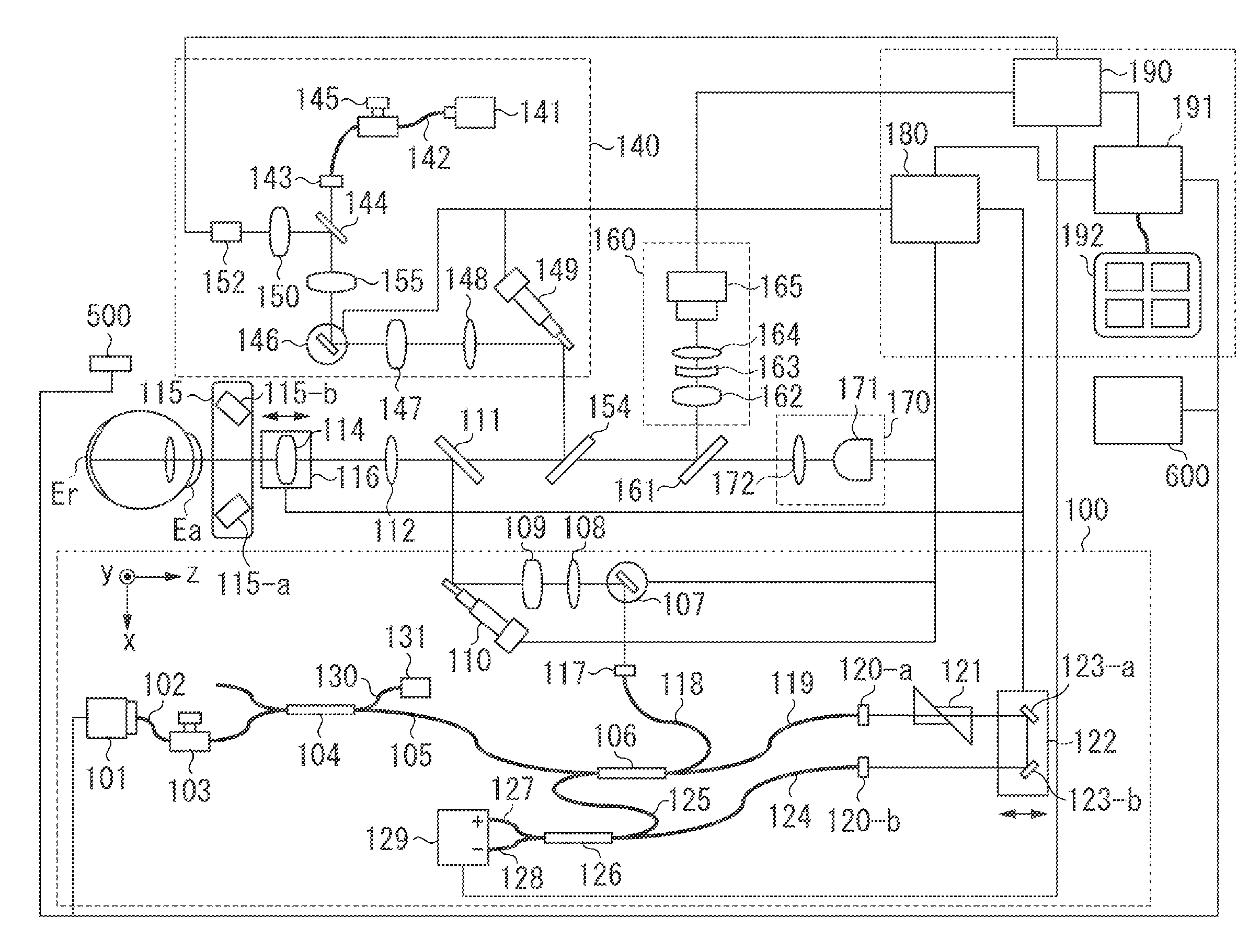
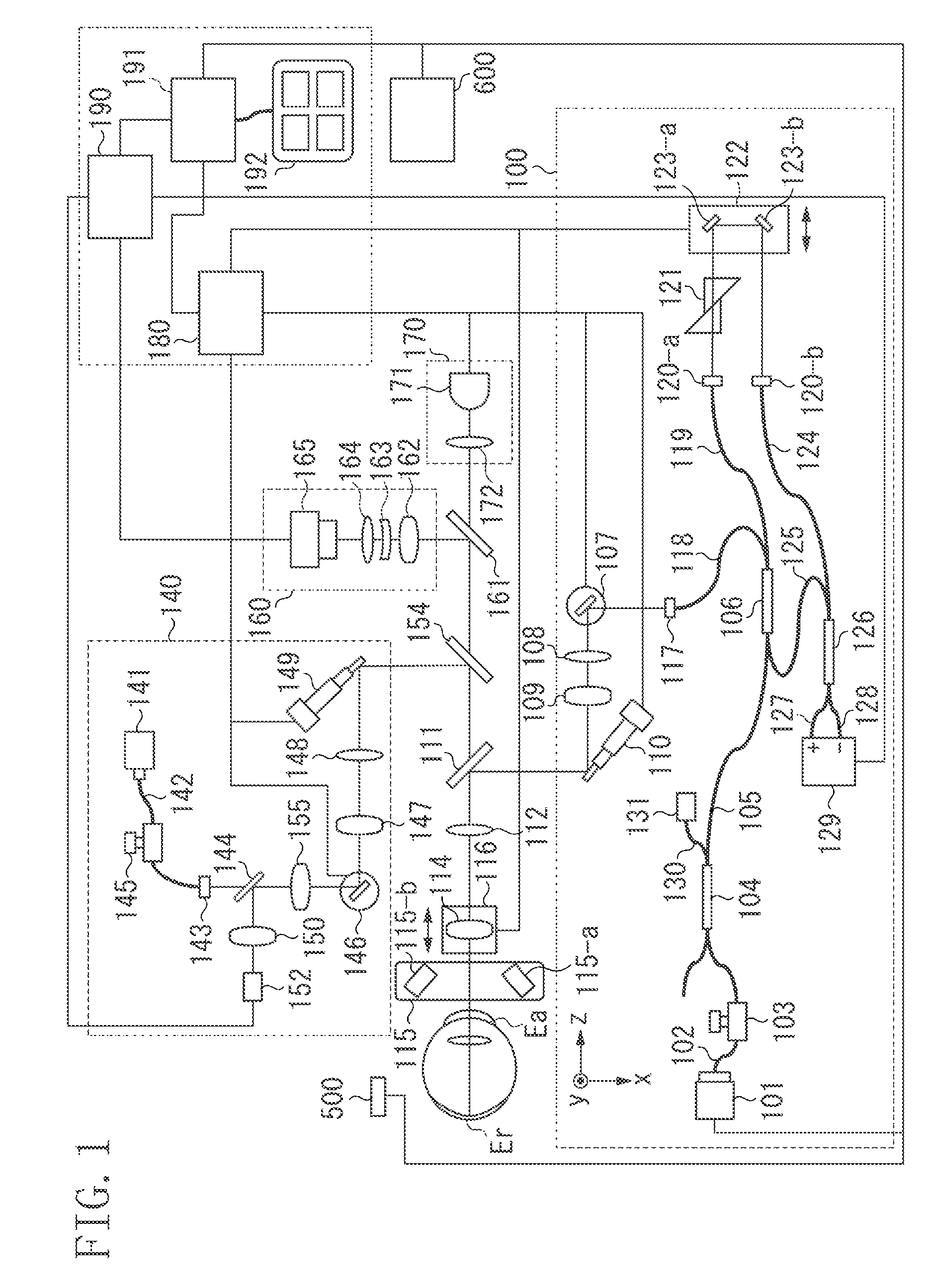
[0031]FIG. 1 is a schematic diagram illustrating an example of a configuration of the ophthalmologic apparatus according to the first exemplary embodiment.

[0032]Referring to FIG. 1, the ophthalmologic apparatus includes a swept source OCT (SS-OCT, or may be simply referred to as OCT) 100, a scanning laser ophthalmoscope (SLO) 140, an anterior segment imaging unit 160, an internal fixation lamp 170, and a control unit 200. The ophthalmologic apparatus illustrated in FIG. 1 thus acquires the unique information of the subject's eye. Further, according to the present exemplary embodiment,...


PUM


Abstract

An ophthalmologic apparatus includes an ophthalmologic apparatus configured to acquire unique information of a subject's eye, an acquisition unit configured to acquire a value indicating brightness of surroundings of the ophthalmologic apparatus, and a recording unit configured to record in a storing unit a value indicating brightness acquired by the acquisition unit, associated with a tomographic image.

Description

BACKGROUND OF THE INVENTION[0001]1. Field of the Invention[0002]The present invention relates to an ophthalmologic apparatus.[0003]2. Description of the Related Art[0004]An optical coherence tomography (OCT) imaging apparatus such as an OCT for an eye part allows three-dimensional observation of a state within retinal layers and a state of an anterior segment such as an angle, a cornea, and an iris. Such an optical coherence tomography imaging apparatus is recently attracting attention as being useful in more accurately diagnosing of diseases. More specifically, Japanese Patent Application Laid-Open No. 2011-072716 discusses using tomographic images in diagnosing glaucoma. Further, tomographic images are used in diagnosing angle closure.[0005]In glaucoma, there is a symptom referred to as an acute glaucoma attack. The acute glaucoma attack occurs when a pupil of a patient having a narrow angle dilates, so that a root of the iris becomes thick and blocks a trabecula in the angle thro...

Claims


Application Information

Patent Timeline
Patent Type & Authority Applications(United States)
IPC IPC(8): A61B3/14
CPCA61B3/102A61B3/14
Inventor GOTO, ATSUSHIMIYATA, KAZUHIDESATO, MAKOTOMAKIHIRA, TOMOYUKIIWASE, YOSHIHIKOYAMADA, KAZUROTOMITA, RITSUYAKIBE, DAISUKESHINBATA, HIROYUKI
Owner CANON KK
Learn More