Image fusion for interventional guidance

a technology of interventional guidance and image fusion, applied in the direction of image enhancement, instruments, catheters, etc., can solve the problems of difficult catheter navigating, poor utilization of splendid complementary nature of tee and fluoro, and inability to capture soft tissue structure. to achieve the effect of minimizing the mapping error

Active Publication Date: 2013-10-03
SIEMENS HEALTHCARE GMBH
View PDF3 Cites 39 Cited by
  • Summary
  • Abstract
  • Description
  • Claims
  • Application Information

AI Technical Summary

Benefits of technology

[0034]According to a further aspect of the invention, minimizing the mapping error further comprises estimating a prior probability for each vertex of a model of pre-operative image I2 by assigning each vertex of a model of the pre-operative image I2 as a positive sample and using random points within a threshold distance as negative samples, reje

Problems solved by technology

However, navigating a catheter inside a patient is challenging, and without direct access or view to the affected anatomy, advanced imaging is required to secure a safe and effective execution of the procedure.
X-ray fluoroscopy is used to visualize the catheter; however, this imaging modality does not capture soft tissue structure.
Nevertheless, the splendid complementary nature of TEE and Fluoro is barely exploited in today's practice where the real-time acquisitions are not synchronized, and images are visualized separately and in misaligned coordinate systems.
However, direct 3D to 2D image registration is challenge to solve, especially within the intra-operative setup that does not allow for user interaction or time consuming processing.
However, clinical guidelines limit the duration and frequency of rapid pacing and the volume of contrast agent that c

Method used


Image

  • Image fusion for interventional guidance
  • Image fusion for interventional guidance
  • Image fusion for interventional guidance

Examples


Embodiment Construction

[0060]Exemplary embodiments of the invention as described herein generally include systems and methods for fusing images for interventional guidance. Accordingly, while the invention is susceptible to various modifications and alternative forms, specific embodiments thereof are shown by way of example in the drawings and will herein be described in detail. It should be understood, however, that there is no intent to limit the invention to the particular forms disclosed, but on the contrary, the invention is to cover all modifications, equivalents, and alternatives falling within the spirit and scope of the invention.

[0061]As used herein, the term “image” refers to multi-dimensional data composed of discrete image elements (e.g., pixels for 2-dimensional images and voxels for 3-dimensional images). The image may be, for example, a medical image of a subject collected by computer tomography, magnetic resonance imaging, ultrasound, or any other medical imaging system known to one of sk...


PUM


Abstract

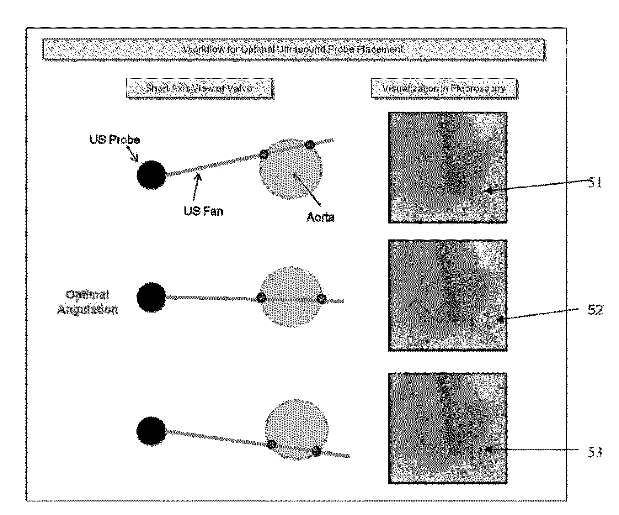
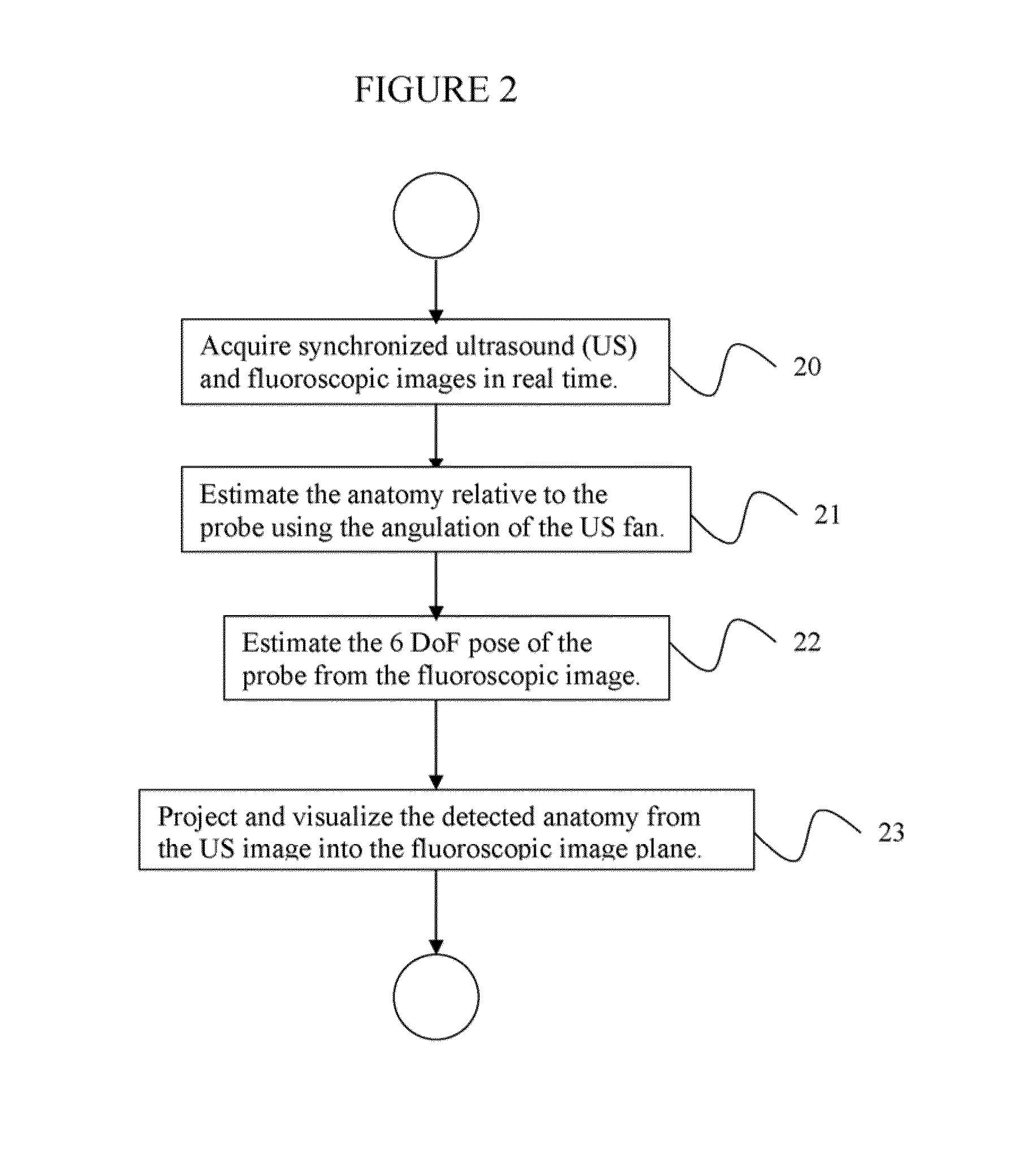
A method for real-time fusion of a 2D cardiac ultrasound image with a 2D cardiac fluoroscopic image includes acquiring real time synchronized US and fluoroscopic images, detecting a surface contour of an aortic valve in the 2D cardiac ultrasound (US) image relative to an US probe, detecting a pose of the US probe in the 2D cardiac fluoroscopic image, and using pose parameters of the US probe to transform the surface contour of the aortic valve from the 2D cardiac US image to the 2D cardiac fluoroscopic image.

Description

CROSS REFERENCE TO RELATED UNITED STATES APPLICATIONS[0001]This application claims priority from “Real-Time TAVI Navigation: Fusing Anatomy from 2D US with Fluoroscopy”, U.S. Provisional Application No. 61 / 602,107 of Mountney, et al., filed Feb. 23, 2012, “Robust Model-based Fusion of Pre- and Intra-Operative Images by Exploiting Data Uncertainties”, U.S. Provisional Application No. 61 / 605,566 of Grbic, et al., filed Mar. 1, 2012, and “Ultrasound and Fluoroscopic images Fusion by Autonomous US Probe Detection”, U.S. Provisional Application No. 61 / 605,573 of Mountney, et al., filed Mar. 1, 2012, the contents of all of which are herein incorporated by reference in their entireties.TECHNICAL FIELD[0002]This disclosure is directed to methods for real-time fusing of 2D and 3D images with 2D fluoroscopic images for interventional guidance.DISCUSSION OF THE RELATED ART[0003]Fluoroscopy guided cardiac interventions such as endovascular stenting, atrial ablation, closure of atrial / ventricula...

Claims


Application Information

Patent Timeline
IPC IPC(8): A61B5/00
CPCA61B5/0035G06T2207/30048A61B6/4441A61B6/487A61B6/503A61B6/5235A61B6/5247A61B8/0883A61B8/12A61B8/5261A61B6/12A61B6/582G06T5/50G06T7/0048G06T2207/10121G06T2207/10132G06T2207/20081G06T2207/20221A61B6/032G06T7/77
Inventor MOUNTNEY, PETERGRBIC, SASAIONASEC, RAZVAN IOANJOHN, MATTHIASBOESE, JANCOMANICIU, DORIN
Owner SIEMENS HEALTHCARE GMBH
Why Eureka
Try Eureka