Methods and apparatus for predicting protein structure

Inactive Publication Date: 2013-11-14
SLOAN KETTERING INST FOR CANCER RES +1
View PDF2 Cites 16 Cited by
  • Summary
  • Abstract
  • Description
  • Claims
  • Application Information

AI Technical Summary

Benefits of technology

[0058]In some embodiments, the polypeptide is a transmembrane protein and wherein the analysis module is configured to identify evolutionary constraints corresponding to residue pairs predicted to be close in 3D space, and eliminate evolutionary constraints for which 3D proximity is unlikely due to presence of a membrane. In some embodiments, the structure is a structure of the entire protein. In some embodiments, the statistical analysis is a pseudolikelihood maximization analysis. In some embodiments, the statistical analysis is an entropy maximization analysis. In some embodiments, the analysis module is configured to identify multiple 3D conformations of the polypeptide. In some embodiments, the analysis mo

Problems solved by technology

However, it is difficult to investigate the 3D structure of proteins that are anchored in biomembranes using standard biochemical methods, even where the sequence of the protein is known.
Pr

Method used


Image

  • Methods and apparatus for predicting protein structure
  • Methods and apparatus for predicting protein structure
  • Methods and apparatus for predicting protein structure

Examples


example 1

Prediction of Three-Dimensional Structures of Membrane Proteins

[0177]Reported here is the development and use of a method, EVfold_membrane, which enables de novo prediction of 3D structures of unknown α-helical transmembrane proteins from evolutionary constraints, using neither fragments, threading, nor homologous 3D structures. The structures of 11 transmembrane proteins of unknown structure were predicted, including six pharmacological targets (FIG. 3, Table 1). To verify that predicted structures were plausible, the structures of a diverse set of 25 transmembrane proteins with known 3D structures (Table 1) were predicted using EVfold_membrane, and an unprecedented level of agreement was found with the cognate crystal structures (TM scores>0.5 for 22 out of 25 of the benchmarked proteins). Functionally important regions of each protein were more accurately predicted than the protein as a whole, and residues that are subject to multiple pair constraints tended to be in substrate bi...


PUM


Abstract

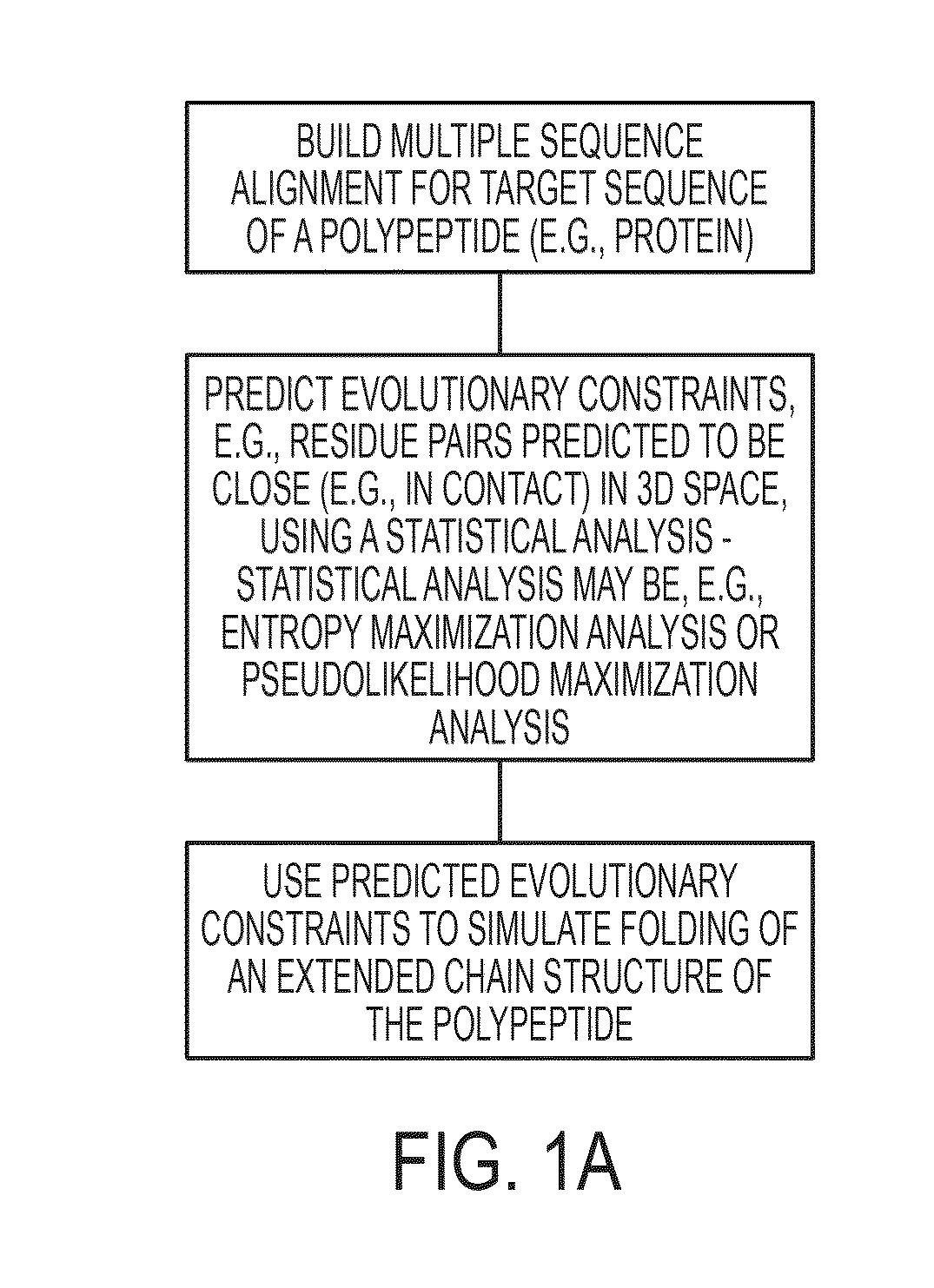
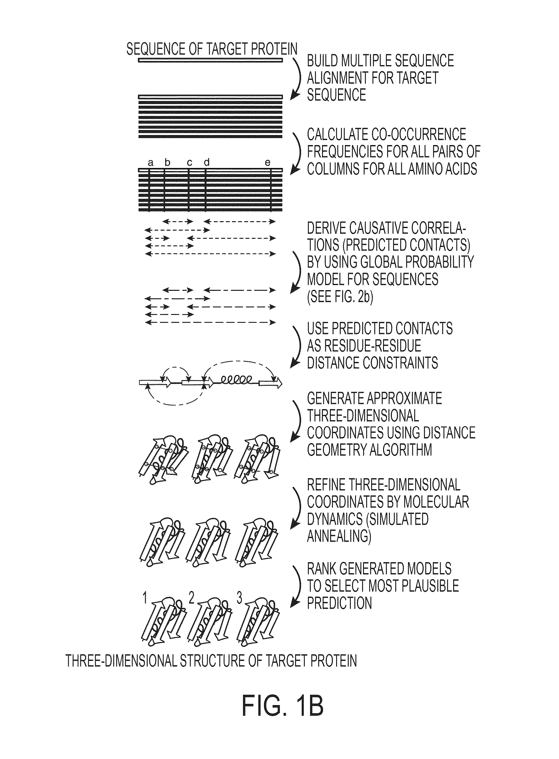
The present invention relates to a method for predicting three-dimensional structure of a protein from its sequence. Three-dimensional structure may be determined by: (a) generating a multiple sequence alignment for a candidate protein having a known sequence; (b) identifying a covariance matrix between all pairs of sequence positions in the multiple sequence alignment; (c) inverting the covariance matrix and identifying predicted evolutionary constraints using a statistical model of the candidate protein; and (d) simulating folding of an extended chain structure of the candidate protein using the predicted constraints.

Description

CROSS REFERENCE TO RELATED APPLICATIONS[0001]This application claims the benefit of U.S. application Ser. No. 13 / 682,703, filed Nov. 20, 2012, of U.S. Provisional Application No. 61 / 645,027, filed May 9, 2012, and of U.S. Provisional Application No. 61 / 645,564, filed May 10, 2012, the contents of all of which are hereby incorporated by reference herein in their entireties.BACKGROUND[0002]Many transmembrane proteins facilitate transport of substances across the biological membrane or function as receptors and are important in the performance of various biological functions. Identifying the 3D structure of such transmembrane proteins is useful, for example, in the pharmacological selection or design of drugs for the treatment of various diseases and disorders. Knowing the 3D structure of such proteins is also important, for example, in the identification of functional genetic variants in normal and disease genomes. However, it is difficult to investigate the 3D structure of proteins t...

Claims


Application Information

Patent Timeline
IPC IPC(8): G06F19/16G16B15/20
CPCG06F19/16G16B15/00G16B15/20
Inventor SANDER, CHRISMARKS, DEBORA S.
Owner SLOAN KETTERING INST FOR CANCER RES
Why Eureka
Try Eureka