Quantitative evaluation of fractional regional ventilation using four-dimensional computed tomography

a computed tomography and fractional region technology, applied in tomography, diagnostic recording/measuring, applications, etc., can solve the problems of severe impairing ventilation, adversely affecting lung function, and uneven distribution of ventilatory disruptions in the lung, so as to reduce data noise.

Inactive Publication Date: 2013-11-14
UNIV OF MARYLAND
View PDF2 Cites 20 Cited by
  • Summary
  • Abstract
  • Description
  • Claims
  • Application Information

AI Technical Summary

Benefits of technology

[0008]An aspect of embodiments of the present invention is to substantially address the above and other concerns, and provide methods and systems for determining regional lung ventila

Problems solved by technology

Disrupting any of these functions can adversely affect lung function.
For example, many diseases, such as lung cancer, asthma, and chronic obstructive pulmonary disease (COPD), can severely impair ventilation.
These ventilatory disruptions, however, are not evenly distributed throughout the lung.
As a result, PFT alone fails to capture these regional variations.
These techniques, however, have limited clinical appli

Method used


Image

  • Quantitative evaluation of fractional regional ventilation using four-dimensional computed tomography
  • Quantitative evaluation of fractional regional ventilation using four-dimensional computed tomography
  • Quantitative evaluation of fractional regional ventilation using four-dimensional computed tomography

Examples


examples

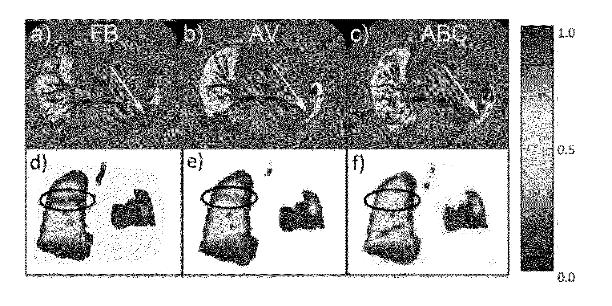
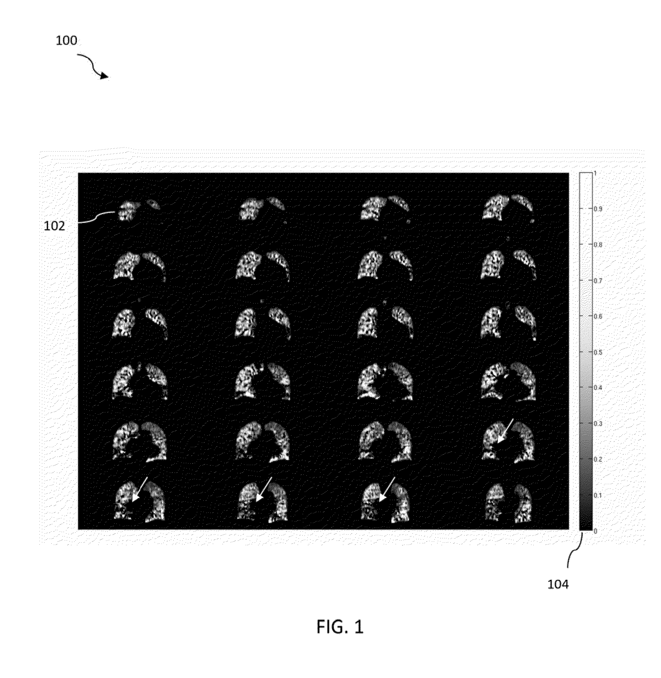
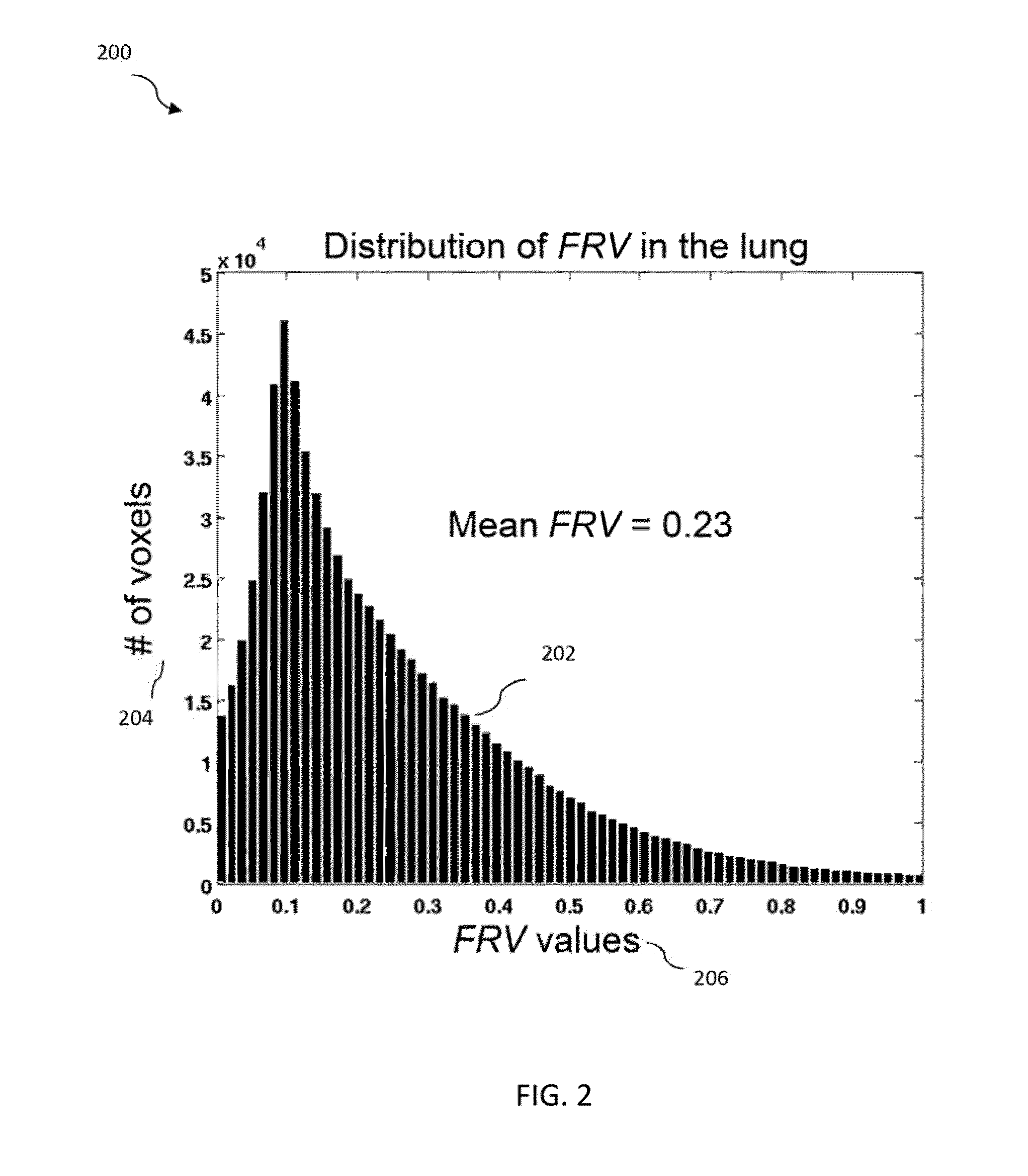
[0052]FIG. 8 shows a method 800 of determining fractional regional ventilation in accordance with an illustrative embodiment of the present invention. The method 800 includes obtaining first lung image data indicative of a first phase of a respiratory cycle, the first lung image data including at least one first voxel, as step 802, obtaining second lung image data indicative of a second phase of a respiratory cycle, the second lung image data including at least one second voxel, at step 804, determining an apparent mass ratio k based on the first lung image data and the second lung image data at step 806, determining first spatially matched lung image data including N voxels and second spatially matched lung image data including N voxels, based on the first lung image data and the second lung image data, at step 808, and determining at least one fractional regional ventilation value (FRV value), in accordance with a first equation FRV(n)=(k·ρ2_n−ρ1_n) / ρ1_n at step 810. The value of ...


PUM


Abstract

Methods and systems for determining fractional regional ventilation are disclosed. A method includes obtaining first and second lung image data indicative of a first phase and a second phase of a respiratory cycle, respectively, determining an apparent mass ratio k based on the first lung image data and the second lung image data, determining first and second spatially matched lung image data, each including N voxels, based on the first lung image data and the second lung image data, and determining at least one fractional regional ventilation value (FRV value), in accordance with a first equation FRV(n)=(k·ρ2_n−ρ1_n)/ρ1_n. The value of n is a voxel index, ρ1_n is indicative of a density of a voxel n of the first spatially matched lung image data, and ρ2_n is indicative of a density of a voxel n of the second spatially matched lung image data.

Description

CROSS-REFERENCE TO RELATED APPLICATION[0001]This application claims the benefit of U.S. Provisional Patent Application No. 61 / 646,554, filed May 14, 2012 in the U.S. Patent and Trademark Office, the disclosure of which is incorporated herein by reference in its entirety.FIELD OF THE INVENTION[0002]The present invention generally relates to systems and methods for medical imaging. More specifically, the present invention relates to systems and methods for evaluating lung ventilation function using four-dimensional computed tomography.BACKGROUND OF THE INVENTION[0003]The primary physiologic function of the lung is to support gas exchange through ventilation (air reaching the alveoli), perfusion (blood reaching the alveoli), and diffusion of gases across the blood-gas interface. Disrupting any of these functions can adversely affect lung function. For example, many diseases, such as lung cancer, asthma, and chronic obstructive pulmonary disease (COPD), can severely impair ventilation.[...

Claims


Application Information

Patent Timeline
IPC IPC(8): A61B6/00
CPCA61B6/5205A61B6/032A61B6/486A61B6/5217A61B6/5288G16H50/30
Inventor MISTRY, NILESHD'SOUZA, WARRENDIWANJI, TEJANFEIGENBERG, STEVEN
Owner UNIV OF MARYLAND
Why Eureka
Try Eureka