In vitro embryo blastocyst prediction methods

Inactive Publication Date: 2013-12-19
PROGYNY
View PDF0 Cites 17 Cited by
  • Summary
  • Abstract
  • Description
  • Claims
  • Application Information

AI Technical Summary

Benefits of technology

[0009]Methods, compositions and kits for determining the likelihood that one or more embryos or pluripotent cells in one or more embryos will reach the blastocyst stage and/or usable blastocyst stage are provided. These methods, compositions and kits find use in identifying embryos and oocytes in vitro that have a likelihood of reaching the blastocyst stage and/or usable blastocyst stage, i.e. the ability or capacity to develop into a blastocyst, which are thus useful in methods of treating infertility in humans, and the like.
[0010]In some aspects of the invention, methods are provided for determining the likelihood that an embryo or a pluripotent cell will reach the blastocyst stage and/or usable blastocyst stage. In some aspects determining the likelihood of reaching the blastocyst stage and/or usable blastocyst stage is determined by selecting with high specificity one or more human embryos that is not likely to reach the blastocyst stage, wherein at least about 70%, 75%, 80%, 85%, 90%, 95% or more or 100% of the human embryos not selected are not likely to reach the blastocyst stage and/or usable blastocyst stage. In such aspects, cellular parameters of an embryo or pluripotent cell are measured to arrive at a cell parameter measurement. The cell parameter is then employed to provide a determination of the likelihood of the embryo or pluripotent cell to reach the blastocyst stage and/or usable blastocyst stage, which determination may be used to guide a clinical course of action. In some embodiments, the cell parameter is a morphological event that is measurable by time-lapse microscopy. In some embodiments, e.g. when an embryo is assayed, the one or more cell parameters is: the duration of a cytokinesis event, e.g. the time interval between cytokinesis I and cytokinesis 2; and the time interval between cytokinesis 2 and cytokinesis 3. In some embodiments, the cell parameter is a morphological event that is measurable by time-lapse microscopy. In some embodiments, e.g. when an embryo is assayed, the one or more cell parameters is: the duration of a cytokinesis event, e.g. the time interval between mitotic cell cycle 1 and mitotic cell cycle 2; and the time interval between mitotic cell cycle 2 and mitotic cell cycle 3. In certain embodiments, the duration of cell cycle 1 is also utilized as a cell parameter. In some embodiments, the duration of the first cytokinesis is not measured. In some embodiments, the cell parameter measurement is employed by comparing it to a comparable cell parameter measurement from a reference embryo, and using the result of this comparison to provide a determination of the likelihood of the embryo to reach the blastocyst stage. In some embodiments, the embryo is a human embryo.
[0011]In some aspects of the invention, methods are provided for ranking embryos or pluripotent cells for their likelihood of reaching the blastocyst stage and/or usable blastocyst stage relative to the other embryos or pluripotent cells in the group. In such embodiments, one or more cellular parameters of the embryos or pluripotent cells in the group is measured to arrive at a cell parameter measurement for each of the embryos or pluripotent cells. The cell parameter measurements are then employed to determine the likelihood of reaching the blastocyst stage and/or usable blastocyst stage for each of the embryos or pluripotent cells in the group relative to one another, which determination may be used to guide a clinical course of action. In some embodiments, the cell parameter is a morphological event that is measurable by time-lapse microscopy. In some embodiments, e.g. when embryos are ranked, the one or more cell parameters are the duration of a cytokinesis event, e.g. the time interval between cytokinesis 1 and cytokinesis 2; and the time interval between cytokines

Problems solved by technology

Infertility is a common health problem that affects 10-15% of couples of reproductive-age.
Multiple gestations have well-documented adverse outcomes for both the mother and fetuses, such as miscarriage, pre-term birth, and low birth rate.
Potential causes for failure of IVF are diverse; however, since the introduction of IVF in 1978, one of the major challenges has been to identify the embryos that are most suitable for transfer and most likely to result in term pregnancy.
The understanding in the art of basic embryo development is limited as studies on human embryo biology remain challenging and often exempt from research funding.
These differences, and many others make it inappropriate to directly extrapolate from one species to another.
In spite of such differences, the majority of studies of preimplantation embryo development derive from model organisms and are difficult to relate t

Method used


Image

  • In vitro embryo blastocyst prediction methods
  • In vitro embryo blastocyst prediction methods
  • In vitro embryo blastocyst prediction methods

Examples


example 1

[0114]This example describes the development of a blastocyst prediction model and its utility in an IVF clinic.

Methods

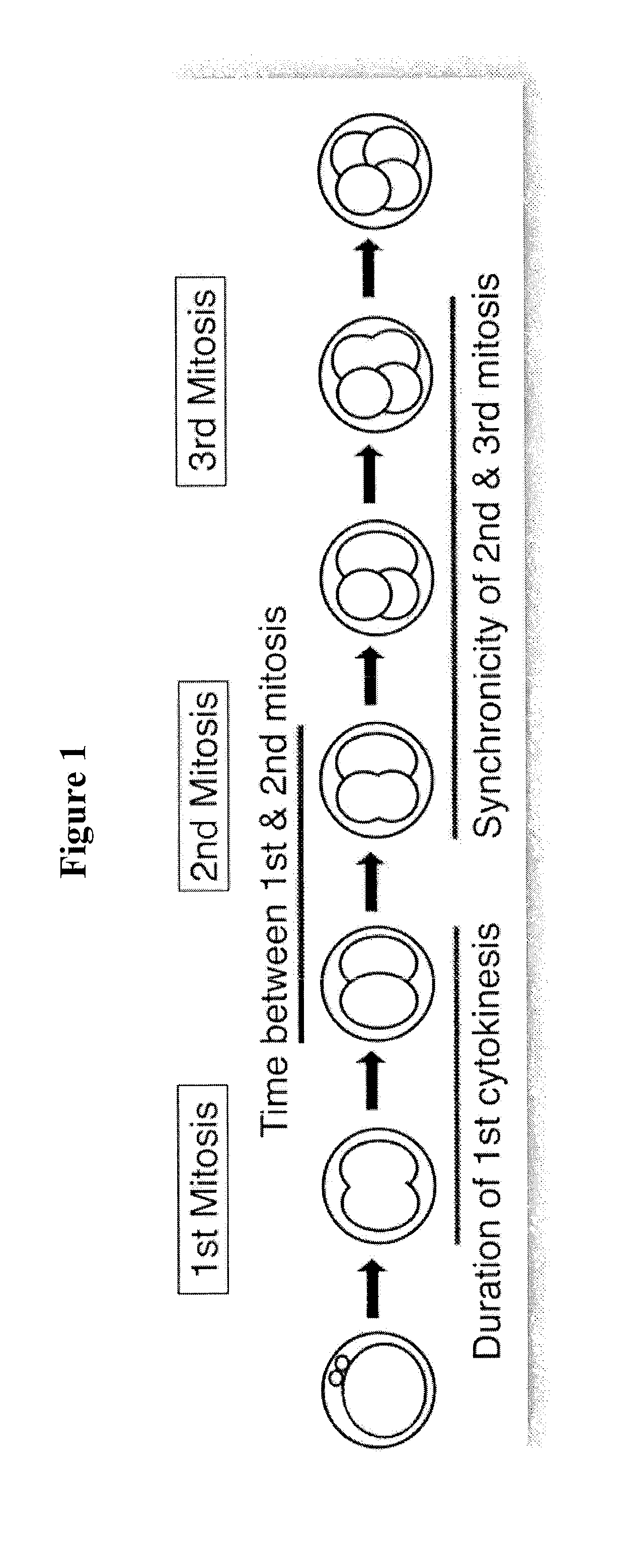
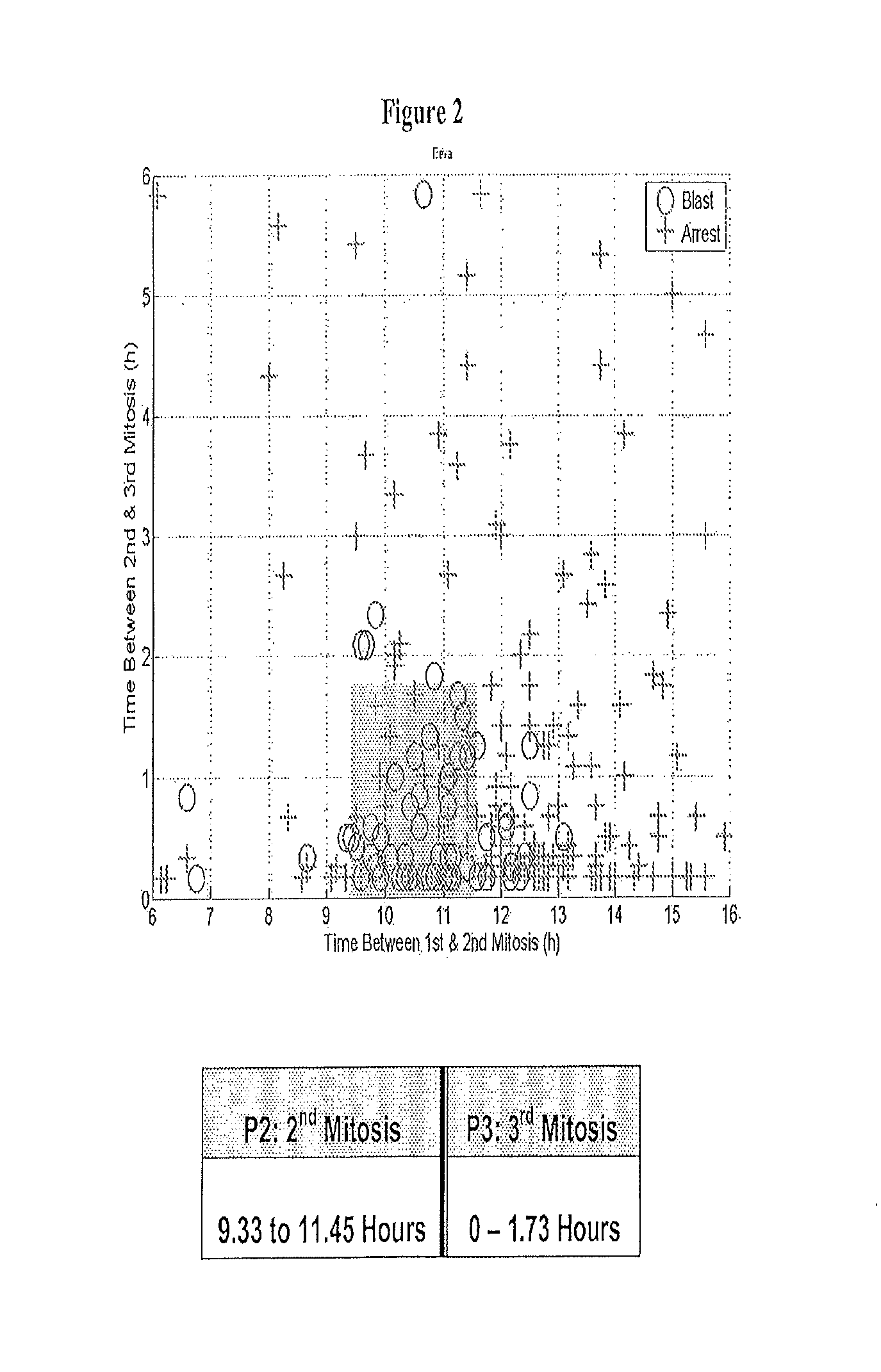
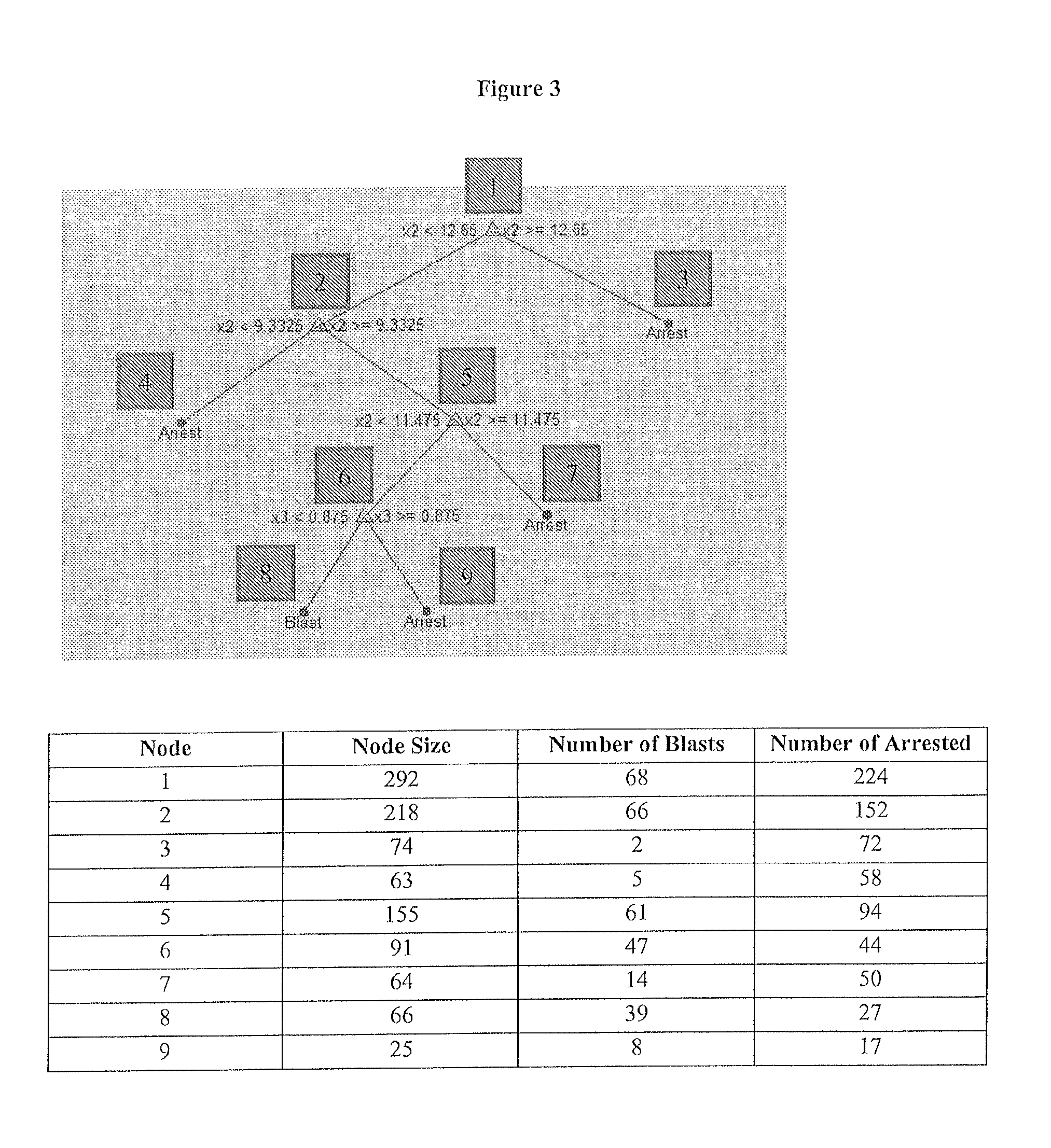
[0115]To develop the blastocyst prediction model, a clinical study was performed to collect data from 3 sites, 54 subjects and 292 embryos. The embryos were cultured using standard procedures in an IVF lab and imaged at 5 minute intervals inside the incubator. By retrospectively analyzing the image data, it was shown that quantification of the timing of early cell division up to approximately 48 hours after fertilization could predict whether an embryo would become a blastocyst on day 5 with a high degree of specificity. During this analysis, it was found that the time between 1st and 2nd mitosis (p2) and the time between 2nd and 3rd mitosis (p3) significantly contributed to the predictive power of the prediction model. Therefore, the blastocyst prediction model was based on the time between 1st and 2nd mitosis (p2) and the time between 2nd and 3rd mitosis (p3).

[0116...

example 2

Purpose

[0122]This example describes the process used to develop statistical classification models for predicting blastocyst formation based on the blastocyst prediction timing parameters.

Model Development

[0123]The clinical study dataset was collected to help build and evaluate different types of statistical classification models for predicting blastocyst formation. The input parameters to these classifiers were the 3 predictive parameters (based on the paper Wong C C, Loewke K E, Bossert N L, Behr B, De Jonge C J, Baer T M, Reijo Pera R A. Non-Invasive Imaging of Human Embryos Before Embryonic Genome Activation Predicts Development to the Blastocyst Stage. Nat. Biotechnol. 2010 October; 28(10):1115-21.): duration of first cytokinesis (P1), time between 1st and 2nd mitosis (P2), and time between 2nd and 3rd mitosis (P3).

[0124]The models were trained on an extensive clinical study dataset The dataset consisted of 292 embryos across 45 patients. The average age of the egg is 33.6±4.8. ...

example 3

[0206]Development and validation of a new test for predicting embryo viability based on time-lapse imaging and automated cell tracking.

Abstract

[0207]The objective of this study was to develop and prospectively validate a new, real-time early embryo viability assessment platform for improving embryo selection in in vitro fertilization (IVF) laboratories.

[0208]The specificity, positive predictive value and overall accuracy of identifying Usable Blastocysts (blastocysts deemed suitable for transfer or freezing) at the cleavage stage are significantly improved when using the new test compared to traditional Day 3 morphology.

[0209]New embryo selection methods are expected to improve IVF success rates and reduce the need for multiple embryo transfer, yet step-by-step approaches to validate new technology for clinical usefulness are lacking. In this study, scientifically-based time-lapse image markers are integrated with cell tracking capabilities to create the first method for quantitativ...


PUM


Abstract

Methods, compositions and kits for determining the likelihood of reaching the blastocyst stage for one or more embryos or pluripotent cells are provided. These methods, compositions and kits find use in identifying embryos and oocytes in vitro that are most useful in treating infertility in humans.

Description

[0001]This application claims priority to U.S. Provisional Application No. 61 / 653,962 filed May 31, 2012 and U.S. 61 / 671,060 filed Jul. 12, 2012, both of which are incorporated by reference herein in their entireties.FIELD OF THE INVENTION[0002]This invention relates to the field of biological and clinical testing, and particularly the imaging and evaluation of zygotes / embryos, oocytes, and stem cells from both humans and animals.BACKGROUND OF THE INVENTION[0003]Infertility is a common health problem that affects 10-15% of couples of reproductive-age. In the United States alone in the year 2006, approximately 140,000 cycles of in vitro fertilization (IVF) were performed (cdc.gov / art). This resulted in the culture of more than a million embryos annually with variable, and often ill-defined, potential for implantation and development to term. The live birth rate, per cycle, following IVF was just 29%, while on average 30% of live births resulted in multiple gestations (cdc.gov / art). M...

Claims


Application Information

Patent Timeline
IPC IPC(8): G01N33/483A61B17/435
CPCG01N33/4833A61B17/435C12M21/06C12M41/48C12M47/04G06T7/0016G06T2207/10056G06T2207/20036G06T2207/30044G06T7/68G06V20/698G01N33/5005C12N5/0604G01N21/75G01N2201/062G06T7/0012G06T2207/30024
Inventor LOEWKE, KEVIN E.SURAJ, VAISHALICHEN KIM, ALICE A
Owner PROGYNY
Why Eureka
Try Eureka