Method for gathering information relating to at least one object arranged on a patient positioning device in a medical imaging device and a medical imaging device for carrying out the method

a technology of patient positioning and information gathering, which is applied in the direction of patient positioning for diagnostics, instruments, applications, etc., can solve the problems of unwanted overhanging of wrongly positioned objects, damage to wrongly positioned objects, crushing of cables and/or injuries to patients, etc., to achieve quick monitoring of the position of objects, improve safety, and improve the effect of measuring procedures

Inactive Publication Date: 2013-12-26
SIEMENS AG
View PDF11 Cites 47 Cited by
  • Summary
  • Abstract
  • Description
  • Claims
  • Application Information

AI Technical Summary

Benefits of technology

[0023]In an advantageous development of the invention, it is proposed that information relating to operating personnel is recorded based on the 3-D image data. For this purpose, a scan range of the 3-D image data recording unit is dimensioned such that the operating personnel is included by the scanning range of the 3-D image data recording unit, in particular during positioning and / or preparation of an object on the patient positioning device. With this embodiment of the invention, a communication of the operating personnel with, for example, a control unit of the medical imaging device and / or of the evaluating unit can advantageously be achieved.
[0024]Particularly advantageously, by means of the information relating to the operating personnel and / or relating to the object positioned on the patient positioning device, at least partial control of a movement of the patient positioning device and / or at least partial control of the medical imaging device is accomplished, so that a quicker and more effective measuring procedure can be achieved. For example, in this way, positioning of the patient positioning device can be controlled in that gestures of the operating personnel are recognized in the 3-D image data by the evaluating unit and said gestures are assigned a control function, for control, for example, of the patient positioning device. The control functions can be made up from raising a patient table of the patient positioning device, lowering of the patient table, displacement of the patient table into or out of a receiving region. In addition, by this means, it is possible to take account, particularly rapidly, of wishes of the patient during the medical imaging examination if, for example, a gesture of the patient recorded in the 3-D image data is recognized by the evaluating unit during the medical imaging examination as a control gesture and said control gesture is assigned to a control signal. For example, lifting a hand by the patient during the medical imaging examination can be assigned to an interruption of the medical imaging examination or to switching on a microphone, or the like.
[0025]It is also proposed that at least one further item of information relating to the object positioned on the patient positioning device is detected by means of at least one marking element arranged at the object. The marking element can be, for example, a colored marking element and / or an infrared marking element and / or a retro-reflective marking element and / or a QR code. In this way, additional information, particularly position information relating to the object positioned on the patient positioning device can advantageously be detected by means of the 3-D image data recording unit, for example, by means of a 2-D camera and / or a color camera and / or an infrared camera, etc., of the 3-D image data recording unit, which is configured for detecting 2-D image data and / or color image data and / or infrared image data.
[0026]Furthermore, the invention relates to a medical imaging device, in particular a magnetic resonance device with a detector unit, a patient positioning device on which a patient is positioned for a medical imaging examination, a receiving region surrounded by the detector unit for receiving the patient positioned on the patient positioning device for the medical imaging examination, and a 3-D image data recording unit for gathering information relating to the patient positioned on the patient positioning device, wherein the 3-D image data recording unit is configured for performing the method according to claims 1 to 12. Particularly time-saving, rapid monitoring of the object positioned on the patient positioning device can be carried out and, consequently, safety can advantageously be enhanced during the medical imaging examination, which is for example a magnetic resonance examination, a computed tomography examination and / or another medical imaging examination which a person skilled in the art deems useful. In addition, particularly the preparation and / or positioning of the patient on the patient positioning device, which takes place chronologically before the medical imaging examination, can be monitored and possible errors during preparation and / or positioning of the patient on the patient positioning device can be detected in good time and thus a fault-laden medical imaging examination can be prevented. Furthermore, clinical personnel supervising the medical imaging examination can be notified rapidly, by means of the output information, of a possible error source during preparation and / or positioning of the patient on the patient positioning device, so that preparation and / or positioning of the patient can be achieved in a particularly time-saving manner
[0027]Preferably, the 3-D image data recording unit is arranged at least partially outside the receiving region, with the result that an advantageous viewing angle and / or recording region over the patient and the patient positioning device, can be achieved for the 3-D image data recording unit in particular for positioning and / or preparation of the patient on the patient positioning device, in particular a patient table of the patient positioning device. Alternatively or additionally, the 3-D image data recording unit can also be arranged at least partially within the receiving region, so that a movement of the patient during the medical imaging examination can be detected. Additionally, a trigger signal, for example, a breathing movement, in particular, can be detected for the medical imaging examination by means of the at least one 3-D image data recording unit which is arranged at least partially within the receiving region.

Problems solved by technology

False positioning can lead to unwanted overhanging of the wrongly positioned objects beyond the patient positioning device and thus, on movement of the patient positioning device, to damaging of the wrongly positioned objects, for example, to crushing of cables and / or injury to the patient, etc.

Method used


Image

  • Method for gathering information relating to at least one object arranged on a patient positioning device in a medical imaging device and a medical imaging device for carrying out the method
  • Method for gathering information relating to at least one object arranged on a patient positioning device in a medical imaging device and a medical imaging device for carrying out the method

Examples


Embodiment Construction

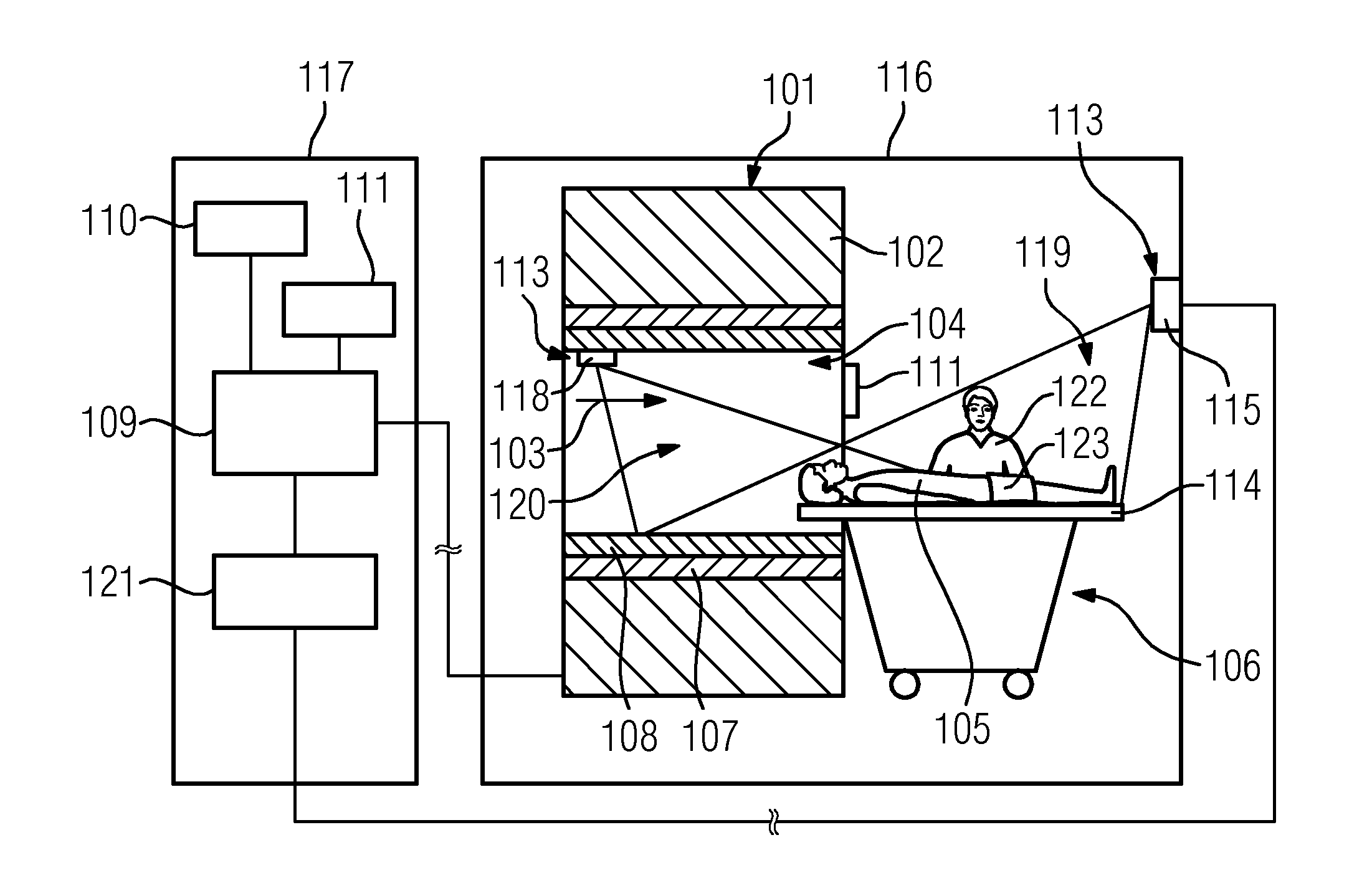
[0031]FIG. 2 shows a schematic representation of an inventive medical imaging device 100 which, in the present exemplary embodiment, consists of a magnetic resonance device. Alternatively, the medical imaging device 100 can also consist of a computed tomography device, a PET device and / or another medical imaging device 100 which a person skilled in the art deems useful.

[0032]The magnetic resonance device comprises a detector unit 101 consisting of a magnet unit, which comprises a main magnet 102 for generating a strong and, in particular, constant main magnetic field 103. The magnetic resonance device also comprises a cylindrical receiving region 104 for receiving a patient 105, wherein the receiving region 105 is surrounded in a peripheral direction by the magnet unit. The patient 105 can be advanced by means of a patient positioning device 106 of the magnetic resonance device into the receiving region. For this purpose, the patient positioning device 106 is movable within the magn...


PUM


Abstract

A method for gathering information relating to at least one object positioned on a patient positioning device of a medical imaging device is provided. The method includes the following steps:
    • gathering by optical means of 3-D image data relating to the object positioned on the patient positioning device by means of a 3-D image data recording unit,
    • transferring the gathered 3-D image data from the 3-D image data recording unit to an evaluating unit,
    • determining information relating to the object positioned on the patient positioning device based on the 3-D image data by means of the evaluating unit,
    • generating output information based on the determined information relating to the object positioned on the patient positioning device, and
    • outputting the output information relating to the object positioned on the patient positioning device.

Description

CROSS REFERENCE TO RELATED APPLICATIONS[0001]The present application claims priority to European Patent Office application No. 102012209190.4 DE filed May 31, 2012, the entire content of which is hereby incorporated herein by reference.FIELD OF INVENTION[0002]The present invention relates to a method for gathering information relating to at least one object arranged on a patient positioning device in a medical imaging device. Firstly, 3-D image data relating to the object arranged on the patient positioning device are gathered optically by means of a 3-D image data recording unit and subsequently the gathered 3-D image data are transferred from the 3-D image data recording unit to an evaluating unit.BACKGROUND OF INVENTION[0003]Investigations with the aid of medical imaging devices, for example, with a magnetic resonance device, a computed tomography device, a PET (Positron Emission Tomography) device, etc. are applied for different medical investigations. When medical diagnostic me...

Claims


Application Information

Patent Timeline
Patent Type & Authority Applications(United States)
IPC IPC(8): G01B11/24
CPCG01B11/24A61B5/0064A61B5/055A61B6/032A61B6/0492G16H40/63A61B6/04A61B6/10
Inventor DRESEL, HOLGERDROBNITZKY, MATTHIASGUHRING, JENSMULLER, EDGARPOPESCU, STEFAN
Owner SIEMENS AG
Why Eureka
Try Eureka