Method for enumerating eukaryotic cell micronuclei with an emphasis on simultaneously acquiring cytotoxicity and mode of action information

a technology of eukaryotic cell micronuclei and cytotoxicity, which is applied in the field of enumerating eukaryotic cell micronuclei with an emphasis on simultaneously acquiring cytotoxicity and mode of action information, can solve the problems of insufficient evaluation of toxicity of natural and industrially manufactured compounds and formulations, laborious traditional toxicity evaluations, and large use costs, and achieves cost savings, fast, reliable and accurate results

Inactive Publication Date: 2014-01-16
LITRON LAB
View PDF1 Cites 12 Cited by
  • Summary
  • Abstract
  • Description
  • Claims
  • Application Information

AI Technical Summary

Benefits of technology

[0023]The methods described herein provide for the enumeration of eukaryotic cell micronuclei while simultaneously providing information about genotoxic MOA using, preferably, flow cytometry technology. The primary advantage of this methodology relative to other flow cytometry-based procedures is that MN scoring, cytotoxicity, and MOA determinations are made simultaneously, even in cells with normal p53 function, for instance TK6 cells. Thus, the present invention identifies procedures that can be employed for an automated in vitro micronucleus assay that can be used to simultaneously evaluate agents (e.g., chemical or physical agents) for genotoxicity, cytotoxicity, and mode of genotoxic activity to eukaryotic cells. The procedure is fast, reliable, and accurate, and can be performed without the need for dosing of animals. Consequently, significant cost savings can be afforded by the present invention in the process of testing agents for genotoxicity and / or cytotoxicity.

Problems solved by technology

In the area of environmental health and safety, many natural and industrially manufactured compounds and formulations have not been adequately evaluated for toxicity.
In both arenas, traditional toxicity evaluations are labor intensive and require extensive use of in vivo assays.

Method used


Image

  • Method for enumerating eukaryotic cell micronuclei with an emphasis on simultaneously acquiring cytotoxicity and mode of action information
  • Method for enumerating eukaryotic cell micronuclei with an emphasis on simultaneously acquiring cytotoxicity and mode of action information
  • Method for enumerating eukaryotic cell micronuclei with an emphasis on simultaneously acquiring cytotoxicity and mode of action information

Examples


example 1

Treatment with Demecolcine

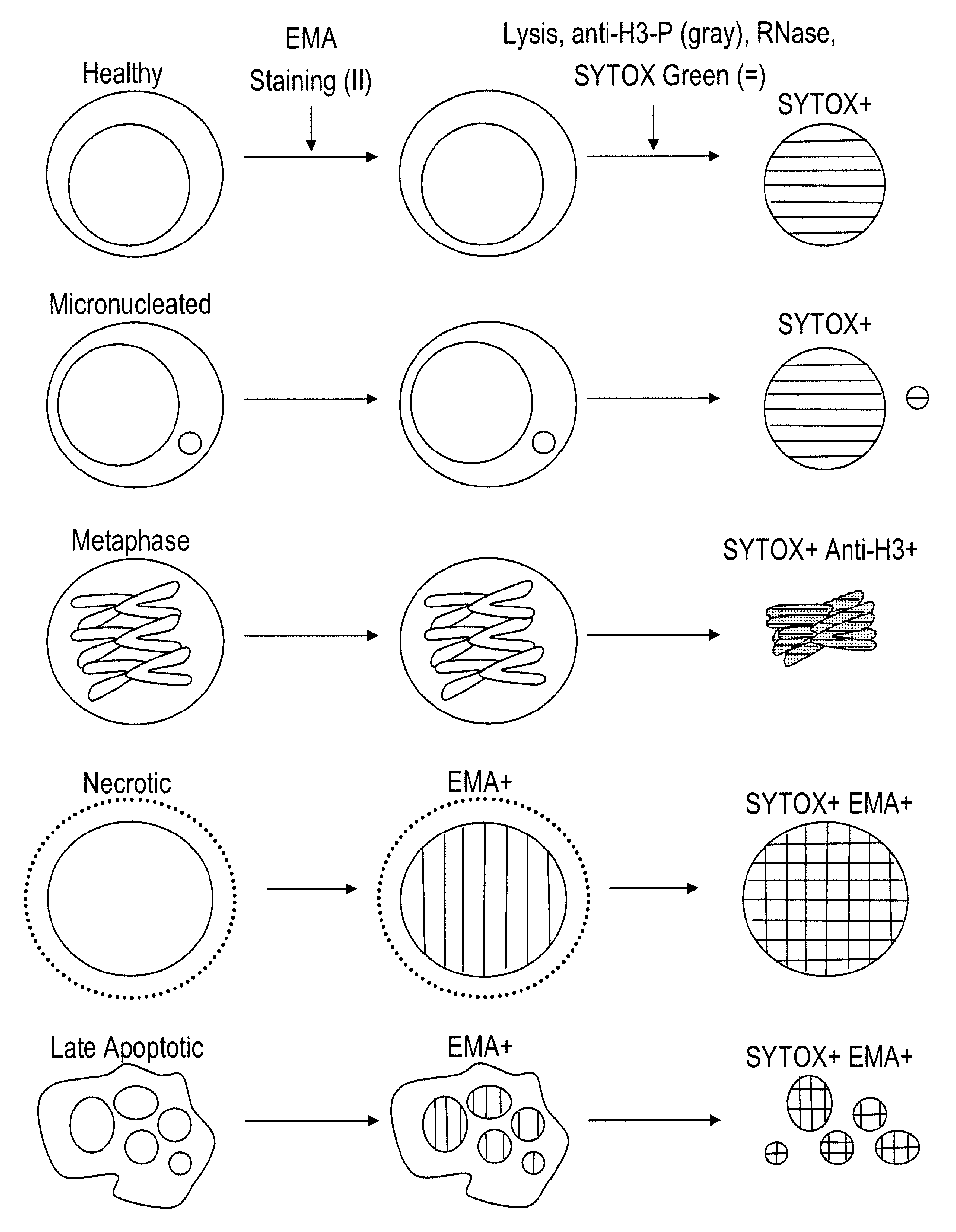
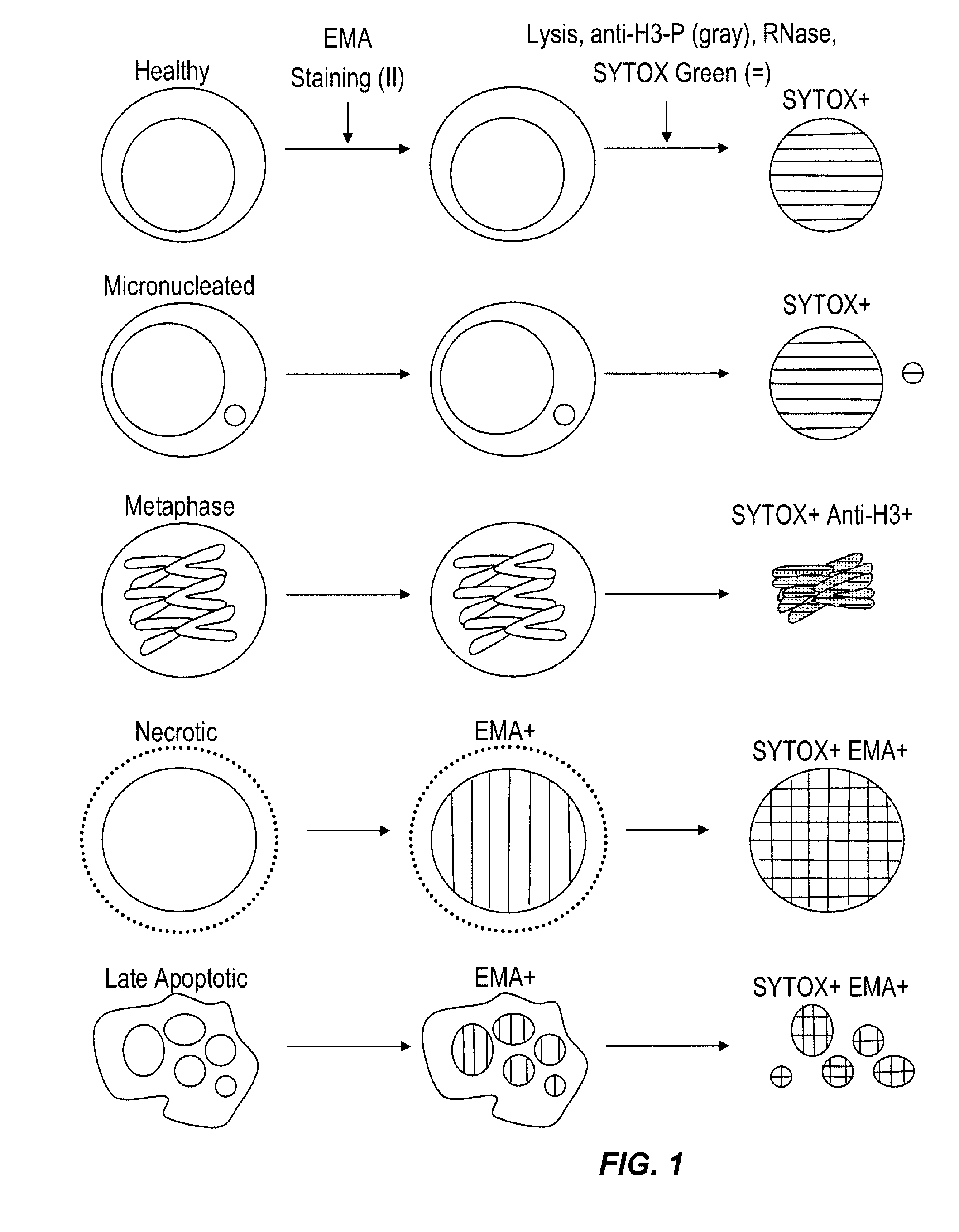
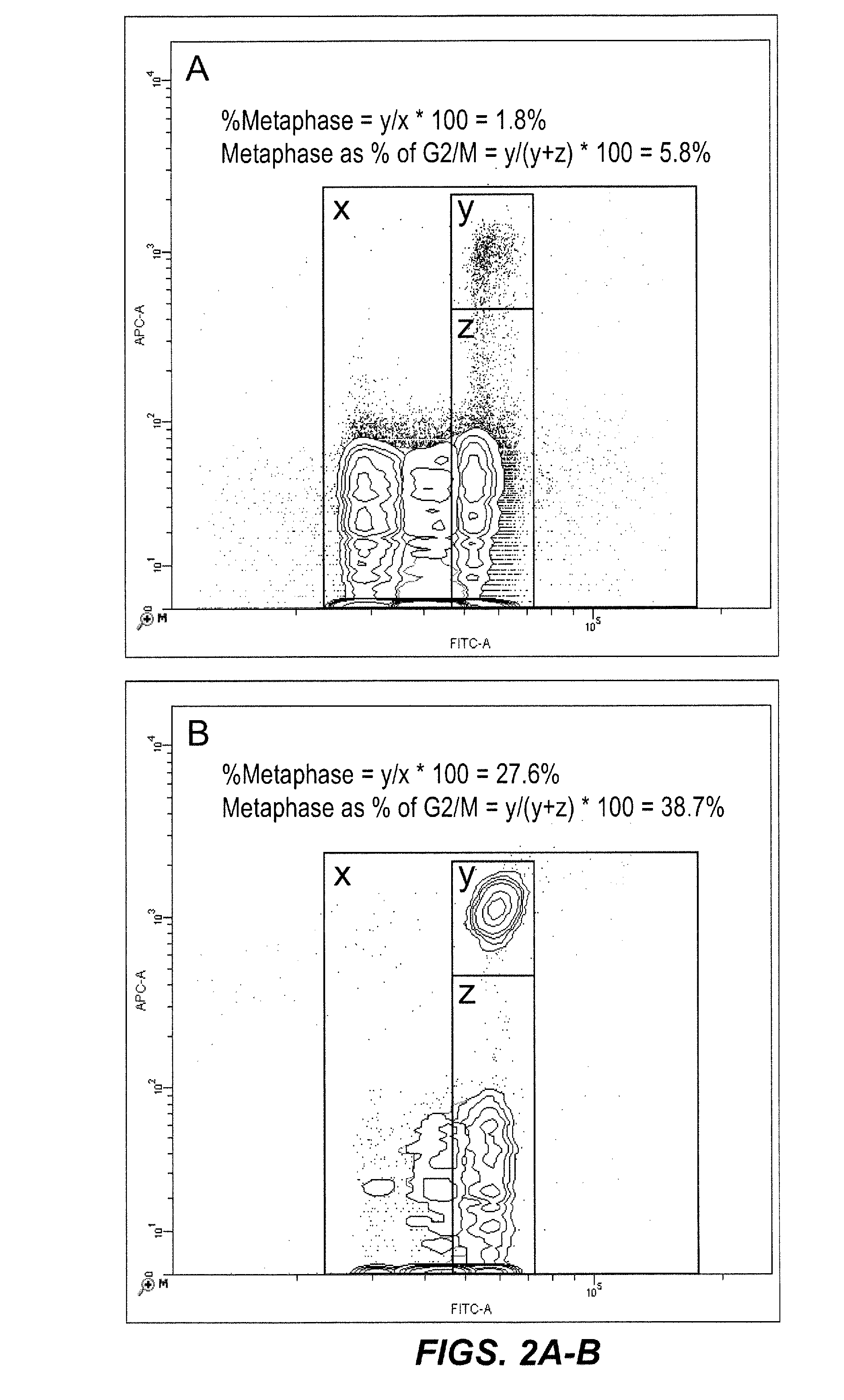
[0084]In this experiment, TK6 cells were treated with 0 or 0.05 ng / ml demecolcine, a metaphase blocking agent. As described above, the cells were then contacted with a first lysis solution that brought them simultaneously in contact with detergent, SYTOX® Green as a pan-DNA fluorescent dye, anti-H3-P (Alexa 647 conjugate) as a metaphase specific fluorochrome, and with RNase. After an appropriate incubation period, the liberated nuclei, MN, metaphase chromosomes, and chromatin debris were contacted with a second lysis solution, and then analyzed on a dual-laser flow cytometer. FIG. 2 shows the resulting bivariate plots of anti-H3-P versus SYTOX® Green fluorescence. Whereas the SYTOX® Green parameter alone is able to differentiate G1, S, and G2 / M cells based on their increasing nucleic acid dye associated fluorescence, it is not capable of differentiating metaphase events from G2 / M, as both exhibit 4n DNA content. However, the anti-H3-P associated fluorescenc...

example 2

Treatment with Reference Genotoxicants

[0085]In these experiment, TK6 cells were exposed to a range of genotoxic chemical concentrations in triplicate wells, with a goal of achieving approximately 50% reduction in relative survival at the termination of the experiment (24 to 27 hours after initiation of treatment). The cell cultures contained equal numbers of counting beads which facilitated the relative survival measurements. As described above, at the termination of the treatment period, cells were contacted with a first fluorescent reagent (EMA) in order to label the chromatin associated with dead and / or dying cells. After photoactivaton and washing steps, the cells were brought into simultaneous contact with detergent to liberate nuclei, MN, metaphase chromosomes, and chromatin debris, a second fluorescent reagent (anti-H3-P Alexa 647 conjugate as a metaphase specific fluorochrome), a third fluorescent reagent (SYTOX® Green as a pan-DNA fluorescent dye), and RNase. The liberated ...

example 3

Polyploidy

[0086]In this experiment, TK6 cells were treated with solvent, the clastogen cisplatin, or the aneugen paclitaxel. Treatments occurred in 96 well plates, demonstrating the ability to scale the invention to smaller vessels and thereby reduce the amount of test article required for testing. As described above, at the termination of the treatment period, cells were contacted with a first fluorescent reagent (EMA) in order to label the chromatin associated with dead and / or dying cells. After photoactivaton and washing steps, the cells were brought into simultaneous contact with detergent to liberate nuclei, MN, metaphase chromosomes, and chromatin debris, a second fluorescent reagent (anti-H3-P Alexa 647 conjugate as a metaphase specific fluorochrome), a third fluorescent reagent (SYTOX® Green as a pan-DNA fluorescent dye), and RNase. The liberated nuclei, MN, metaphase chromosomes, and chromatin debris were subsequently contacted with a second lysis solution and were then ana...


PUM

PropertyMeasurementUnit
exposure timeaaaaaaaaaa
concentrationaaaaaaaaaa
depthaaaaaaaaaa

Abstract

The present invention relates a method for the enumeration of eukaryotic cell micronuclei, while simultaneously acquiring cytotoxicity and mode of action information. The method utilizes differential labeling of chromatin from dead and dying cells to distinguish the chromatin from micronuclei, nuclei, and metaphase chromosomes, and differential labeling of metaphase events to provide additional information regarding cytotoxicity and genotoxic modes of action. Counting of micronuclei events relative to the number of nuclei and quantifying perturbations to the proportion of metaphase events can be used to assess the DNA-damaging potential of a chemical agent, the DNA-damaging potential of a physical agent, the effects of an agent which can modify endogenously-induced DNA damage, the effects of an agent which can modify exogenously-induced DNA damage, and genotoxic mode of action.

Description

[0001]This application claims the priority benefit of U.S. Provisional Patent Application Ser. No. 61 / 670,394, filed Jul. 11, 2012, which is hereby incorporated by reference in its entirety.FIELD OF THE INVENTION[0002]This invention relates to a method for enumerating eukaryotic cell micronuclei while simultaneously acquiring information that characterizes treatment-related cytotoxicity and, in the case of micronucleus induction, provides evidence for whether the genotoxic activity is the result of an aneugenic or clastogenic mode of action.BACKGROUND OF THE INVENTION[0003]The induction of DNA damage and the resulting sequelae of mutations and chromosomal rearrangements are primary mechanisms by which cancers arise. These types of events have also been implicated in diseases such as atherosclerosis, processes such as aging, and the development of birth defects such as Down syndrome. Therefore, there is an important need for sensitive methods which are capable of identifying chemical...

Claims


Application Information

Patent Timeline
Patent Type & Authority Applications(United States)
IPC IPC(8): G01N33/68C12Q1/68
CPCG01N33/6875C12Q1/68G01N33/5005G01N33/5014
Inventor DERTINGER, STEPHEN D.BRYCE, STEVEN M.
Owner LITRON LAB
Why Eureka
Try Eureka