Method for identifying breast cancer status and kit
a breast cancer and kit technology, applied in the field of breast cancer status identification kits, can solve the problems of inability to show good results in early breast cancer screening with imaging technology, lack of typical symptoms and signs of early breast cancer, and no molecular marker with high sensitivity and specificity found so far. , the effect of fast, reliable and accurate new
- Summary
- Abstract
- Description
- Claims
- Application Information
AI Technical Summary
Benefits of technology
Problems solved by technology
Method used
Image
Examples
example 1
ction
[0170]The DNA extraction reagent is composed of a lysis buffer, a binding buffer, a washing buffer, and an elution buffer. The lysis buffer is composed of a protein denaturant, a detergent, a pH buffering agent and a nuclease inhibitor. The binding buffer is composed of a protein denaturant and a pH buffering agent. The washing buffer is divided into washing buffer A and washing buffer B. Washing buffer A is composed of a protein denaturant, a nuclease inhibitor, a detergent, a pH buffering agent and ethanol; washing buffer B is composed of a nuclease inhibitor, a pH buffering agent and ethanol. The elution buffer is composed of a nuclease inhibitor and a pH buffering agent. The protein denaturant is guanidine hydrochloride; the detergent is TWEEN®20; the pH buffering agent is Tris-HCl; and the nuclease inhibitor is EDTA.
[0171]In this example, a plasma sample of a breast cancer patient is taken as an example to extract plasma DNA. The extraction method comprises the following s...
example 2
[0184]Treatment of DNA with bisulfite is to treat the extracted DNA sample with the bisulfite reagent. The bisulfite reagent is composed of a bisulfite buffer and a protection buffer. The bisulfite buffer is a mixed liquid of sodium bisulfite and water; the protective buffer is a mixed liquid of oxygen radical scavenger hydroquinone and water.
[0185]The DNA extracted in Example 1 is used as the processing object in this Example, and the DNA is treated with bisulfite. It comprise:
[0186](1) prepare the bisulfite buffer: weigh 1 g of sodium bisulfite powder, and add water to it to obtain 3 M buffer solution;
[0187](2) prepare the protection buffer: weigh 1 g of hydroquinone reagent, and add water to it to obtain 0.5M protection buffer;
[0188](3) mix together 100 μL of the DNA solution, 200 μL of the bisulfite buffer and 50 μL of the protection solution, and mix by shaking;
[0189](4) thermal treatment: 95° C. for 5 minutes, 80° C. for 60 minutes, and 4° C. for 10 minute...
example 3
Fluorescent PCR Detection of DNA Methylation and Verification of Primer Sets
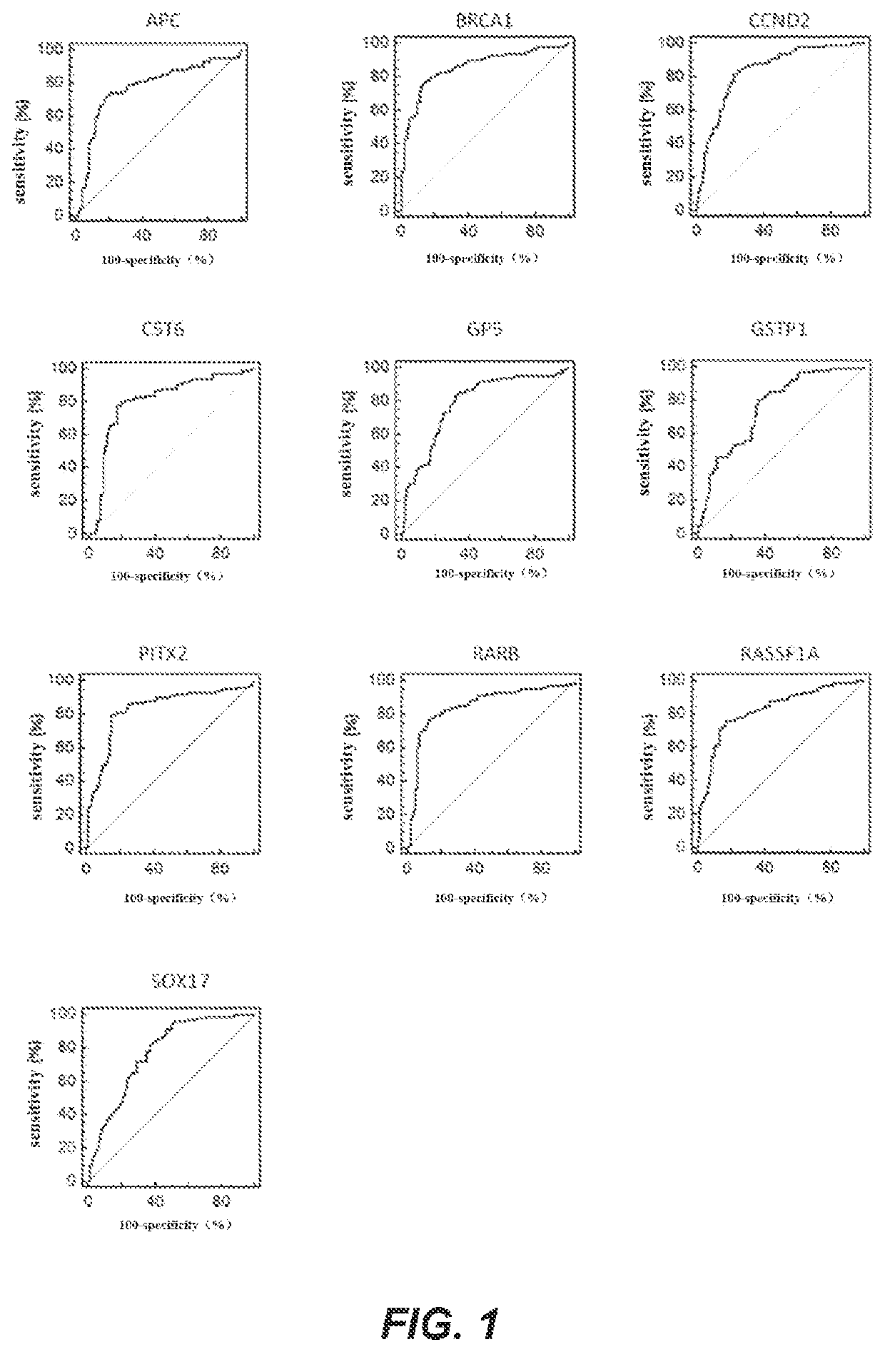
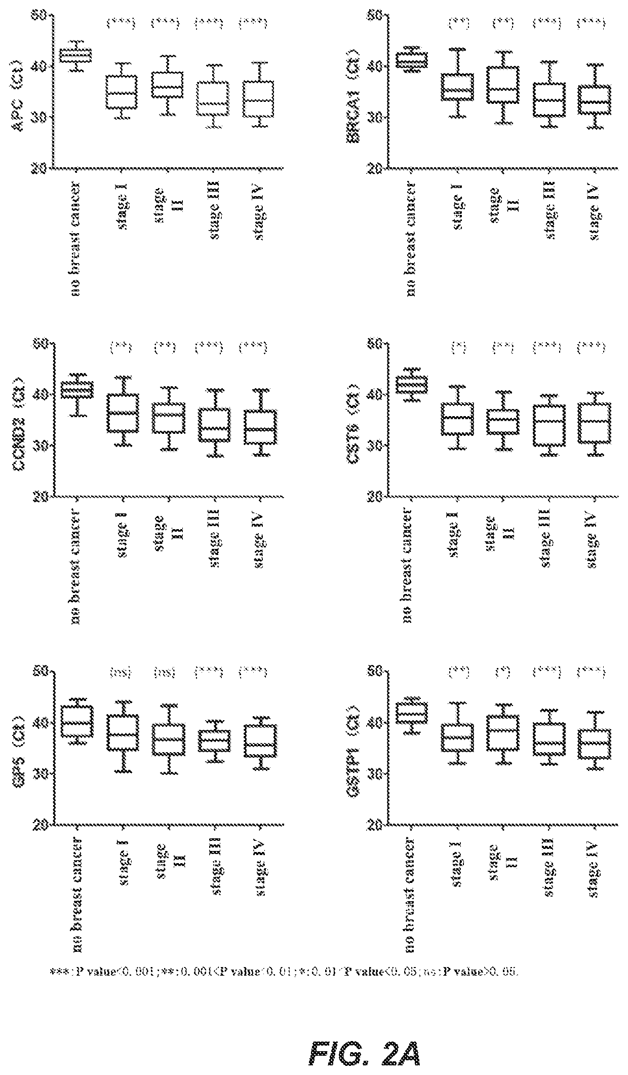
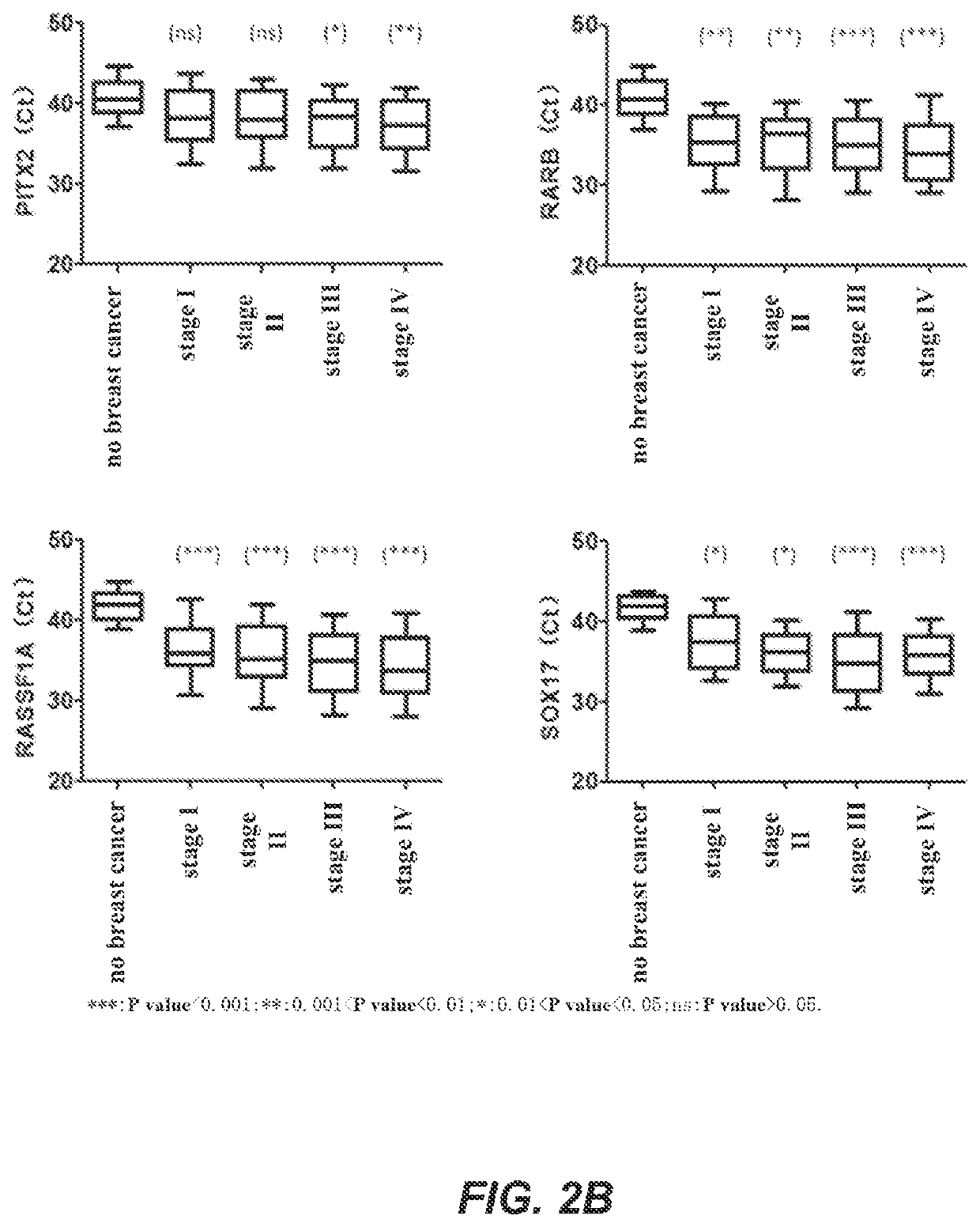
[0200]In this example, a real-time fluorescent PCR was used as an example to detect the methylation levels of biomarker genes. The genes to be detected were APC, BRCA1, CCND2, CST6, GP5, GSTP1, PITX2, RARB, RASSF1A and SOX17 genes, and the internal reference gene was ACTB. In this example, the bisulfate-treated DNA of Example 2 was used as a template for real-time fluorescent PCR amplification. The DNA samples to be detected, a negative quality control product, a positive quality control product and no template controls were all detected in three replicates. The negative quality control product and the positive quality control product were, respectively, prepared as follows: take 400 μL of human leukocyte DNA with a concentration of 10 ng / μL and add it to TE buffer solution containing 1% BSA, mix, and dilute the solution to 200 mL to obtain a negative control substance with a concentration of 0.02 ng / μL; tak...
PUM
| Property | Measurement | Unit |
|---|---|---|
| concentration | aaaaa | aaaaa |
| concentration | aaaaa | aaaaa |
| volume | aaaaa | aaaaa |
Abstract
Description
Claims
Application Information
 - R&D
- Intellectual Property
- Life Sciences
- Materials
- Tech Scout
- Unparalleled Data Quality
- Higher Quality Content
- 60% Fewer Hallucinations
Browse by: Latest US Patents, China's latest patents, Technical Efficacy Thesaurus, Application Domain, Technology Topic, Popular Technical Reports.
© 2025 PatSnap. All rights reserved.Legal|Privacy policy|Modern Slavery Act Transparency Statement|Sitemap|About US| Contact US: help@patsnap.com


