
Device and Method for Heart Valve Repair

a heart valve and device technology, applied in the field of medical devices, can solve the problems of heart failure and abnormal heart rhythm, enlargement and redundancy and enlargement of leaflets and chorda

Inactive Publication Date: 2014-01-30
THE MEDICAL RES INFRASTRUCTURE AND HEALTH SERVICES FUND OF TEL AVIV MEDICAL CENT
View PDF11 Cites 48 Cited by
  • Summary
  • Abstract
  • Description
  • Claims
  • Application Information

AI Technical Summary

Benefits of technology

The present invention provides a device and system for treating heart valves, specifically mitral or tricuspid valves, by deploying an anchor and attaching one or more sutures to the anchor. The anchor can be inserted into the heart through a needle and a catheter, and the sutures can be attached to the anchor while still in its undeployed configuration. The anchor can be released from the catheter and brought to its deployed configuration on the valve leaflet to treat the valve. The invention allows for precise placement of the anchor and sutures, and can be easily guided using echocardiography. The device and system can be used to treat heart valves with mitral or tricuspid valve prolapse.

Problems solved by technology

In mitral valve prolapse, the chordaes have become elongated due to myxomatous degeneration in which collagen in the heart structures forms abnormally and causes thickening, enlargement, and redundancy of the leaflets and chordae.
In addition this process may causes rupture of chordae.
When severe, mitral regurgitation leads to heart failure and abnormal heart rhythms.
These methods require reliable determination of the required length of the neochordae to be introduced, which can be difficult to obtain in a beating heart.
In most systems of this type it is difficult to adjust the lengths of the neochordae after deployment.

Method used


Image

  • Device and Method for Heart Valve Repair
  • Device and Method for Heart Valve Repair
  • Device and Method for Heart Valve Repair

Examples


Embodiment Construction

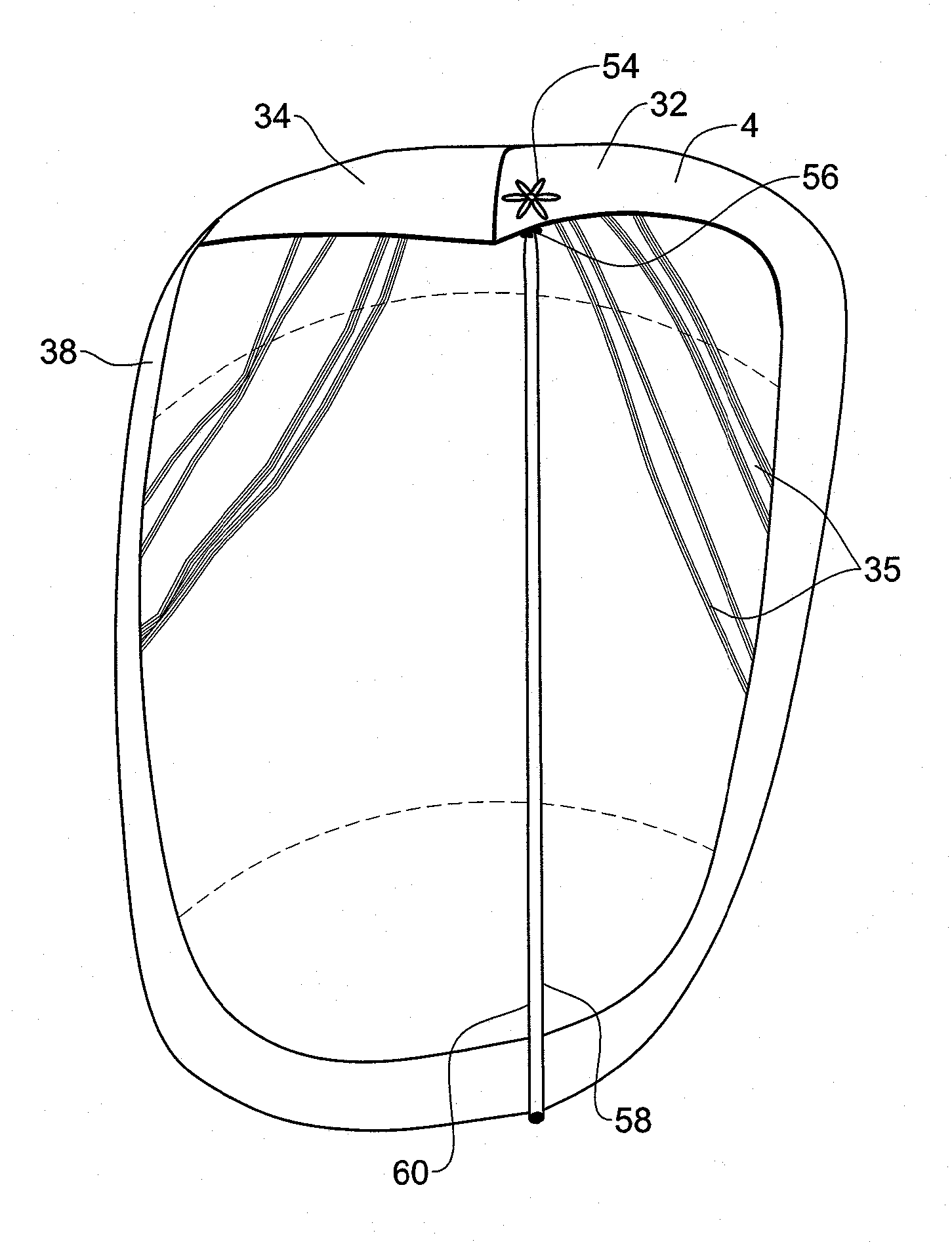
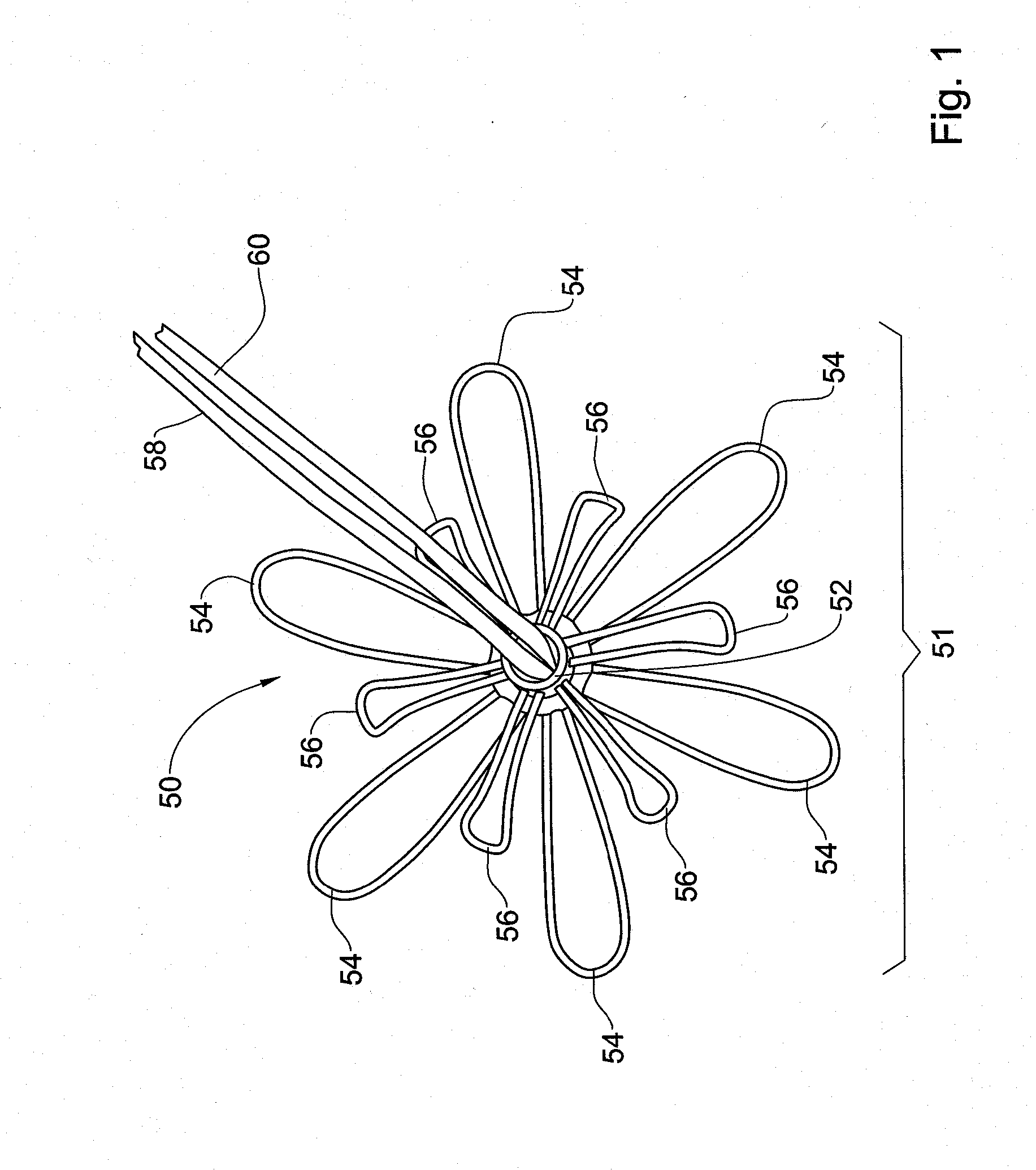
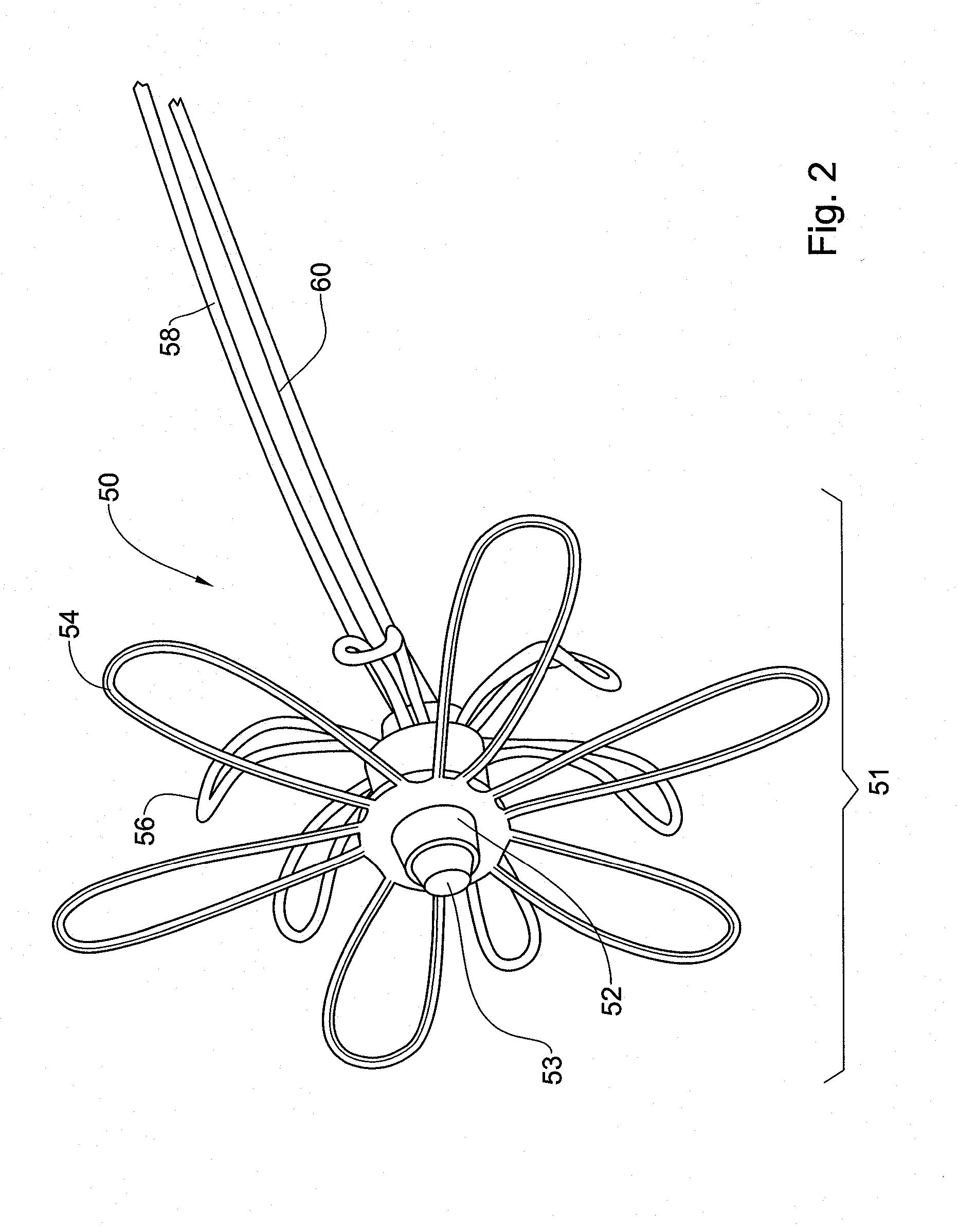
[0057]FIGS. 1 to 3 show a device 50 for treating a heart valve in accordance with one embodiment of the invention. The device 50 has an expanded configuration shown from different perspectives in FIGS. 1 and 2 in which the device 50 is deployed in a heart chamber, as explained below. The device 50 also has a low caliber undeployed configuration, shown in FIG. 3, which is used during delivery of the device 2 to a heart valve.

[0058]The device 50 has an anchor portion 51 comprising a central hub 52 from which extend a plurality of loops 54 and 56. The hub 52 is a tube that is completely closed at the distal end of the tube, for example, by plugging the distal end of the tube with an adhesive 53. In the embodiment of FIGS. 1 to 3, there are 12 loops. This is by way of example only, and the device 50 may have any number of loops are required in any application. The device 50 includes six coplanar loops 54 and another six loops 56 located below the plane of the loops 54 and which curve up...


PUM


Abstract

The invention provides a device (50) and system for treating a heart valve having an anchor (54, 56) and one or more sutures attached to the anchor. The anchor has an expanded deployed configuration and a low caliber undeployed configuration. A delivery in a heart includes catheter and a needle slidable in the lumen of the catheter. The distal end of the catheter is inserted through the myocardium of the heart until the catheter tip is juxtaposed to the underside of the valve leaflet to be treated. The leaflet is then pierced with tip of the needle. The needle is pushed until the anchor in its undeployed configuration passes through the needle tip and is released from the catheter. The anchor is then brought to its deployed configuration on one or both surfaces of the valve leaflet.

Description

FIELD OF THE INVENTION[0001]This invention relates to medical devices, and more specifically to such devices for treating a heart valves.BACKGROUND OF THE INVENTION[0002]In the heart, the mitral valve is located between the left atrium and the left ventricle, while the tricuspid valve is located between the right atrium and the right ventricle. Each valve consists of thin leaflets, located between the atrium and the ventricle. The valve leaflets are attached to the inner wall of the ventricle by a series of fibers called chordae. In a healthy heart, when the ventricles contract during systole, the valve leaflets are apposed and thus prevent backflow of blood from the ventricle into the atrium. When the ventricles relax during diastole, the valve opens to allow blood to flow from the atrium into the ventricle.[0003]In mitral valve prolapse, the chordaes have become elongated due to myxomatous degeneration in which collagen in the heart structures forms abnormally and causes thickenin...

Claims


Application Information

Patent Timeline
Patent Type & Authority Applications(United States)
IPC IPC(8): A61F2/24
CPCA61F2/2466A61F2/2454A61B17/0401A61B2017/0464A61F2/2457A61F2/2463A61B2017/00349A61B2017/00557A61B2017/00862A61B2017/00867A61B2017/0417A61B2090/3912A61B2090/3925
Inventor KUDLIK, NACHMANPEVNAY, DEMITRY
Owner THE MEDICAL RES INFRASTRUCTURE AND HEALTH SERVICES FUND OF TEL AVIV MEDICAL CENT
Learn More