
Contrast enhanced magnetic resonance angiography with chemical shift encoding for fat suppression

a technology of enhanced magnetic resonance and chemical shift, applied in the field of contrast enhanced magnetic resonance angiography with chemical shift encoding for fat suppression, can solve the problem of selective depiction of arterial vasculatur

Active Publication Date: 2014-02-13
KONINKLIJKE PHILIPS ELECTRONICS NV
View PDF2 Cites 11 Cited by
  • Summary
  • Abstract
  • Description
  • Claims
  • Application Information

AI Technical Summary

Benefits of technology

The invention allows for a fast and reliable way to do CE-MRA with fat suppression. It uses a single- or multi-echo data acquisition technique to collect magnetic resonance images of blood vessels only, without needing a separate baseline scan and effectively eliminates fat signals. The technique also uses a Generalized Dixon water-fat separation technique to produce a high-quality image data set. Overall, the invention provides a more efficient and accurate method for performing CE-MRA.

Problems solved by technology

However, this was performed during the steady state 50 minutes after a gadofosveset injection and did not aim to depict the arterial vasculature selectively.

Method used


Image

  • Contrast enhanced magnetic resonance angiography with chemical shift encoding for fat suppression
  • Contrast enhanced magnetic resonance angiography with chemical shift encoding for fat suppression
  • Contrast enhanced magnetic resonance angiography with chemical shift encoding for fat suppression

Examples


Embodiment Construction

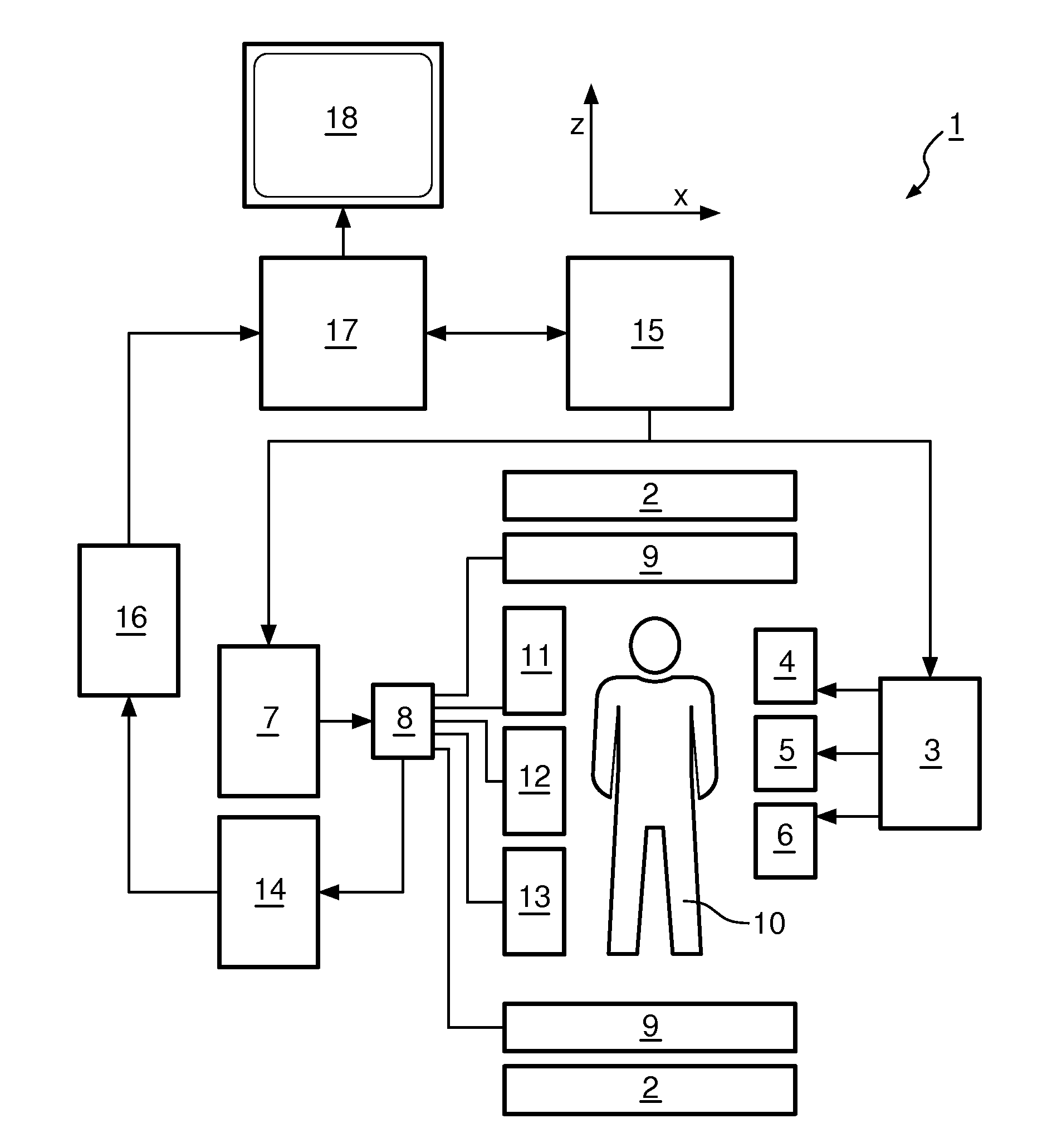
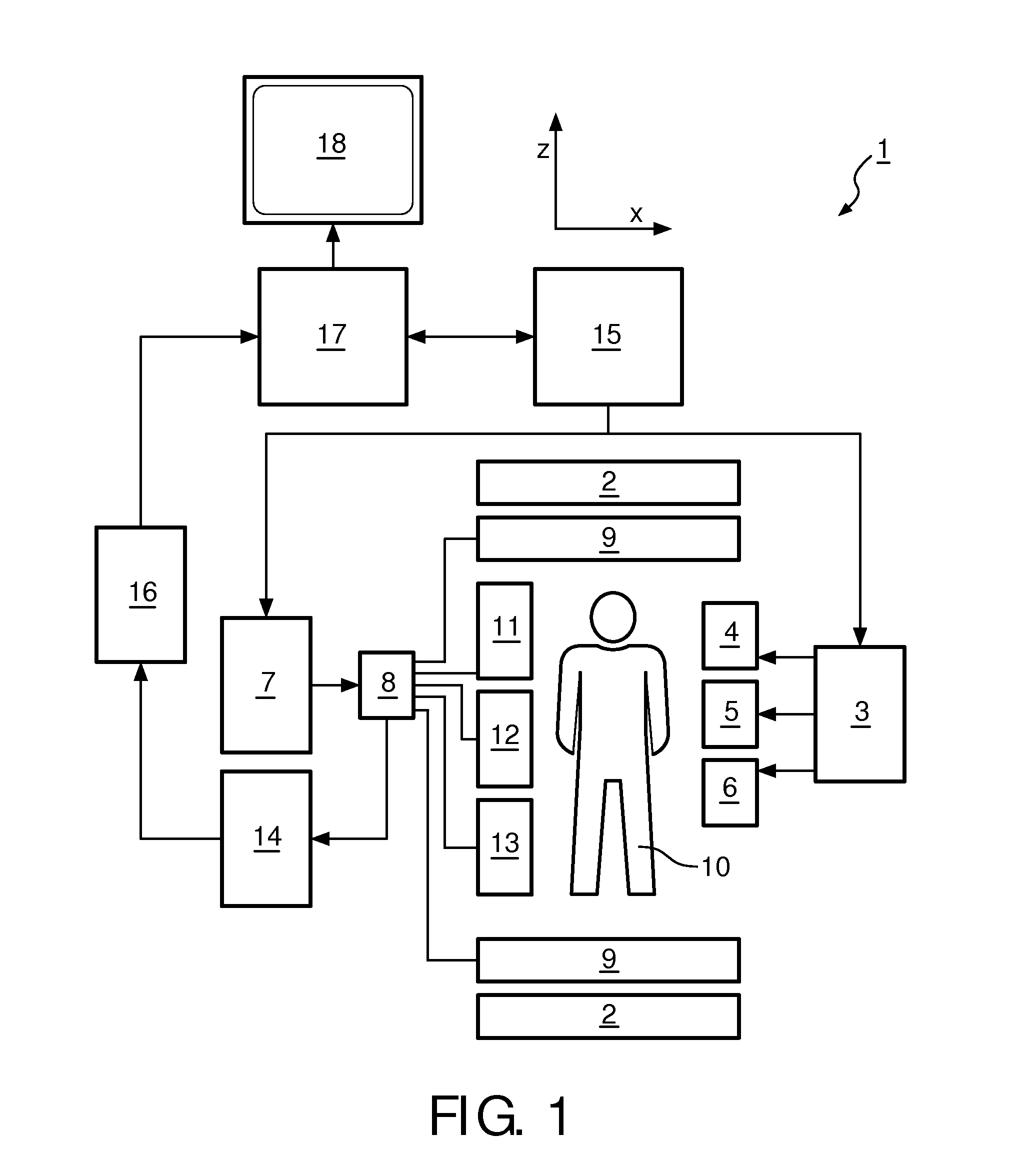
[0053]With reference to FIG. 1, an MR imaging system 1 is shown. The system comprises superconducting or resistive main magnet coils 2 such that a substantially uniform, temporarily constant main magnetic field B0 is created along a z-axis through an examination volume.

[0054]A magnetic resonance generation manipulation system applies a series of RF pulses and switched magnetic field gradients to invert or excite nuclear magnetic spins, induce magnetic resonance, refocus magnetic resonance, manipulate magnetic resonance, spatially or otherwise encode the magnetic resonance, saturate spins and the like to perform MR imaging.

[0055]More specifically, a gradient pulse amplifier 3 applies current pulses to selected ones of whole body gradient coils 4, 5 and 6 along x, y and z-axes of the examination volume. An RF transmitter 7 transmits RF pulses or pulse packets, via a send / receive switch 8 to an RF antenna 9 to transmit RF pulses into the examination volume. A typical MR imaging sequenc...


PUM


Abstract

The invention relates to a method of performing contrast enhanced first pass magnetic resonance angiography, the method comprising: acquiring (302) magnetic resonance datasets of a region of interest using a single- or multi-echo data acquisition technique, wherein the echo times of the one or multiple echoes are flexible, wherein at the time of the data acquisition the region of interest comprises fat, water and a contrast agent, processing (304) the datasets using a generalized Dixon water-fat separation technique to eliminate the signal originating from the fat from the background for reconstruction of an image data set.

Description

TECHNICAL FIELD[0001]The invention relates to a method of performing contrast enhanced first pass magnetic resonance angiography, a computer program product, as well as a magnetic resonance imaging apparatus for performing contrast enhanced first pass magnetic resonance angiography.[0002]Image-forming MR methods which utilize the interaction between magnetic field and nuclear spins in order to form two-dimensional or three-dimensional images are widely used nowadays, notably in the field of medical diagnostics, because for the imaging of soft tissue they are superior to other imaging methods in many respects and do not require ionizing radiation and they are usually not invasive.BACKGROUND OF THE INVENTION[0003]According to the MR method in general, the body of a patient or in general an object to be examined is arranged in a strong, uniform magnetic field B0 whose direction at the same time defines an axis, normally the z-axis, of the coordinate system on which the measurement is b...

Claims


Application Information

Patent Timeline
IPC IPC(8): G01R33/48G01R33/563
CPCG01R33/5635G01R33/4828G01R33/5601G01R33/5615G01R33/56308G01R33/56563A61B5/055A61B5/02007
Inventor GEERTS-OSSEVOORT, LIESBETHKOOIJMAN, HENDRIKALBERTS, EVELINEEGGERS, HOLGER
Owner KONINKLIJKE PHILIPS ELECTRONICS NV
Learn More