System for Perfusion and Diffusion MR Imaging

a technology of diffusion and imaging, applied in the field of perfusion and diffusion mr imaging, can solve the problems of inability to automatically evaluate the known system, the variability of stroke diagnosis,

Active Publication Date: 2014-03-06
NORTHWESTERN UNIV
View PDF8 Cites 21 Cited by
  • Summary
  • Abstract
  • Description
  • Claims
  • Application Information

AI Technical Summary

Problems solved by technology

Perfusion-diffusion mismatch is poorly standardized among imaging facilities, causing variability when used for stroke diagnosis.
However, known systems lack capability to automatically evaluate the degree of perfusion-diffusion mismatch between MR images and additionally need to acquire perfusion-weighted and diffusion-weighted MR images separately, requiring an acquisition time of typically ten minutes or more.

Method used


Image

  • System for Perfusion and Diffusion MR Imaging
  • System for Perfusion and Diffusion MR Imaging
  • System for Perfusion and Diffusion MR Imaging

Examples


Embodiment Construction

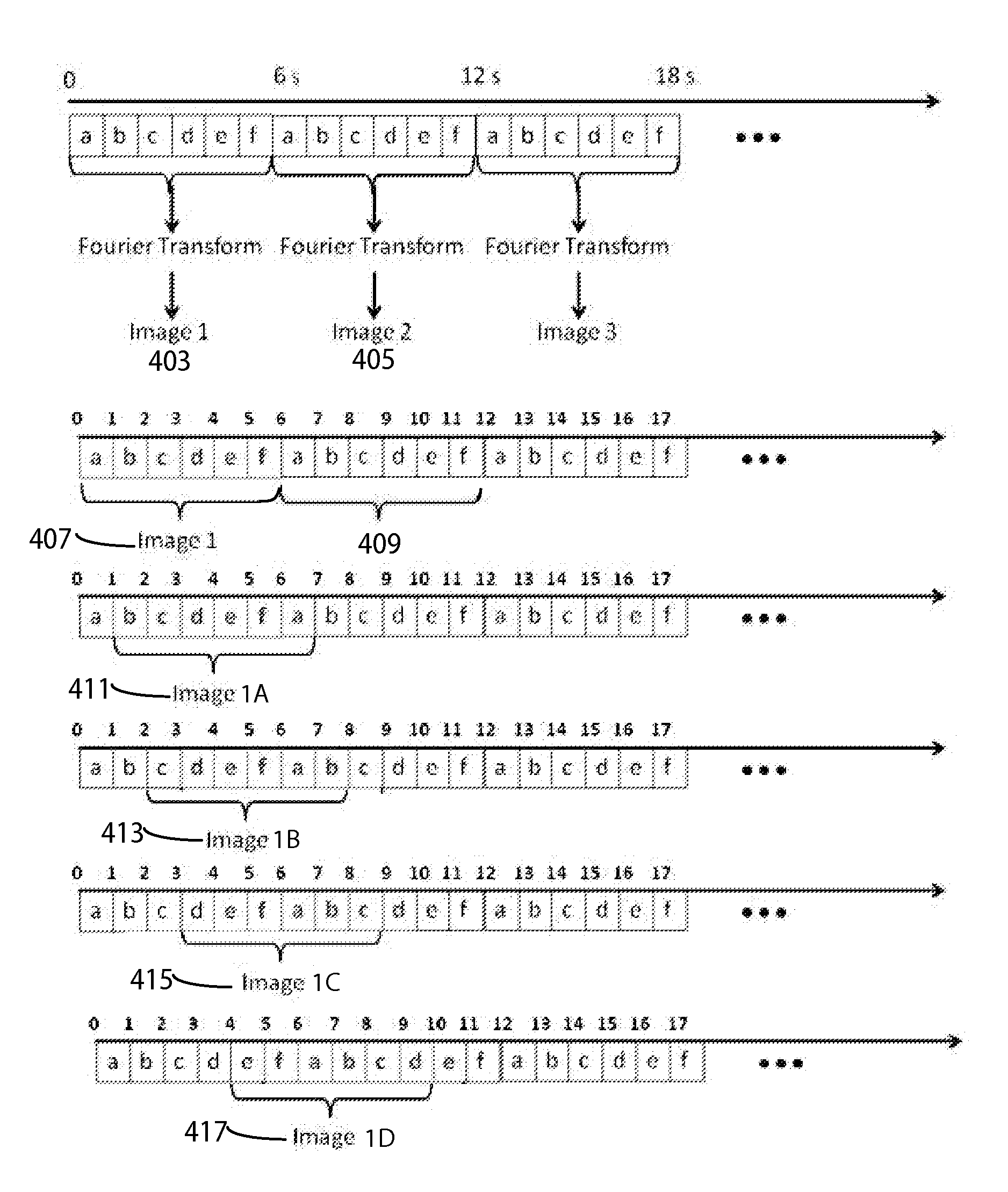
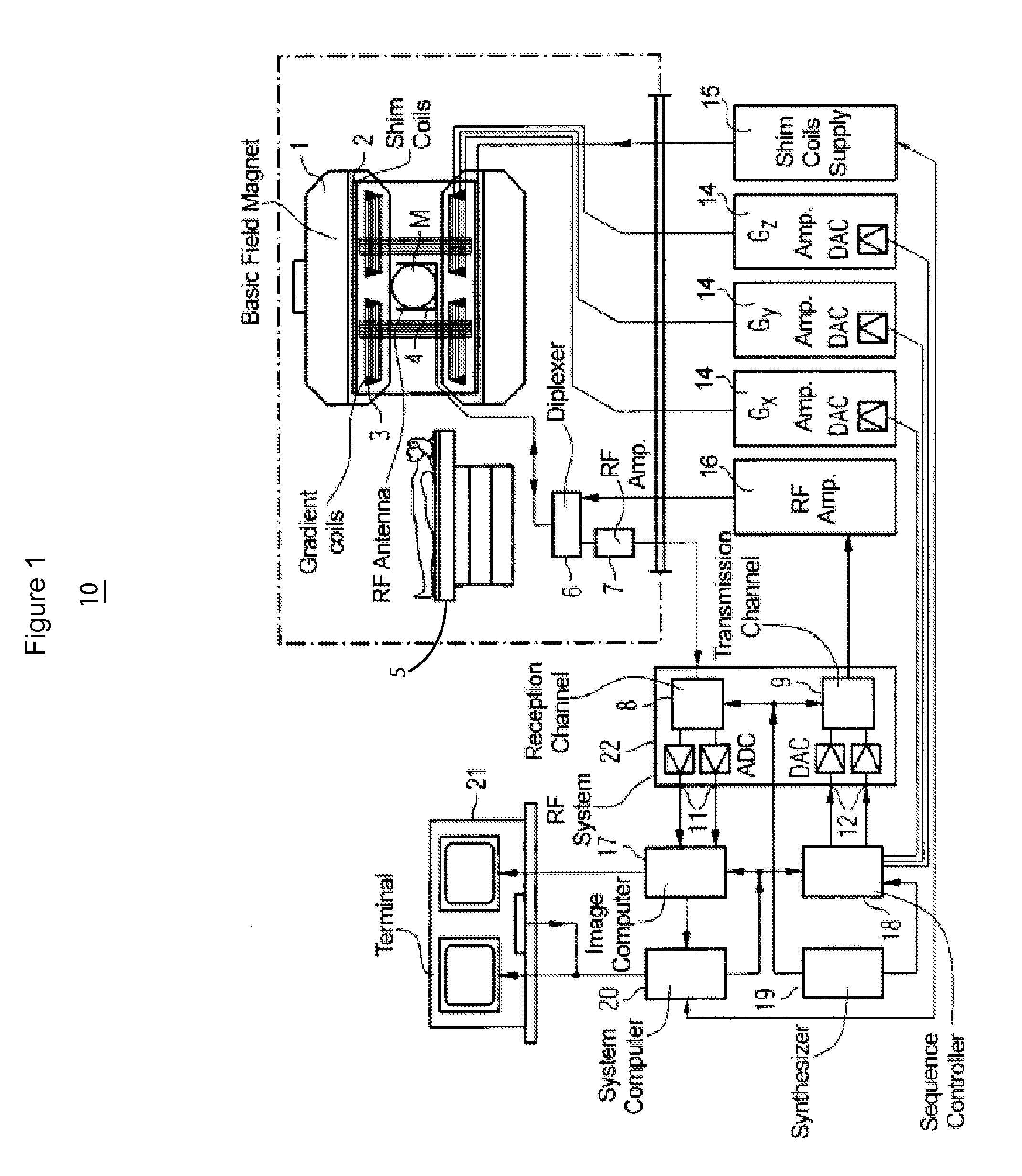
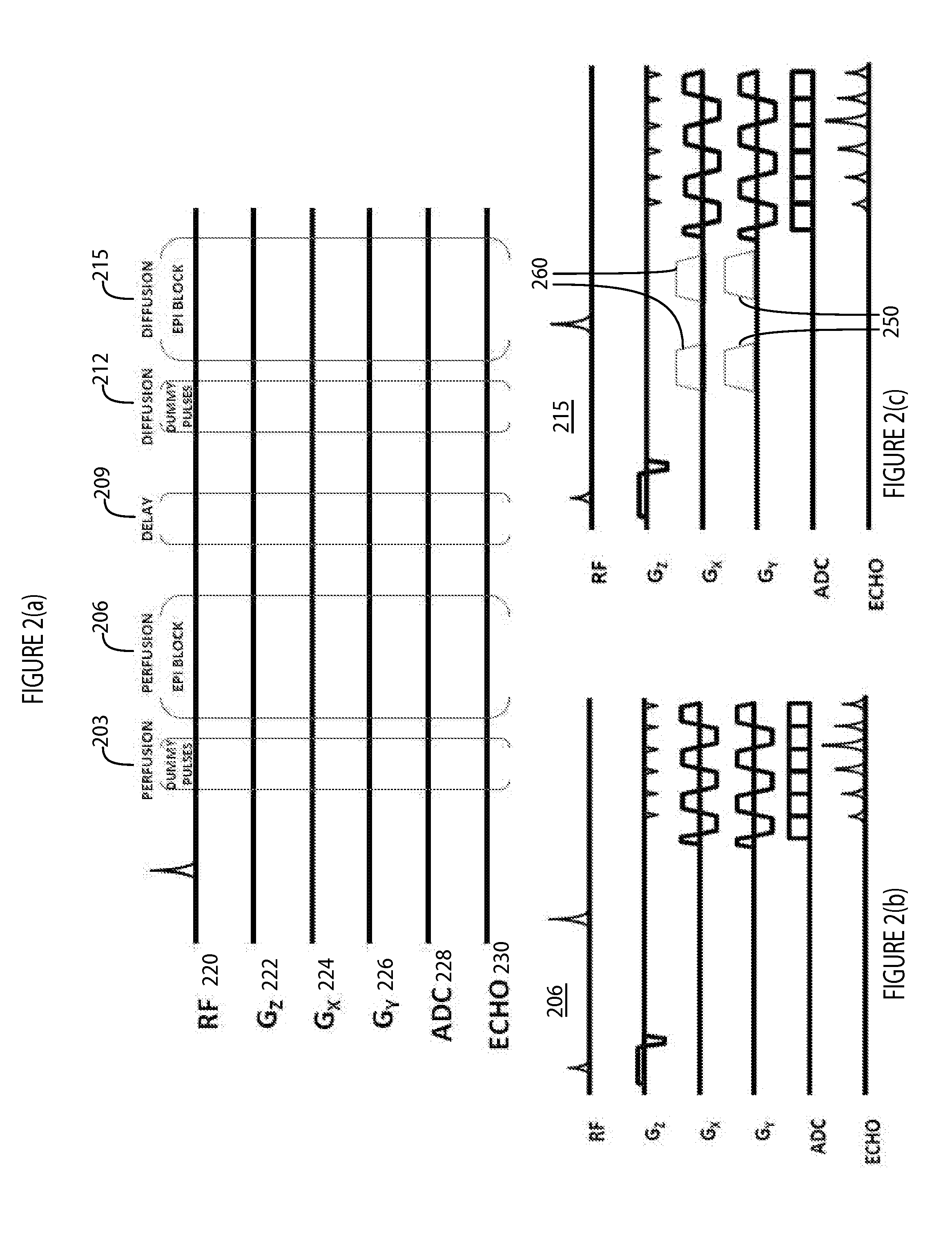
[0011]A system provides a non-invasive, diagnostic imaging examination concurrently acquiring perfusion-weighted and diffusion-weighted MR images for patients with acute ischemic stroke. In one embodiment, the examination uses a 3D-based MR pulse sequence with radial gradient trajectories that acquires high resolution perfusion-weighted and diffusion-weighted images of a brain and quantifies the ischemic penumbra (e.g. in less than two minutes). The system addresses the need to minimize time between diagnosis and intervention in stroke and other conditions using a single, comprehensive imaging examination that provides concurrent acquisition of perfusion-weighted and diffusion-weighted MR images to quantify ischemic penumbra, i.e. the residual viable neurons which are the target of intervention. The system performs a non-invasive, diagnostic imaging examination by concurrently acquiring perfusion-weighted and diffusion-weighted MR images for patients with acute ischemic stroke (e.g....


PUM


Abstract

A system for perfusion and diffusion MR imaging of a portion of patient anatomy includes an RF (Radio Frequency) signal generator for generating RF excitation pulses in anatomy and enabling subsequent acquisition of associated RF echo data. A magnetic field gradient generator generates anatomical volume select magnetic field gradients for phase encoding and readout RF data acquisition in a three dimensional (3D) anatomical volume. The RF signal generator and the gradient generator acquire within a single MR imaging scan, perfusion image data of the 3D volume, at least partially in the presence of contrast agent, and diffusion image data of the 3D volume.

Description

CROSS-REFERENCE TO RELATED APPLICATIONS[0001]This application is a non-provisional of, and claims priority to, U.S. provisional patent application Ser. No. 61 / 694,373 by T. J. Carroll et al. filed on 29 Aug. 2012.[0002]This invention was made with government support under grant numbers 5R01HL088437-04 and 5T32EB005170-05 awarded by the National Institutes of Health. The government has certain rights in the invention.FIELD OF THE INVENTION[0003]This invention concerns a system for perfusion and diffusion MR imaging of a portion of patient anatomy, by acquiring, within a single MR imaging scan, perfusion image data of a 3D volume, at least partially in the presence of contrast agent, and diffusion image data of the 3D volume.BACKGROUND OF THE INVENTION[0004]Ischemic stroke is the third leading cause of death and disability in the industrialized world. Known systems perform magnetic resonance imaging (MRI) scans on a patient suffering from ischemic stroke to identify the degree of mism...

Claims


Application Information

Patent Timeline
Patent Type & Authority Applications(United States)
IPC IPC(8): G01R33/34G01R33/54
CPCG01R33/54G01R33/34G01R33/4826G01R33/56341G01R33/56366
Inventor CARROLL, TIMOTHY J.JEONG, YONGJONATHAN, SUMEETH VIJAYVAKIL, PARMEDE
Owner NORTHWESTERN UNIV
Why Eureka
Try Eureka