
Method and apparatus for image-centric standardized tool for quality assurance analysis in medical imaging

a quality assurance analysis and image-centric technology, applied in image analysis, healthcare informatics, healthcare resources and facilities, etc., can solve the problems of ineffective standardized methods for quantitative and qualitative quality assessment, stagnation of quality deliverables in healthcare, and often being sacrificed (or subservient). achieve the effect of improving medical imaging

Inactive Publication Date: 2014-03-13
REINER BRUCE
View PDF3 Cites 54 Cited by
  • Summary
  • Abstract
  • Description
  • Claims
  • Application Information

AI Technical Summary

Benefits of technology

The present invention improves medical imaging by standardizing image quality analysis, continuously tracking and recording image quality data, and taking into account a variety of clinical, technical, and patient-specific variables. The computer system performs image quality analysis of a plurality of images from an imaging examination, displaying the examination on a computer system and allowing the user to review and comment on the images. The system then saves the annotated image data and transfers it to a quality assurance database for review and analysis. The invention also includes a biometrics authentication system and a database of comparative analysis for comparison with future imaging examinations. The technical effects of the invention include improved image quality, improved quality assurance, and improved medical diagnostics.

Problems solved by technology

While almost all healthcare participants view quality as a high priority, standardized methods for quantitative and qualitative quality assessment are essentially non-existent.
This lack of standardization in medical quality assessment produces an environment where quality deliverables are often left to the discretion of individual healthcare providers, who at the same time are being challenged to maintain revenue and profitability in an environment of declining economic reimbursements.
The net result is that quality often becomes sacrificed (or subservient) to concerns over productivity and workflow.
In the absence of standardized quality metrics and tools for analysis, quality deliverables in healthcare will continue to stagnate (and potentially deteriorate), with the potential for adverse clinical outcomes and economic inefficiency.
Further, while all medical disciplines are affected to some degree by the relative lack of objective standards and methods of analysis, medical imaging is perhaps more prone to deficiencies and variation in quality due to its dependence on imaging (i.e., pictorial) data, as opposed to clinical (i.e., numerical) data.
In the absence of an objective methodology to analyze image quality, the net result is that quality assessment in everyday practice remains somewhat elusive and undefined.
As a result, many consumers of medical imaging services (e.g., referring clinicians, patients, third party payers) currently evaluate quality in these easily defined operational efficiency measures and largely ignore the intrinsic “clinical” quality of the imaging dataset, which ultimately determines the ability to render an accurate and reproducible diagnosis.
This process is almost always “off the record” and results in no recorded data for longitudinal analysis.
Unfortunately, in the current practice environment, productivity often tends to supersede quality, and as a result many imaging exams lacking in quality are accepted “as-is”.
While the interpreting radiologist in theory serves as a “second line of defense” in maintaining quality standards, there is little incentive for the radiologist to override the decision of the technologist once the exam has been completed, the patient has left the department, and the imaging dataset has been transmitted to the imaging archive for interpretation.
This has the undesired effect of increased radiation, reducing patient throughout, creating a backlog in the queue, and delaying diagnosis.
As a result of these negative pressures, many imaging service providers tend to “make do” and accept image quality deficiencies, which in turn has the potential to result in equivocal or erroneous diagnoses, additional follow-up imaging exams, and additional consultations.
This results in incomplete imaging records, lack of traceable quality data, and lost educational opportunities.
In doing so however, the broad spectrum of images is never really evaluated and this can mask existing quality deficiencies.
Since this accreditation / review process only takes place every 3-4 years, there is little incentive to critically review quality deliverables on a regular basis.
The end result is that quality assessment in its current form is inherently flawed, performed in a piecemeal fashion, devoid of data, and lacks documentation.

Method used


Image

  • Method and apparatus for image-centric standardized tool for quality assurance analysis in medical imaging
  • Method and apparatus for image-centric standardized tool for quality assurance analysis in medical imaging
  • Method and apparatus for image-centric standardized tool for quality assurance analysis in medical imaging

Examples


Embodiment Construction

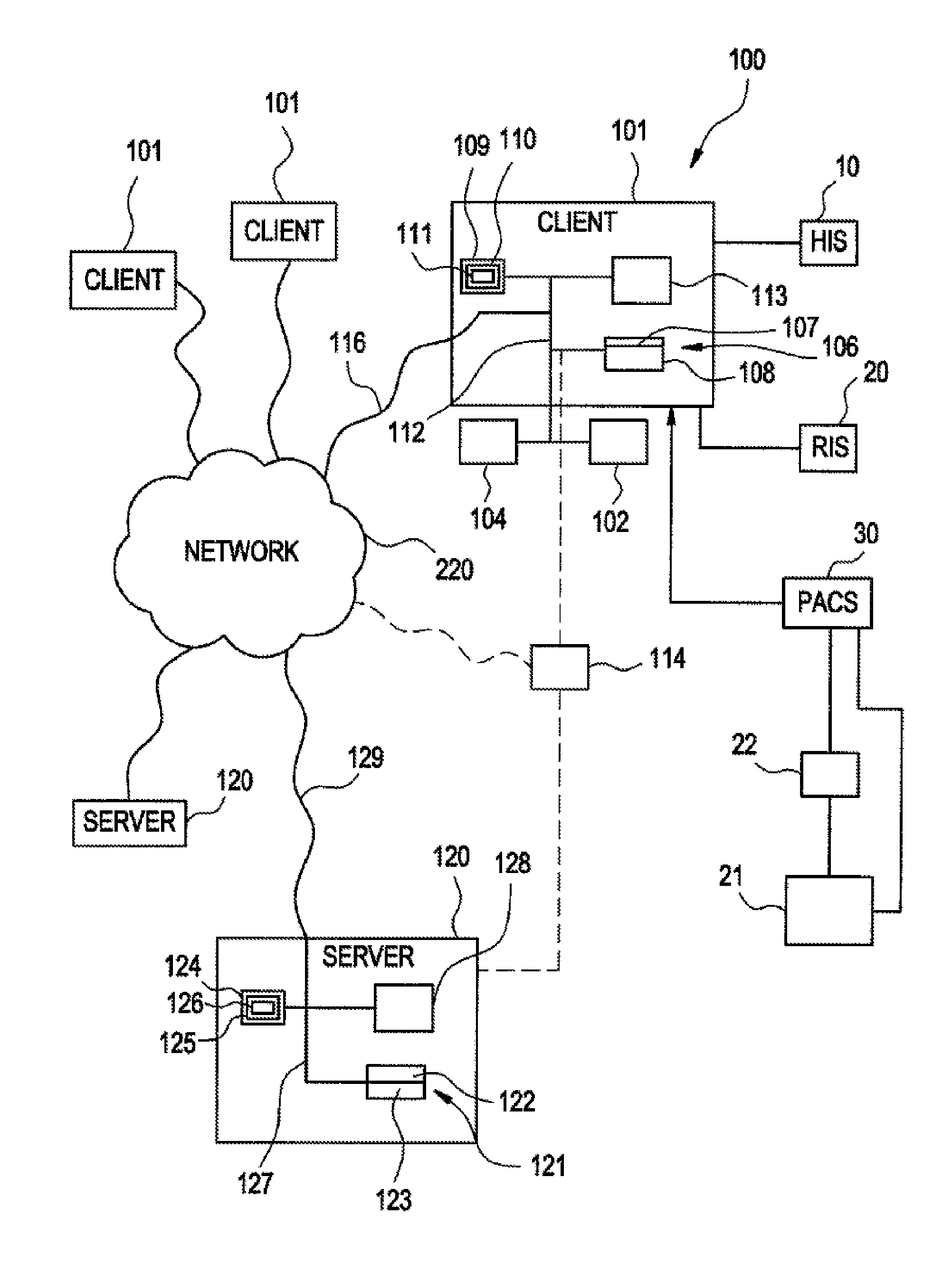
[0041]The present invention improves medical imaging by standardizing image quality analysis, directly links the quality data with the imaging dataset (i.e., image-centric), continuously tracks and records image quality data for longitudinal analysis, and analyzes all imaging datasets while taking into account a myriad of clinical, technical, and patient-specific variables affecting quality measures.

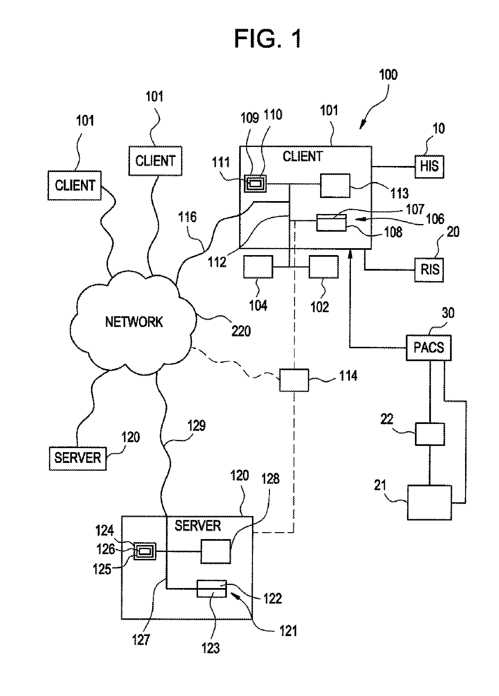
[0042]According to one embodiment of the invention illustrated in FIG. 1, medical (radiological) applications may be implemented using the system 100. The system 100 is designed to interface with existing information systems such as a Hospital Information System (HIS) 10, a Radiology Information System (RIS) 20, a radiographic device 21, and / or other information systems that may access a computed radiography (CR) cassette or direct radiography (DR) system, a CR / DR plate reader 22, a Picture Archiving and Communication System (PACS) 30, and / or other systems. The system 100 may be designed...


PUM


Abstract

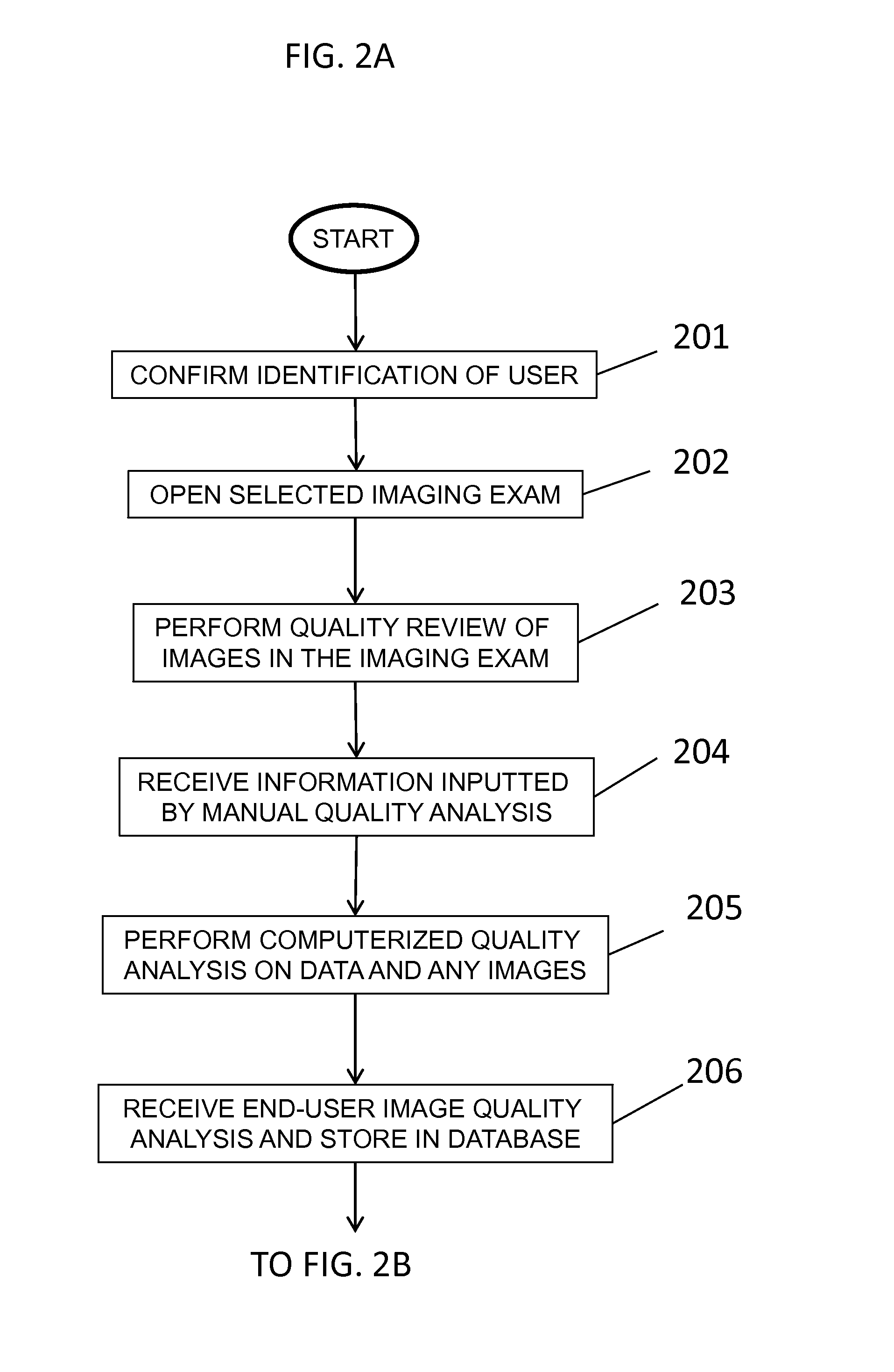
The present invention relates to a computer-implemented method of performing image quality analysis on images from an imaging examination, including: displaying an imaging examination; performing a quality review of the images; performing a quality analysis on data from the imaging examination and on the images; receiving image quality analysis from the user on the data and the images and storing the image quality analysis in at least one database; saving data from the image quality analysis on at least one key image of the images, using computer-generated standardized annotation and mark-up, or graphical data input, or speech data, using a computerized tool, and saving the data as annotated image data in the at least one database; and transferring the annotated image data to at least one quality assurance database with linking to images from the imaging examination.

Description

CROSS-REFERENCE TO RELATED APPLICATIONS[0001]The present application claims priority from U.S. Provisional Patent Application No. 61 / 743,789, filed Sep. 12, 2012, the contents of which are herein incorporated by reference in its entirety.BACKGROUND OF THE INVENTION[0002]1. Field of the Invention[0003]The present invention relates a method and apparatus to improve medical imaging by standardizing image quality analysis, which directly links the quality data with the imaging dataset (i.e. image-centric), continuously tracks and records image quality data for longitudinal analysis, and analyzes all imaging datasets while taking into account a myriad of clinical, technical, and patient-specific variables affecting quality measures.[0004]2. Description of the Related Art[0005]In current medical practice, assessment of quality is highly subjective and personalized, personifying the old adage “beauty (i.e., quality) is in the eyes of the beholder”. While almost all healthcare participants ...

Claims


Application Information

Patent Timeline
IPC IPC(8): G06T7/00G16H30/20G16H30/40
CPCG06T7/0012G16H40/20G16H30/40G16H30/20
Inventor REINER, BRUCE
Owner REINER BRUCE
Learn More