Method for increasing the intraocular pressure in an animal

a technology of intraocular pressure and an animal, applied in the field of increasing the intraocular pressure in an animal, can solve the problems of difficult and restrictive work, trabecular pathophysiology study with this model is rendered impossible, and the effect of the model is only effective in monkeys

Inactive Publication Date: 2014-06-26
CENT NAT DE LA RECHERCHE SCI +2
View PDF1 Cites 3 Cited by
  • Summary
  • Abstract
  • Description
  • Claims
  • Application Information

AI Technical Summary

Benefits of technology

[0015]The inventors have shown that injection of benzalkonium chloride (BAK) in animal eyes increases the intra-ocular pressure, reduces outflow facility and increases trabecular cell apoptosis (see Example 2). BAK is a chemical commonly used as a stabilizer or a disinfectant in ocular medicines. However, the inventors have surprisingly shown that, when used at higher concentrations than usual, BAK induces trabeculum apoptosis and obstruction leading to intra-ocular pressure increase and thus to ocular hypertonia.
[0031]As used herein, the term intra-ocular pressure means the fluid pressure inside the eye. The intra-ocular pressure is a function of the production of liquid aqueous humor by the ciliary processes of the eye and its drainage through the trabecular meshwork. A modification of the intra-ocular pressure may be measured in the animals' eyes by methods well-known by the skilled person using a handheld tonometer. Typically, ocular administration of BAK may increase the intra-ocular pressure by at least 1.5-fold, at least 2-fold, by at least 3-fold or by at least 4-fold.
[0042]The compound to be screened for therapeutic use against ocular hypertension or glaucoma may be used for preventing or treating ocular hypertension or glaucoma. Such compound may be any kind of compound that may act on ocular hypertension or glaucoma symptoms. It may for instance decrease secretion of aqueous humor (such as e.g. beta-blockers eyedrops, carbonic anhydrase inhibitors, or alpha2 adrenergic agonist eyedrops), or increase the elimination of aqueous humor (such as e.g. adrenal derivatives, parasympathetic miotic eyedrops, or prostaglandins analogues). The compound to be screened for therapeutic use against ocular hypertension or glaucoma should preferably display a low toxicity.

Problems solved by technology

However, this model has been effective only in monkeys, which are difficult and restrictive to work with due to legal restrictions, ethical requirements and the complex facilities needed to perform experiments in primates.
Moreover, studying trabecular pathophysiology with this model is rendered impossible due to direct physical destruction (Ghasterland et al.
Consequently, the trabecular meshwork is obstructed by the beads, leading to intra-ocular pressure increase.
Occlusion of the latter prevents aqueous humour drainage and lead to intra-ocular pressure increase.
However, such a model is surgically not easy to set up, and requires many animals to be checked for long-term ocular hypertension.
This injection results in episcleral trabecular obstruction and destruction.
However, this model is associated with significant morphological alterations, such as e.g. enlargement of the anterior chamber (AC) and destruction of the ciliary.
Moreover, the resulting modifications are only temporary and do not provide a long-term model of glaucoma (Morrison et al, exp eye Res 1997; Guo et al, IOVS 2005).
Thus validated models known in the art require complex and poorly reproducible surgical procedures, and show variable efficacy rates depending on the experimenter.
Moreover, these models are not always relevant to the pathology of glaucoma.

Method used


Image

  • Method for increasing the intraocular pressure in an animal
  • Method for increasing the intraocular pressure in an animal
  • Method for increasing the intraocular pressure in an animal

Examples


example 1

Materials and Methods

[0051]Animals and Intra-Ocular Pressure Monitoring

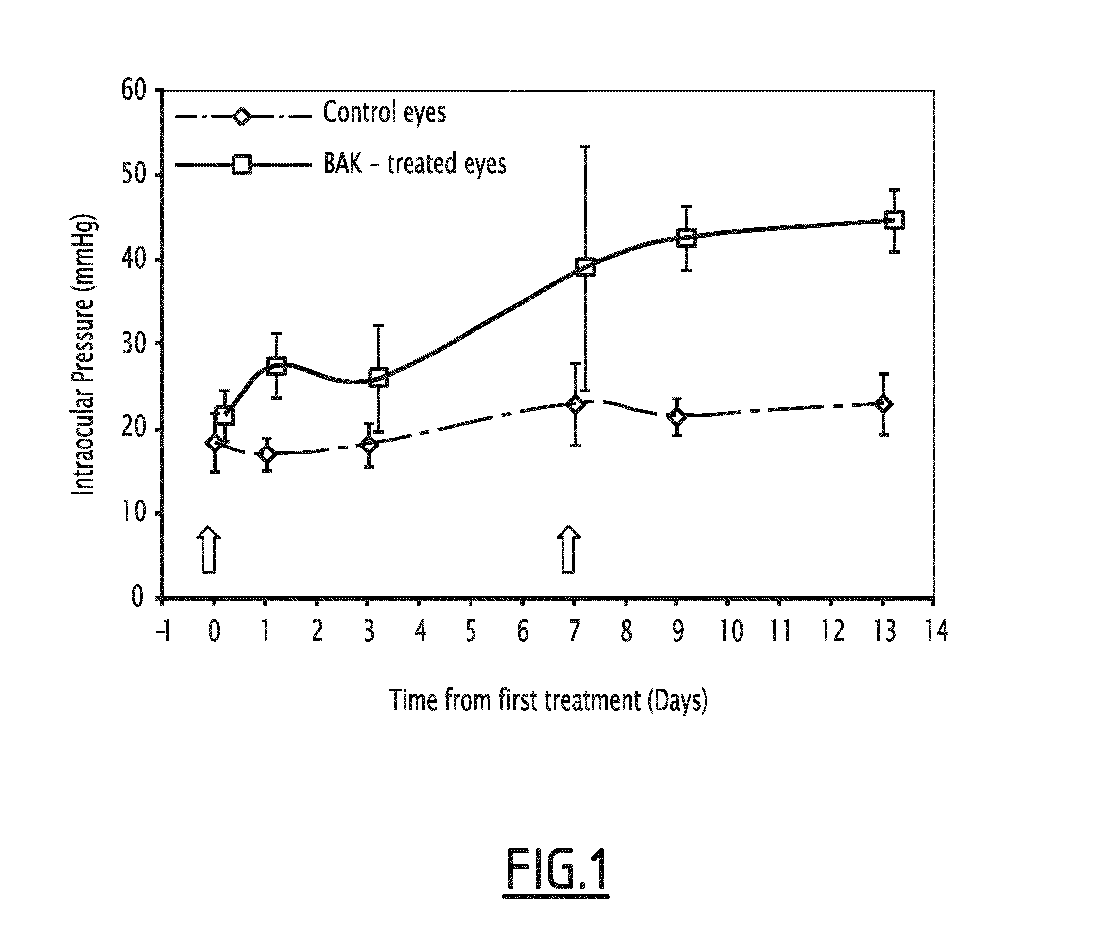
[0052]Six male 8-week-old Long-Evans rats weighing 300-350 g were used. Animals were kept in pathogen-free conditions with food and water available ad libitum and housed in a 12-h light / 12-h dark cycle. Ocular integrity was checked using the slit lamp biomicroscope. At D0, first subconjunctival injection of 0.1% BAK (100 μL) was performed in the right eye whereas the left eye received PBS only as control. A second injection was performed seven days after following the same protocol. Animals were daily monitored for intraocular pressure using a handheld tonometer (TonoLab, Medtronics, Jacksonville, Fla., USA) without sedation. All experiments were conducted in accordance with the Association for Research in Vision and Ophthalmology for the Use of Animals in Ophthalmic research.

[0053]In vivo Outflow Facility Measurement

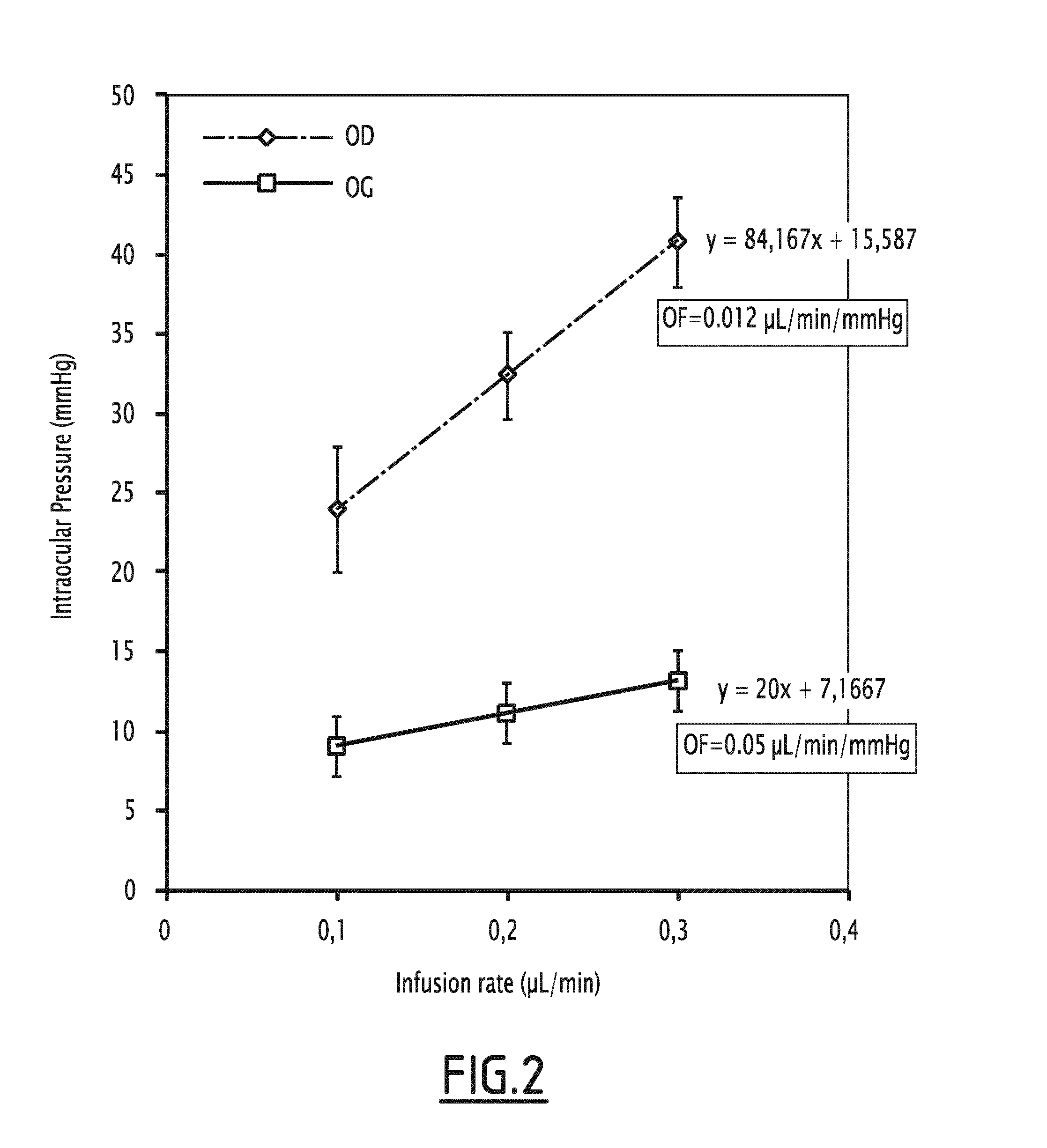
[0054]Six days after the second injection of BAK, trabecular outflow facility was measured in viv...

example 2

Results

[0062]Subconjunctival Injection of BAK Increases the Intra-Ocular Pressure

[0063]First injection of 0.1% BAK induced a slight increase in intra-ocular pressure at D7. Interestingly, the increase in intra-ocular pressure became significant after a second administration of 0.1% BAK, as compared to control eyes receiving the vehicle only. Such significant ocular hypertension remained significant until the end of the experiments (FIG. 1).

[0064]Subconjunctival Injection of BAK Reduces Outflow Facility

[0065]Important increase in intra-ocular pressure with infusion rate revealed a low trabecular outflow facility in BAK-injected eyes compared to control eyes, further confirming that BAK-induced ocular hypertension resulted from an impaired trabecular function (FIG. 2).

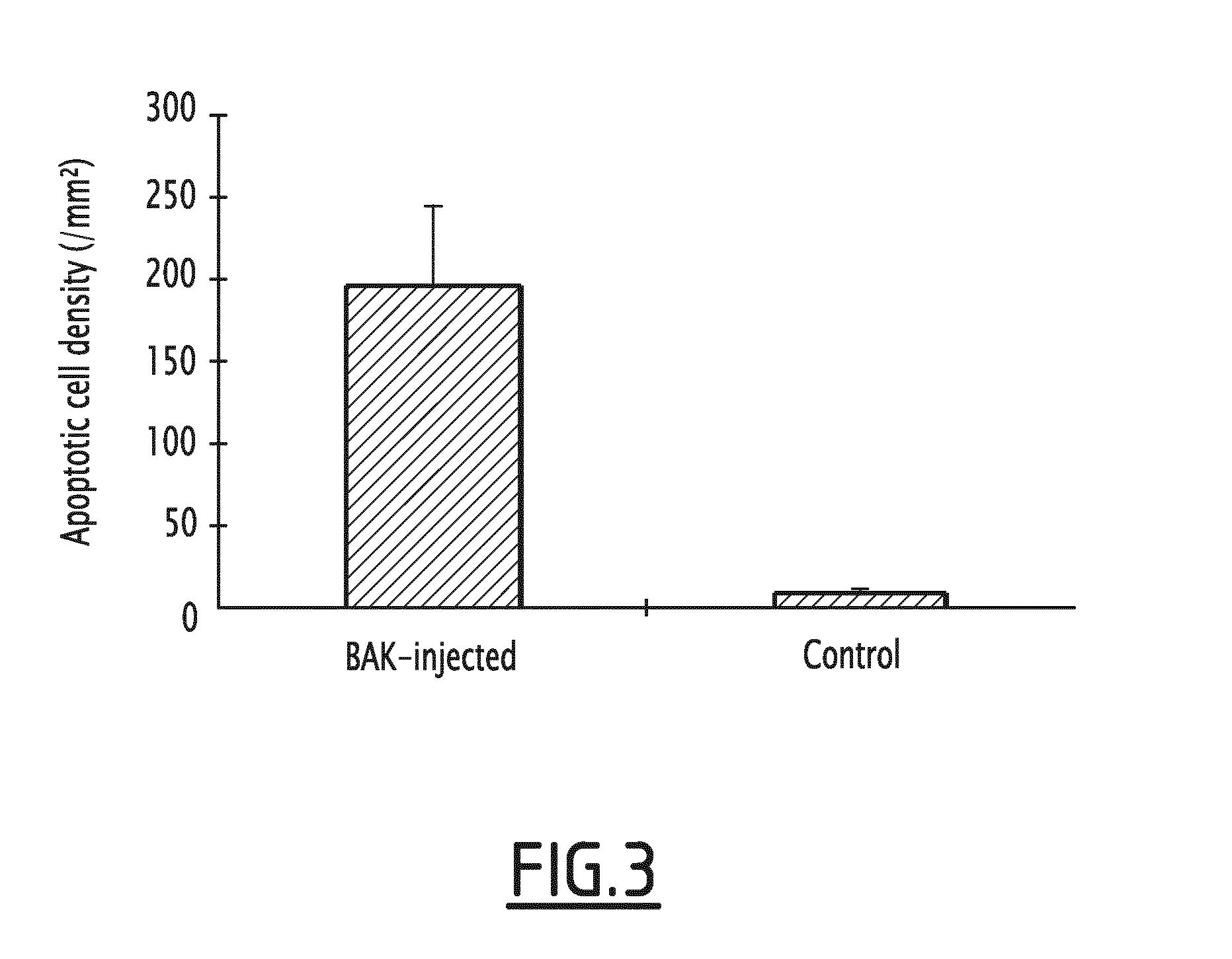
[0066]Subconjunctival Injection of BAK Increases Trabecular Cell Apoptosis

[0067]Trabecular density of apoptotic cells was increased in BAK-injected eyes compared to controls as assessed by TUNEL labelling.

[0068]BAK Subco...


PUM

PropertyMeasurementUnit
pressureaaaaaaaaaa
pressureaaaaaaaaaa
timeaaaaaaaaaa

Abstract

The present invention concerns a method for inducing an increase in the intraocular pressure in an animal, said method comprising ocular administration of a solution comprising benzalkonium chloride (BAK). The present invention further relates to an animal having ocular hypertension, said animal being obtained by the method according to the invention. Said animal provides a convenient animal model for better understanding of the patho-physiological mechanisms involved in ocular hypertension and glaucoma. The present invention also relates to the use of said animal for screening a compound for therapeutic use against ocular hypertension or glaucoma, and / or for assessing potential side-effects of treatment of ocular hypertension and glaucoma.

Description

[0001]The present invention concerns a method for inducing an increase in the intraocular pressure in an animal, said method comprising ocular administration of a solution comprising benzalkonium chloride (BAK). The present invention further relates to an animal having ocular hypertension, said animal being obtained by the method according to the invention. Said animal provides a convenient animal model for better understanding of the patho-physiological mechanisms involved in ocular hypertension and glaucoma. The present invention also relates to the use of said animal for screening a compound for therapeutic use against ocular hypertension or glaucoma, and / or for assessing potential side-effects of treatment of ocular hypertension and glaucoma.BACKGROUND OF THE INVENTION[0002]Glaucoma is an eye disorder in which the optic nerve suffers damage, permanently impacting vision in the affected eye and progressing to complete blindness if untreated. It is often associated with increased ...

Claims


Application Information

Patent Timeline
Patent Type & Authority Applications(United States)
IPC IPC(8): A61K49/00A01K67/027A61K31/14
CPCA61K31/14A01K67/027A61K49/0008A61B3/16A61B2503/40A61B2503/42A61K9/0019A61K9/0048
Inventor ROSTENE, WILLIAMBAUDOUIN, CHRISTOPHEGODEFROY, DAVIDDENOYER, ALEXANDRE
Owner CENT NAT DE LA RECHERCHE SCI
Why Eureka
Try Eureka