Method of dissecting tissue layers

a tissue layer and tissue technology, applied in the field of tissue layer dissection, can solve the problems of inability to monitor the pressure being applied to the body tissues, necrosis or tissue death, edema or swollen tissue, etc., and achieve the effects of expanding deep tissues, high pressure, and opening wounds

Inactive Publication Date: 2000-08-15
KYPHON
View PDF38 Cites 231 Cited by
  • Summary
  • Abstract
  • Description
  • Claims
  • Application Information

AI Technical Summary

Benefits of technology

Typically, the height of the bladder, that is, its width or transverse dimension, is significantly greater than its length (axial or longitudinal dimensions). The bladder expands primarily in width only, not in length, although the shape will vary with the application. This is in accordance with the purpose of the device which is to enable the separation of adjacent tissue layers to, for example, allow improved visualization in front of a scope inserted between the tissue layers.
The present invention also provides a new type of self retaining retractor which is more compact and is disposable for use in open surgery. The retractor is used to hold tissues laterally, pushing tissue edges apart. For such an application, the retractor includes a U-shaped bladder which is a tube that acts like a spring and under high pressure attempts to straighten itself out when inflated. The deflated tube-like structure is first properly positioned within the wound. As the retractor is inflated, it tries to straighten out, and thus opens the wound. As it presses on the edges of the wound, it compresses tissue edges, giving a hemostatic effect. For example, if the retractor is put vertically into a wound, it expands the deep tissues. The retractor can also use the inflatable bladder to push apart metal or plastic plates to push apart tissue.

Problems solved by technology

Problems with the water method include fluid extravasation including into and through the tissue itself.
Increased pressure and swelling result in the area, resulting in edematous or swollen tissue.
Excess pressure from mechanical refractors may cause necrosis or tissue death.
With these methods, it is impossible to monitor the pressure being applied to the body tissues, and tissue damage or necrosis can result.
While operating from within the body, i.e., fiber optic assisted surgery as opposed to open surgery, there is no known way to selectively move or retract tissue, either hard tissue such as bone or soft tissue, out of the way to improve visualization.
No device in use adequately allows a surgeon to create an actual space or expand a potential space in the body, by separating adjacent layers of tissue.
The prior art does not disclose a retractor that is powerful enough and made of a material that is strong and resilient enough to, for example, separate tissue planes from within.

Method used


Image

  • Method of dissecting tissue layers
  • Method of dissecting tissue layers
  • Method of dissecting tissue layers

Examples


Embodiment Construction

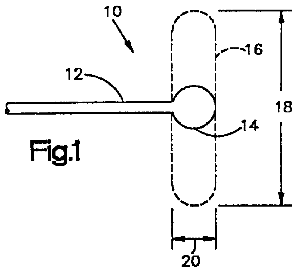
FIG. 1 illustrates schematically a-retractor 10 in accordance with the present invention. The retractor 10 includes a fluid supply structure 12 and an expandable balloon or bladder 14 located at or near the end of the structure 12. The bladder is expandable, under the force of fluid under pressure, from an unexpanded condition as indicated in full lines at 14 to an expanded condition as shown in broken lines at 16. In the expanded condition, the transverse dimension 18 of the bladder 14 is significantly greater than its transverse dimension before expansion the longitudinal dimension 20. Also, in the expanded condition, the transverse dimension 18 of the bladder 14 is significantly greater than its longitudinal dimension 20.

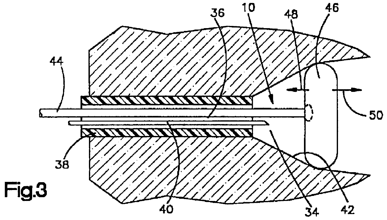
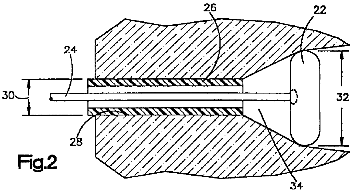
When the bladder of the retractor is expanded inside the body, it retracts tissue. As soon in FIG. 2, a bladder 22 is mounted on the end of a separate shaft 24 within a cannula or scope 26. The cannula or scope 2G has been inserted into the body through an openin...


PUM


Abstract

A fluid operated retractor for use in surgery. The retractor has a portion that is expandable upon the introduction of fluid under pressure. The expandable portion is made of a material strong enough, and is inflated to enough pressure, to spread adjoining tissues within the body. The retractor is especially useful in fiber optic surgery because it can be inserted percutaneously through a small opening then expanded to a much larger dimension when in the desired location, to retract tissue from within. The retractor may be used to spread a joint such as a knee joint or a shoulder joint, or may be used to separate tissue planes generally to spread a joint such as a knee joint or a should joint or may be used to separate tissue planes generally, to improve visualization and create a working space for the surgeon.

Description

The present invention relates to tissue retractors for use in medicine. More particularly, the present invention relates to fluid operated apparatus for use in moving apart abutting tissues, during surgery, in order to create a space between the tissues to improve visualization and for increased working space during open surgery and fiberoptic assisted surgery.DESCRIPTION OF THE PRIOR ARTCurrent methods used for retracting tissue and improving visualization are mechanical separation using metal retractors during open surgery, or the direct pressure of an unconfined flow of fluid such as water or CO.sub.2 during fiberoptic surgery. A typical mechanical external fixator has pins driven through the bones and mechanically distracts the elements of the joint. Problems with the water method include fluid extravasation including into and through the tissue itself. Increased pressure and swelling result in the area, resulting in edematous or swollen tissue. Excess pressure from mechanical r...

Claims


Application Information

Patent Timeline
Patent Type & Authority Patents(United States)
IPC IPC(8): A61B17/02A61B17/22A61B17/32A61B19/00A61M25/10A61B17/00A61F2/958
CPCA61B17/0218A61B17/02A61B17/025A61B17/320036A61B17/320725A61B2017/00535A61B2017/00557A61B2017/0256A61B2017/320048A61B2019/462A61M25/1002A61M2025/006A61B2090/062
Inventor BONUTTI, PETER M.
Owner KYPHON
Why Eureka
Try Eureka