Method for detecting a target particle in biosample containing pancreatic juice

Inactive Publication Date: 2014-06-26
OLYMPUS CORP
View PDF8 Cites 3 Cited by
  • Summary
  • Abstract
  • Description
  • Claims
  • Application Information

AI Technical Summary

Benefits of technology

[0013]The inventor of the present invention and others have conducted earnest studies to solve the above-mentioned problems, which resulted in the following findings. When indirectly detecting a particle dispersed and moving at random in a sample solution with, as an indicator, light emitted from fluorescent probes bound to the particle, it is possible by detecting the particles bound to the fluorescent probes with use of the scanning molecule counting method, to sensitively detect the particles bound to the fluorescent probes even if the concentration of the target particles in

Problems solved by technology

However, in the case where an attempt is made to detect target particles in a biosample by the photoanalysis technique after artificially labeling with a

Method used


Image

  • Method for detecting a target particle in biosample containing pancreatic juice
  • Method for detecting a target particle in biosample containing pancreatic juice
  • Method for detecting a target particle in biosample containing pancreatic juice

Examples


reference example 1

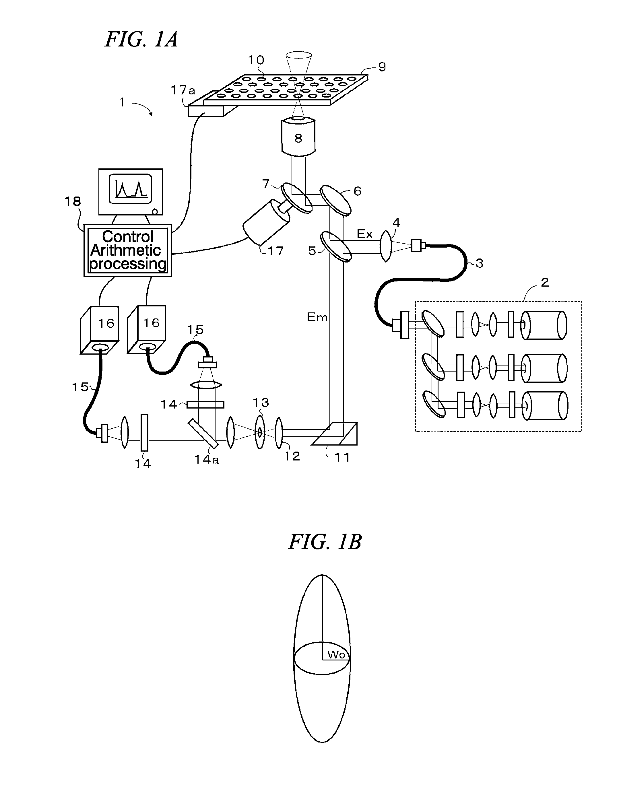
[0114]Autofluorescent substances existing in duodenal juice collected from three examinees were measured with a detection wavelength by the scanning molecule counting method.

[0115]Of the three duodenal juice specimens used herein, one showed a pink color, another one showed a relatively strong yellow color, and the remaining one showed a very weak yellow color. Considering the difference in colors, these duodenal juice specimens were suggested to be largely different in types and amounts of autofluorescent substances contained therein.

[0116]10 μL of each duodenal juice specimen was diluted 10-fold with Tris buffer (10 mM Tris-HCl, 1 mM EDTA, 400 mM NaCl, and pH 8.0), and this diluted solution was used as a sample solution. Each sample solution was measured with excitation light from 488 nm to 670 nm by the scanning molecule counting method. Moreover, Tris buffer, used as a reference, was also measured in the same manner by the scanning molecule counting method.

[0117]Concretely, in t...

example 1

[0120]Samples made by adding single stranded nucleic acid molecules as the target particles to the three duodenal juice specimens used in Reference Example 1 were adopted as the biosample containing pancreatic juice to be subjected to the method for detecting a target particle of the present invention.

[0121]A single stranded nucleic acid molecule consisting of a base sequence represented by the SEQ NO: 1 was used as the target particle. Hereinunder, in this Example, this nucleic acid is referred to as the target nucleic acid. Moreover, a molecular beacon probe 1 prepared by adding TAMRA (registered trademark) to the 5′-end and BHQ-2 to the 3′-end of an oligonucleotide consisting of the base sequence represented by the SEQ NO: 2, or a molecular beacon probe 2 prepared by adding ATTO (registered trademark) 647N to the 5′-end and BHQ-2 to the 3′-end of an oligonucleotide consisting of the base sequence represented by the SEQ NO: 2, was used as the luminescent probe to be bound to this ...


PUM

PropertyMeasurementUnit
Wavelengthaaaaaaaaaa

Abstract

Provided is a method for detecting a target particle in a biosample containing pancreatic juice, the method enabling the detection in a solution that has a lower concentration or number density of the target particles than the level possible for conventional photoanalysis techniques. This method comprises: a probe-binding step for preparing a sample solution, which contains a biosample containing pancreatic juice and a fluorescent probe capable of binding to a target particle, and binding the fluorescent probe to the target particle in the biosample; and a calculation step for calculating the number of molecules of the target particles bound to the fluorescent probes by the scanning molecule counting method. A light emission property of emitted light is different between a state where the fluorescent probe is bound to the target particle and a state where the fluorescent probe is present alone. In a state where the fluorescent probe is bound to the target particle, the fluorescent probe emits fluorescence having a wavelength of 600 nm or longer.

Description

[0001]Priority is claimed on Japanese Patent Application No. 2011-187601, filed Aug. 30, 2011, the content of which is incorporated herein by reference. The present application is a U.S. continuation application based on the PCT International Patent Application, PCT / JP2012 / 071331, filed on Aug. 23, 2012; the content of which is incorporated herein by reference.BACKGROUND OF THE INVENTION[0002]1. Field of the Invention[0003]The present invention relates to a method for detecting a target particle in a biosample containing pancreatic juice, with use of an optical system which can detect light from a micro region in a solution, such as an optical system of a confocal microscope and a multiphoton microscope.[0004]2. Background Art[0005]According to the developments in photometric measurement techniques in recent years, detection and measurement of faint light at a single photon or single fluorescent molecule level have become possible by using an optical system of a confocal microscope ...

Claims


Application Information

Patent Timeline
IPC IPC(8): C12Q1/68
CPCG01N21/6408G01N21/6428G01N21/6452G01N2021/6419G01N2021/6421G01N2021/6432G01N15/1456G01N21/6458G01N2015/1006G01N21/645G02B21/0076C12Q1/6825
Inventor NISHIKAWA, KAZUTAKA
Owner OLYMPUS CORP
Why Eureka
Try Eureka