Dual-modality mammography

a mammography and dual-modal technology, applied in the field of dual-modal mammography, can solve the problems of potential x-ray signal attenuation, geometric blurring, and excessive compression, and achieve the effect of constant volum

Active Publication Date: 2014-06-26
CAPERAY MEDICAL
View PDF5 Cites 28 Cited by
  • Summary
  • Abstract
  • Description
  • Claims
  • Application Information

AI Technical Summary

Benefits of technology

[0018]the scanning surface has an acoustic impedance resembling that of the tissue and material properties enabling it to withstand compression forces applied to the tissue without substantial deformation,

Problems solved by technology

However, due to the discomfort generally associated during significant compression of the breast tissue, excessive compression is not desirable.
A disadvantage of this configuration is that it necessitates the impending X-ray radiation and ultrasonic waves to move through regions of air before reaching the X-ray detector, compression platform or ultrasound transducer, as the case may be.
This results in impedance mismatches and acoustic reflection of the ultrasonic waves at the compression platform interface, which in turn negatively affects the ultrasound images obtained, as well as potential X-ray signal attenuation and geometric blurring due to the finite X-ray focal point.
In addition to the impedance mismatch at the compression platform interface, the problem for the ultrasound imaging is exacerbated at the outermost extremities (most distal parts) of the breast between the compression surfaces.
This results in a peripheral volume of the breast not being accessible, or being poorly accessible, to the ultrasound beams, yielding inferior imaging of the breast in these regions.

Method used


Image

  • Dual-modality mammography
  • Dual-modality mammography
  • Dual-modality mammography

Examples


Embodiment Construction

[0053]Reference will now be made in detail to various exemplary embodiments of the invention. It is to be understood that the following discussion of exemplary embodiments is not intended as a limitation on the invention. Rather, the following discussion is provided to give the reader a more detailed understanding of certain aspects and features of the invention.

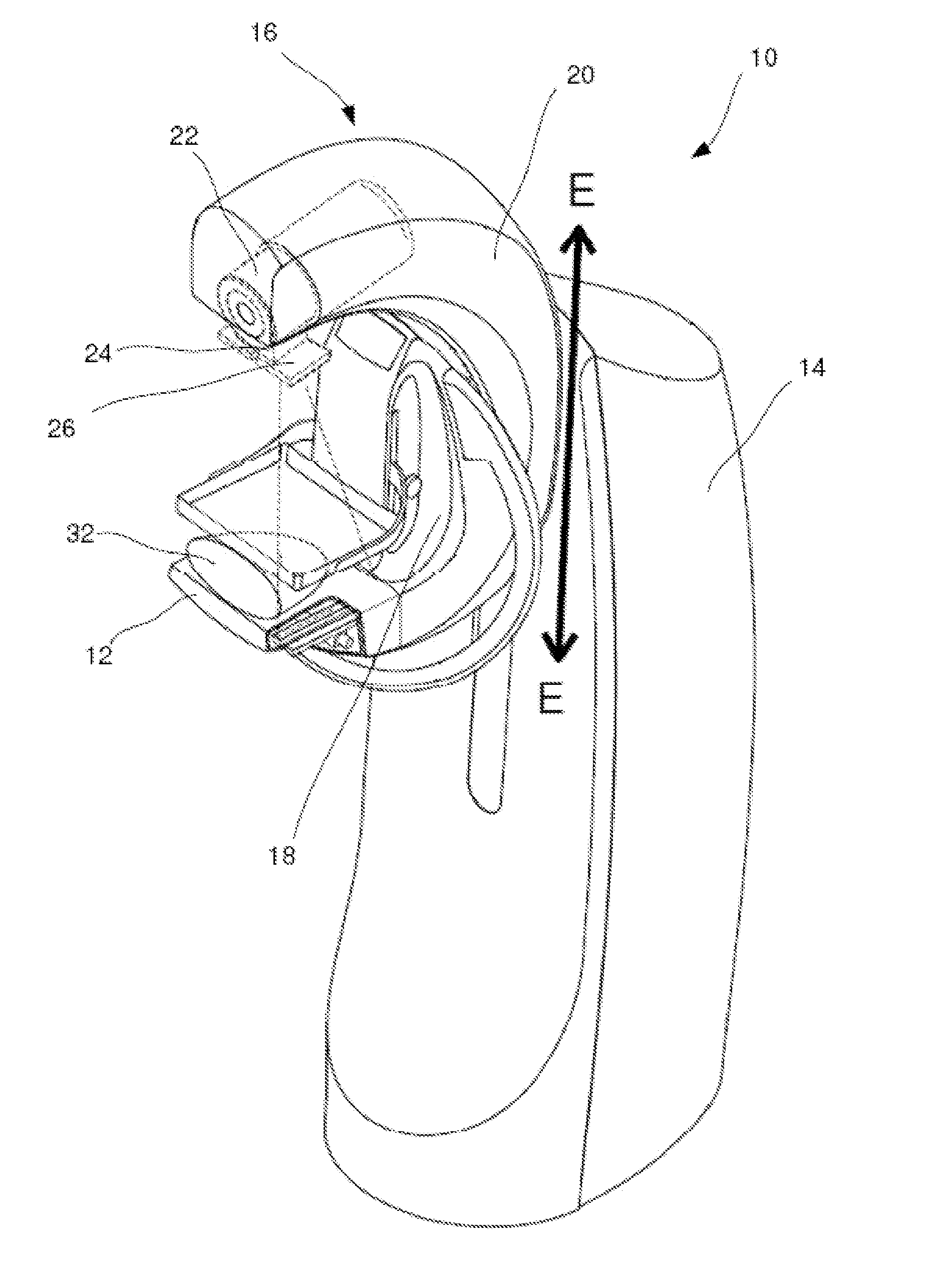
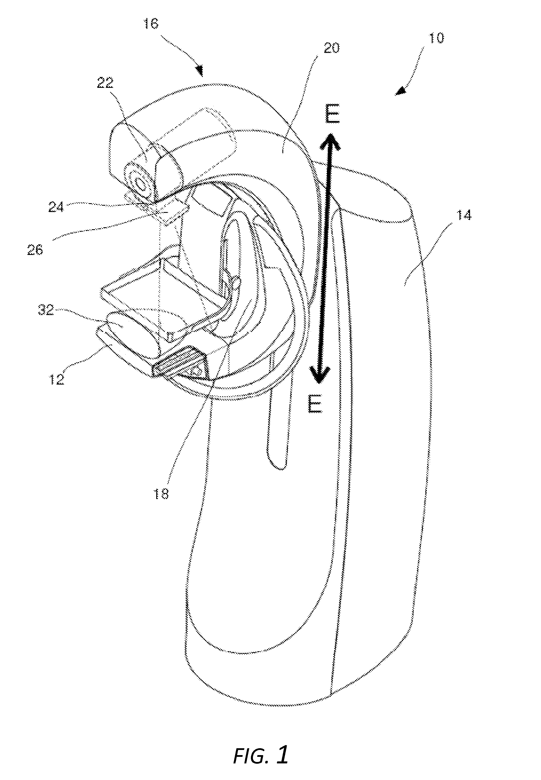
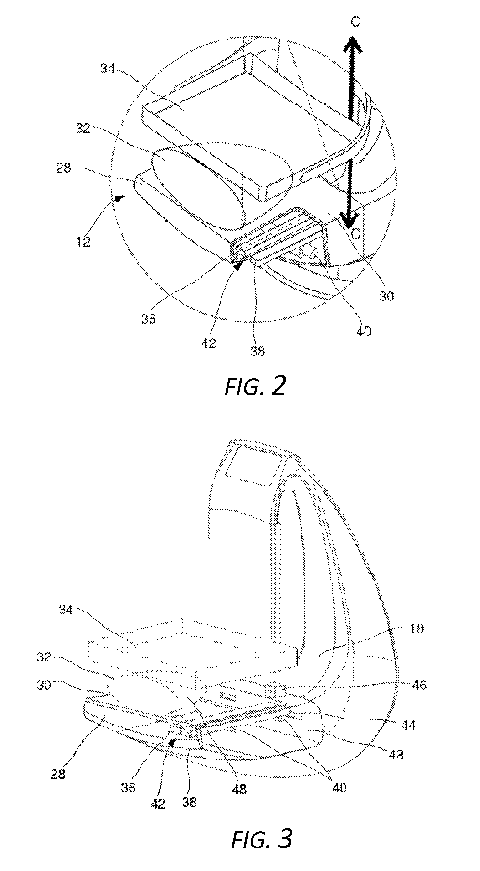
[0054]In accordance with one embodiment of the invention, a dual-modality mammography system (10) utilising a scanning assembly (12) is shown in FIG. 1. The system (10) may include a support pillar (14) on which is mounted a C-arm (16). The C-arm (16) may include a base member (18), which is rotatably mounted to the support pillar (14), and an overhanging arm (20) mounted so as to be rotatable relative to both the base member (18) and the support pillar (14). The arm (20) may include an X-ray source (22) with an X-ray beam-shaper (24) and pre-collimator (26).

[0055]The C-arm (16) may be adjustable in its entirety in a directi...


PUM

PropertyMeasurementUnit
surface areaaaaaaaaaaa
cone angleaaaaaaaaaa
widthaaaaaaaaaa

Abstract

A scanning assembly for a dual-modality automated biological tissue imaging device having first and second compression surfaces is provided. The system comprises a housing defining a scanning / compression surface, an ultrasonic transducer mounted within the housing adjacent the scanning surface for movement in a plane parallel to the scanning surface and imaging the tissue through the scanning surface, an X-ray detector mounted within the housing for forming an X-ray image of the tissue based on X-ray radiation passed through the tissue and scanning surface from an X-ray source, and a drive for moving the transducer across the scanning surface so that the transducer generates a plurality of two-dimensional ultrasound tissue images. The housing is hermetically sealed and filled with non-conductive fluid with acoustic impedance resembling that of the tissue. The scanning surface has acoustic impedance resembling that of the tissue and can substantially withstand compression forces applied to the tissue.

Description

CROSS-REFERENCE TO RELATED APPLICATIONS[0001]The present application relies on the disclosure of and claims priority to and the benefit of the filing date of South African Application No. ZA201209726 filed on Dec. 21, 2012, and U.K. Application No. GB1311124.0 filed on Jun 21, 2013, the disclosures of which are hereby incorporated by reference herein in their entireties.BACKGROUND OF THE INVENTION[0002]1. Field of the Invention[0003]This invention relates to an apparatus and method for imaging human tissue which use geometrically registered X-ray and ultrasound imaging techniques. In particular, the invention relates to improvements to existing dual-modality mammography equipment which uses a combination of such techniques, as well as to apparatus for augmenting and improving conventional mammography equipment.[0004]2. Description of the Related Art[0005]Dual-modality imaging systems that use a combination of full-field digital mammography and automated breast ultrasound imaging in ...

Claims


Application Information

Patent Timeline
Patent Type & Authority Applications(United States)
IPC IPC(8): A61B6/00A61B8/00
CPCA61B6/502A61B8/4416A61B5/708A61B6/488A61B6/5247A61B8/0825A61B8/5261A61B5/0037A61B5/4312A61B5/0035A61B5/004A61B6/025A61B6/0414A61B8/403A61B8/4281A61B8/483Y10T29/49002
Inventor EVANS, MICHAEL DAVIDSMITH, RAPHAEL V.VAUGHAN, CHRISTOPHER LEONARD
Owner CAPERAY MEDICAL
Why Eureka
Try Eureka