

miRNA-BASED UNIVERSAL SCREENING TEST (UST)
a universal screening and mirror technology, applied in the field of early non-invasive or minimally invasive detection of pathological changes, can solve the problems that the screening of each particular disease is principally impossible and the ust cannot be based on the inducing factor, so as to improve the diagnosis and treatment of disease, reduce medical expenses, and high disease specificity
- Summary
- Abstract
- Description
- Claims
- Application Information
AI Technical Summary
Benefits of technology
Problems solved by technology
Method used
Image

Examples
example 1
Comparison of Different Methods Used for miRNA Purification from Serum and Plasma
[0300]There are many commercial kits for miRNA isolation, including the miRNeasy kit (Qiagen), the MirVana RNA isolation kit (Ambion / ABI), miRACLE (Agilent), High Pure miRNA isolation kit (Roche), and miRNA Purification kit (Norgen Biotek Corp.). In addition, the in-house techniques based on the use of Trizol (Invitrogen) are commonly used. After Trizol deproteinization, RNA is precipitated with isopropyl alcohol or additionally purified on silica columns. In some experiments, purified RNA is treated with RNAse-free DNAse (Qiagen, ABI, Invitrogen or other). miRNA preparations obtained by different methods are compared using RT PCR.
[0301]miRNA was purified from plasma and serum samples obtained from the same 5 healthy donors. 107 copies of Arabidopsis thaliana miR-159a (ath-miR-159a) were spiked per 1 ml plasma or serum after addition of guanidine-containing solution for evaluation of miRNA yield. Two te...
example 2
Selection of miRNA for Testing
[0302]Potential miRNA biomarkers (Table 1) were initially selected based on literature data on their enrichment in various organs and tissues (See, e.g., Hua et al., BMC Genomics 2009, 10:214; Liang et al., BMC Genomics. 2007, 8:166; Landgraf et al., Cell. 2007, 129:1401-1414; Lee et al., RNA. 2008, 14:35-42; http: / / ferrolab.dmi.unict.it / miro / ; http: / / mips.helmholtz-muenchen.de / phenomir / ). Then miRNA common for several organs of the same organ system were selected as potential biomarkers for the respective system (Table 2, column 2). Those miRNA that are enriched in one organ but not in other organs of the system were identified as potential biomarkers for more precise pathology localization (Table 2, column 3). For normalization, in addition to spiked synthetic non-human miRNA, e.g., ath-mir-159a, and ubiquitous miRNA, such as miR-16 and miR-30e-3p, miRNA expressed in numerous tissues but not in a target tissue were selected. For example, miR-10b and m...
example 3
Detection of an Increase in Levels of Brain-Enriched miRNA in Serum / Plasma of Patients Diagnosed with Neurological Diseases
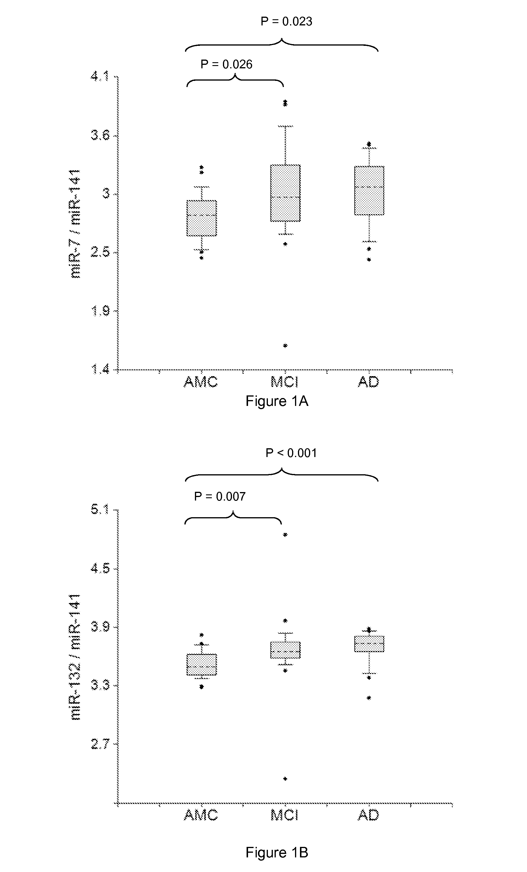
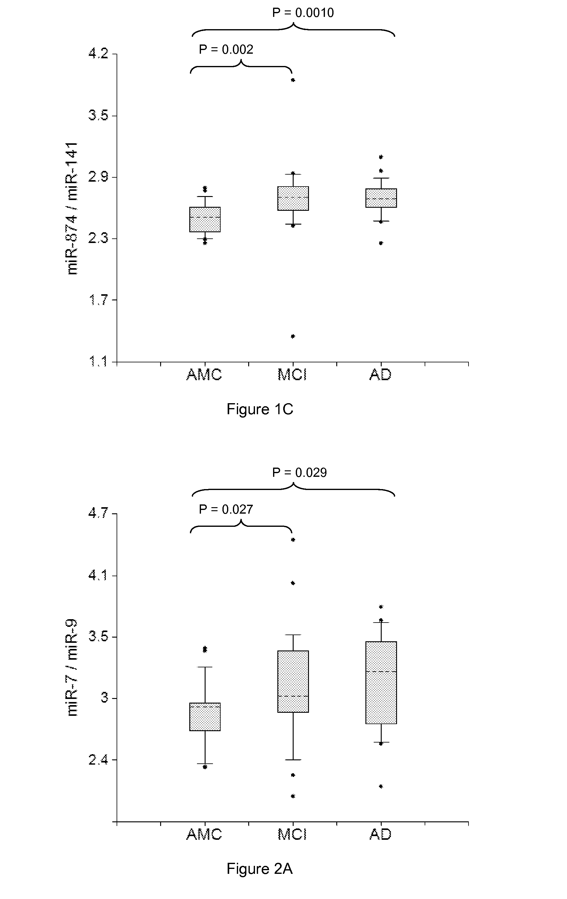
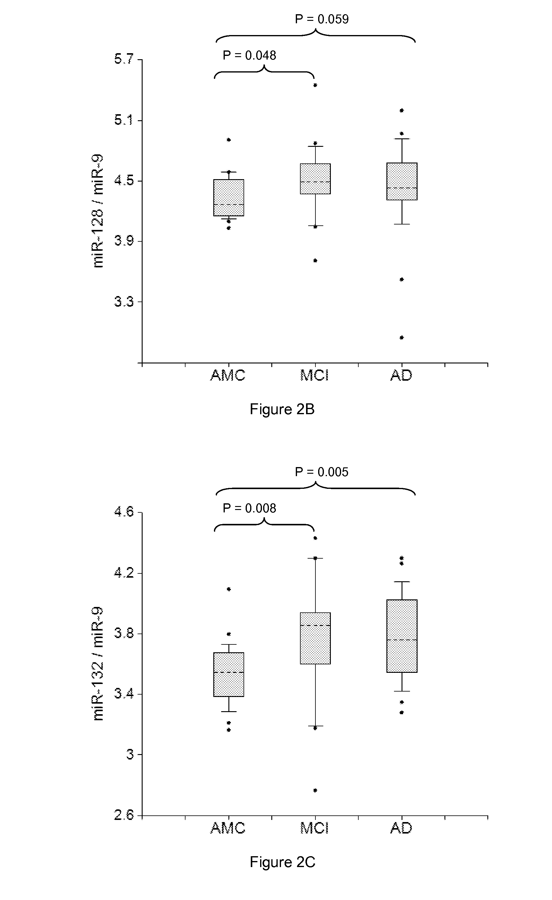
[0304]Plasma samples from amnestic MCI and AD patients and AMC, 20 in each group, were used in the study. RNA was isolated from two 200 μl aliquots of plasma samples by the Trizol-silica method according to an Asuragen procedure. Single target qRT-PCR was performed using the TaqMan® Reverse Transcription Kit and miRNA-specific stem-loop primers (Applied Biosystems). RT step was performed in triplicate and 2 μl plasma equivalents were present in final PCR.
[0305]Data presented in FIGS. 1-6 demonstrate the 2-5 times increase in median concentrations of neurite / synapse miRNAs (miR-7, miR-125b, miR-128, miR-132, miR-134, miR-323-3p, miR-382, miR-874) in plasma of MCI and AD patients when compared to age-matched controls. The effect is more prominent when normalization is performed per brain-enriched miRNA, such as miR-9, miR-127, miR-181a, miR-370, and miR-491-5p.
[03...
PUM
| Property | Measurement | Unit |
|---|---|---|
| time | aaaaa | aaaaa |
| PSA | aaaaa | aaaaa |
| nucleic acid detection | aaaaa | aaaaa |
Abstract
Description
Claims
Application Information