Angular multiplexed optical coherence tomography systems and methods

a tomography and coherence technology, applied in the field of angular multiplexed optical coherence tomography systems and methods, can solve the problems of affecting the accuracy of axial length measurement, spectral domain methods that do not afford the depth of range desirable for axial length measurement, and the time domain measurement techniques used to measure axial length may be prone to such errors, so as to improve or supplement other optical evaluation modalities, improve diagnostic accuracy, and improve the effect of accuracy

Active Publication Date: 2014-10-23
AMO DEVMENT
View PDF5 Cites 18 Cited by
  • Summary
  • Abstract
  • Description
  • Claims
  • Application Information

AI Technical Summary

Benefits of technology

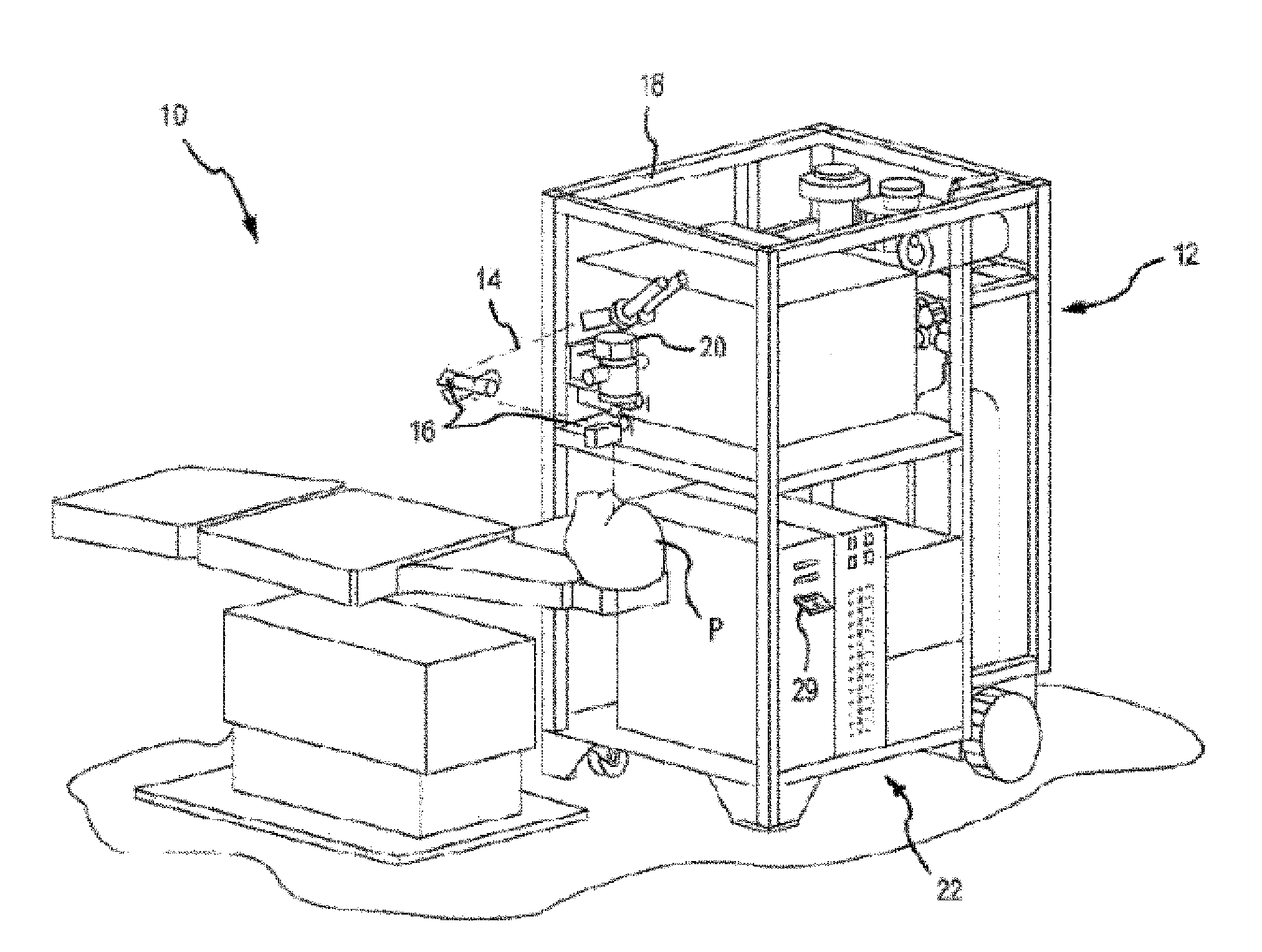
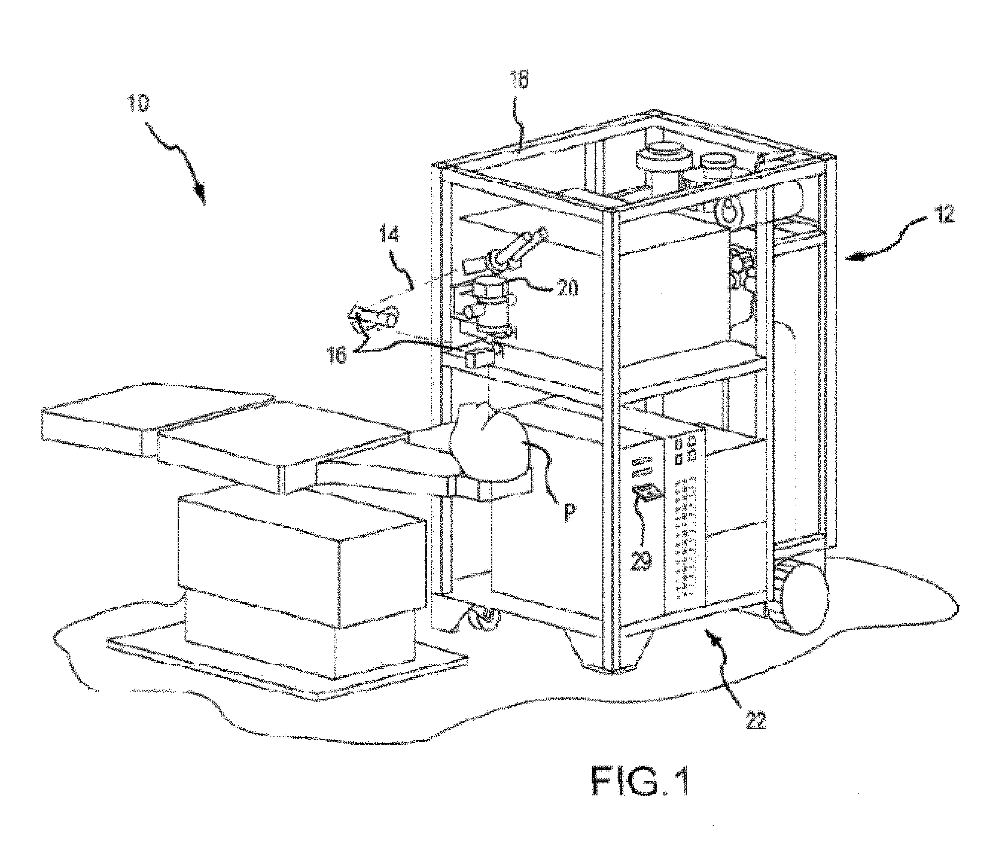
[0006]Embodiments of the present invention encompass the use of highly precise OCT techniques, which are particularly useful for noninvasive evaluation of the optical characteristics of the human eye or other tissues, and can be used to enhance or supplement other optical evaluation modalities such as topography and / or wavefront sensing. For example certain OCT approaches can be used to ascertain a range distance for use with topography procedures, and / or to measure internal distance features present within the eye. In some cases, OCT techniques as discussed herein can be used to precisely evaluate the distance between an optical element (e.g. final lens) of a optical system device and an optical feature (e.g. corneal apex) of the eye. Accordingly, embodiments of the present invention encompass systems and methods for obtaining extremely low surface uncertainties, for example on the order of a fraction of a micron. Topography measurements obtained in conjunction with such techniques can provide a highly accurate diagnostic evaluation for a patient eye, and such approaches are well suited for use in evaluating corneal morphology and / or pathology, at times before, during, or after surgery.
[0007]Embodiments of the present invention encompass systems and methods for evaluating optical tissue in an eye of a patient and for other medical imaging purposes. Exemplary techniques involve simultaneously monitoring a wide depth of range across patient tissue structure. In some cases, opthalmological evaluation techniques as discussed herein can be used to obtain volumetric three dimensional images of the human eye. Exemplary techniques involve simultaneously monitoring multiple depth ranges with offsets, optionally with a single detector. Such approaches can ensure time coincidence and also lower hardware costs. In some cases, exemplary techniques do not involve the use of complicated IR detectors or swept light sources. In some cases, exemplary techniques do not involve the use of devices requiring a complex assembly of moving parts or complicated data acquisition and processing routines. According to some embodiments, angularly multiplexed signals from different depths or spatial locations can be combined on a single detector to provide simultaneous homodyne detection. According to some embodiments, such combination methods can provide a depth span for each angularly encoded signal, making it possible to simultaneously measure over a large effective depth of range or to simultaneously measure at different spatial locations.

Problems solved by technology

Currently known OCT systems involving axial measurements may be negatively impacted by axial or transverse eye movement during the measurement.
For example, existing time domain measurements techniques used to measure axial length may be prone to such errors where a mirror must be physically scanned to scan the depth range.
Relatedly, many currently known spectral domain methods typically do not afford the depth of range desirable for axial length measurements because the depth range is determined by the spectral resolution of the spectrometer / detector combination.
In addition, existing spectral domain OCT techniques may be prone to ghost images.
However, many time such proposals suffer from high costs.

Method used


Image

  • Angular multiplexed optical coherence tomography systems and methods
  • Angular multiplexed optical coherence tomography systems and methods
  • Angular multiplexed optical coherence tomography systems and methods

Examples


Embodiment Construction

[0012]Embodiments of the present invention encompass systems and methods for the tomographical monitoring of optical or other patient tissue across a wide depth of range in a simultaneous fashion. According to some embodiments, angular multiplexed optical coherence tomography (AMOCT) techniques can use non-collinear beams to produce a spatial interference pattern with a unique carrier frequency. Such a frequency can be determined by the angle between the signal and reference beams. Multiple carrier frequencies can be supported simultaneously on a single linear array detector by assigning a unique angle to each unique signal beam and combining them with a single, common reference beam.

[0013]According to some embodiments, a detector can receive multiple signal frequencies simultaneously, and the readout of a signal assigned to a specific carrier frequency can be achieved by frequency filtering the composite signal. Having resolved the specific signals, the composite echo signal can be...


PUM


Abstract

Angle multiplexed optical coherence tomography systems and methods can be used to evaluate ocular tissue and other anatomical structures or features of a patient.

Description

CROSS-REFERENCES TO RELATED APPLICATIONS[0001]This application claims priority to U.S. Provisional application No. 61 / 794,276, filed on Mar. 15, 2013. The entire contents of which are incorporated herein by reference.BACKGROUND OF THE INVENTION[0002]Optical Coherence Tomography (OCT) is used in the ophthalmic industry in pachymetry, anterior chamber depth, axial length, and retinal imaging applications. Known OCT systems typically use either a time domain or a spectral domain technology to provide line of sight length optical analogs of the echoes in ultrasonic imaging via homodyne detection. With the use of broadband sources, there is homodyne signal amplitude when the reference and signal beams are sufficiently close in optical delay. Time domain and spectral domain OCT use collinear reference and signal beams to achieve a detectable interference that includes the depth information.[0003]Currently known OCT systems involving axial measurements may be negatively impacted by axial o...

Claims


Application Information

Patent Timeline
Patent Type & Authority Applications(United States)
IPC IPC(8): A61B3/10
CPCA61B3/102A61B3/107A61B5/0066G01B9/0205G01B9/02087G01B9/02091G01N21/4795A61B3/1005
Inventor RAYMOND, THOMAS D.REID, DEVON E.
Owner AMO DEVMENT
Why Eureka
Try Eureka