Method Of Determining The Viability Of At Least One Cell

a cell and viability technology, applied in the field of determining the viability of at least one cell, can solve the problems of limited number of techniques, each with significant drawbacks, and cost effectiveness, and achieve the effect of accurate preservation of histology, cost saving and limited number of techniques

Active Publication Date: 2014-12-11
RGT UNIV OF MICHIGAN
View PDF1 Cites 9 Cited by
  • Summary
  • Abstract
  • Description
  • Claims
  • Application Information

AI Technical Summary

Benefits of technology

[0005]The instant disclosure provides a method of determining the viability of at least one cell via quantification of nicotinamide adenine dinucleotide (phosphate) (NAD(P)H), flavin adenine dinucleotide (FAD), and/or collagen. The method includes the steps of contacting the at least one cell with light having a wavelength of from 700 to 900 nm, using two photon

Problems solved by technology

Currently, a limited number of techniques, each with significant drawbacks, are utilized.
Histology accurately preserves a cross sectional slice of the tissue, but is limited because the technique is destructive, ti

Method used


Image

  • Method Of Determining The Viability Of At Least One Cell
  • Method Of Determining The Viability Of At Least One Cell
  • Method Of Determining The Viability Of At Least One Cell

Examples


examples

First Set of Examples

[0113]This first set of Examples is independent from the second set of examples set forth below.

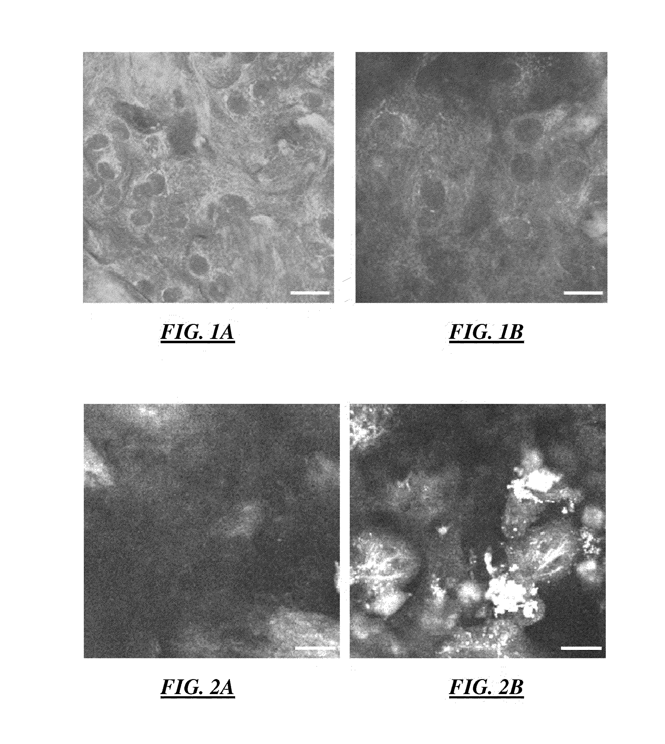
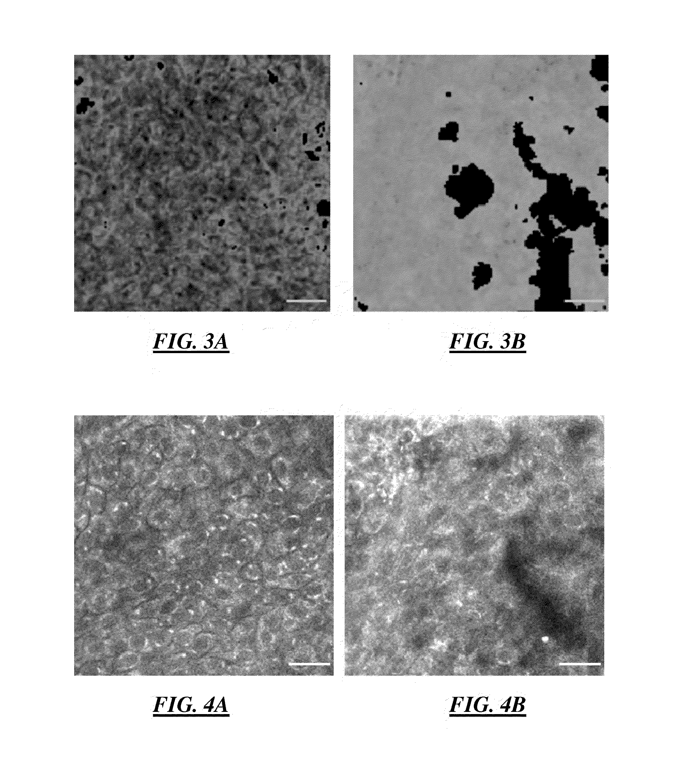
[0114]In various embodiments, phasor plot analysis combined with error propagation analysis was developed to advance the applications of fluorescence lifetime imaging microscopy (FLIM). The developed algorithm was applied to label-free one-channel FLIM images from the living engineered tissue EVPOME to automatically indentify and segment different tissue constituents. The result was compared to two-channel fluorescence intensity images and a fast FLIM map.

[0115]In these embodiments, phasor analysis has the advantages of rapid and direct interpretation and visualization of FLIM images without prior knowledge of the samples. The algorithm was explored and extended to develop the phasor error analysis. Phasor error analysis quantitatively identifies the size and the shape of the uncertainty areas of the fluorescence lifetimes of interest on a phasor plot. The developed p...


PUM


Abstract

This disclosure provides a method of determining the viability of at least one cell via quantification of nicotinamide adenine dinucleotide (phosphate) (NAD(P)H), flavin adenine dinucleotide (FAD), and/or collagen. The method includes the steps of contacting the at least one cell with light having a wavelength of from 700 to 900 nm, using two photon excitation, or from 335 to 400 nm, using one photon excitation, to induce an optical response from the NAD(P)H, FAD, and/or collagen and measuring the optical response. The method also includes the step of quantifying one or more of an amount, spatial localization, and/or time-dependent response of the NAD(P)H, FAD, and/or collagen utilizing the optical response.

Description

CROSS-REFERENCE TO RELATED APPLICATION[0001]This application is a Continuation-In-Part of International Application No. PCT / US2013 / 021268, filed on Jan. 11, 2013, which claims priority to U.S. Ser. No. 61585827, filed on Jan. 1, 2012, wherein the disclosures of each are expressly incorporated herein by reference in various non-limiting embodiments.GOVERNMENT LICENSE RIGHTS[0002]This invention was made with government support under DE019431 awarded by the National Institutes of Health. The government has certain rights in the invention.[0003]This disclosure relates to a method of determining the viability of at least one cell. More specifically, this disclosure relates to quantifying nicotinamide adenine dinucleotide (phosphate) (NAD(P)H), flavin adenine dinucleotide (FAD), and / or collagen.[0004]The fields of tissue-engineering and regenerative medicine have evolved such that tissue burns, removed tumors, or open wounds can be replaced with tissue developed from patients' primary cel...

Claims


Application Information

Patent Timeline
IPC IPC(8): G01N33/483
CPCG01N2333/46G01N33/4833G01N21/6408G01N21/6456G01N33/5005G01N33/5735G01N33/582G01N33/6887G01N2333/78
Inventor MYCEK, MARY-ANNFEINBERG, STEPHENMARCELO, CYNTHIALLOYD, WILLIAMCHEN, LENG-CHUNKUO, SHIUHANGKIM, HYUNGJIN MYRA
Owner RGT UNIV OF MICHIGAN
Why Eureka
Try Eureka