Method for determining the glycosylation of an antibody

a glycosylation and antibody technology, applied in the field of antibody glycosylation methods, can solve the problems of expensive equipment that requires a certain level of expertise, little is known about the manner of action of antibodies in patients, and the current methods available for studying the binding of antibodies to fc receptors are relatively tedious

Active Publication Date: 2015-01-22
CISBIO BIOASSAYS
View PDF1 Cites 17 Cited by
  • Summary
  • Abstract
  • Description
  • Claims
  • Application Information

AI Technical Summary

Problems solved by technology

Although their clinical efficacy no longer requires any proof, still little is known about their manner of action in patients.
The techniques currently available for studying the binding of antibodies to the Fc receptors are relatively tedious.
This approach has several drawbacks, notably a critical step of fixation of one of the binding partners on the surface of the microchip, the need for expensive equipment that requires a certain level of expertise, and the fact that the technique cannot be used for working with living cells expressing the Fc receptor under investigation.
Moreover, this approach also does not supply information regarding possible binding of the antibody tested to its antigen in its cellular conformation.
This technique does not allow direct measurement of the binding of the antibody to the FcR.
No method exists at present for direct measurement of the binding of an antibody to an Fc receptor expressed on the surface of an intact cell.
The existing techniques have several drawbacks: indirect assays of cytokines secreted by the cells, centrifugation steps that are rather unsuitable for automated use, or else the use of approaches that require tedious preliminary steps and take a relatively long time.
Moreover, these techniques do not allow information to be obtained easily and quickly concerning the level of glycosylation of a given antibody and its effect on binding to the Fc receptors.

Method used


Image

  • Method for determining the glycosylation of an antibody
  • Method for determining the glycosylation of an antibody
  • Method for determining the glycosylation of an antibody

Examples


example 1

Material and Method

Reagents Used:

[0114]Culture Medium: DMEM / Glutamax+10% FCS+streptomycin (50 μg / ml), penicillin 50 U / ml, HEPES 2 mM, nonessential amino acids 1% (InVitrogen)

[0115]OptiMEM: culture medium used for transfection, Invitrogen

[0116]Lipofectamine 2000: Invitrogen

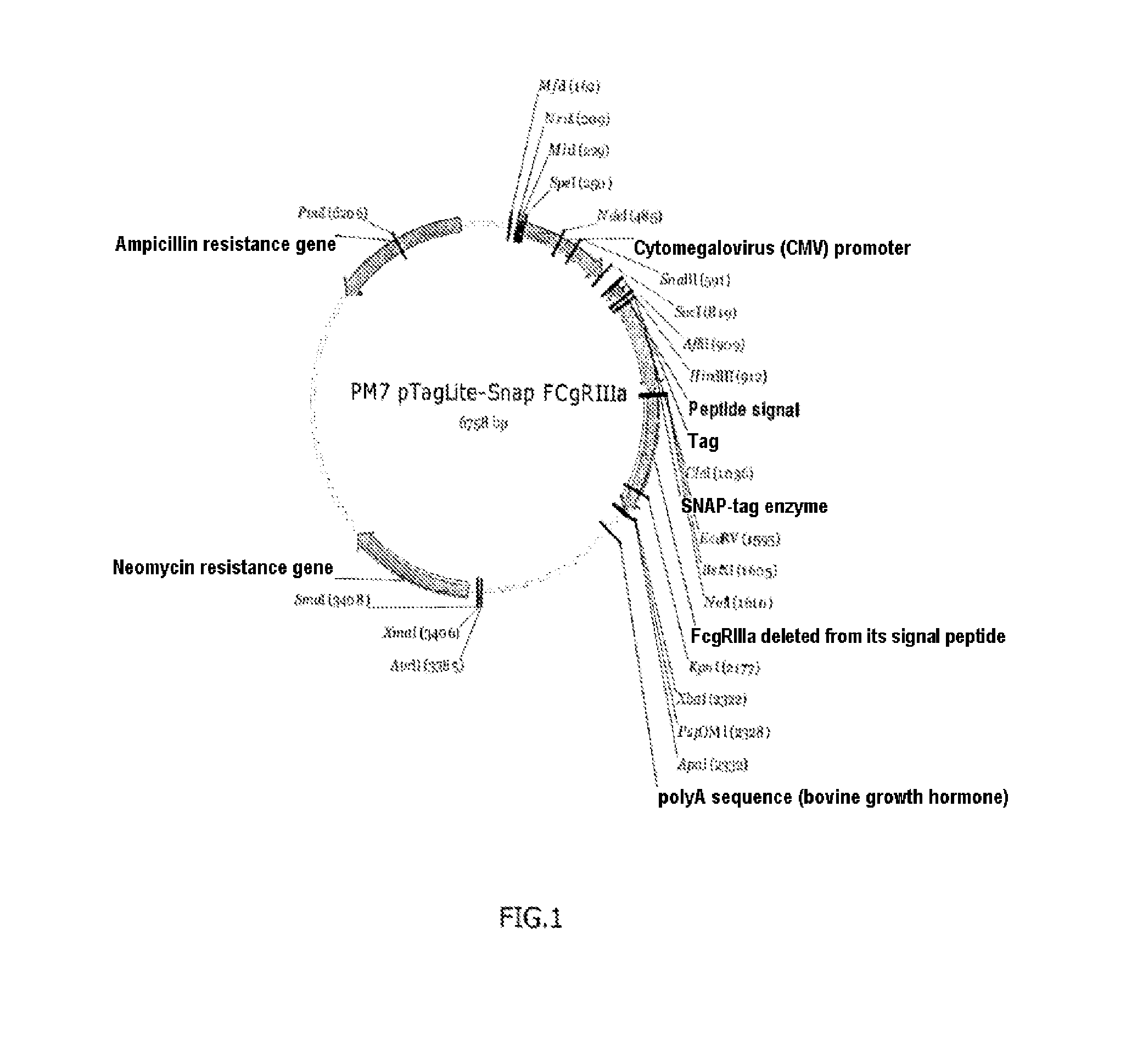
[0117]SNAP-CD16A plasmid: prepared by inserting the nucleic acid sequence (SEQ ID No. 4) coding for the CD16a receptor (variant V158 (or V176) ref. Genbank NM—000569.6, protein sequence NP—000560.5) in the plasmid SNAP-tag® pT8-SNAP-Neomycin (Cisbio Bioassays), see FIG. 1.

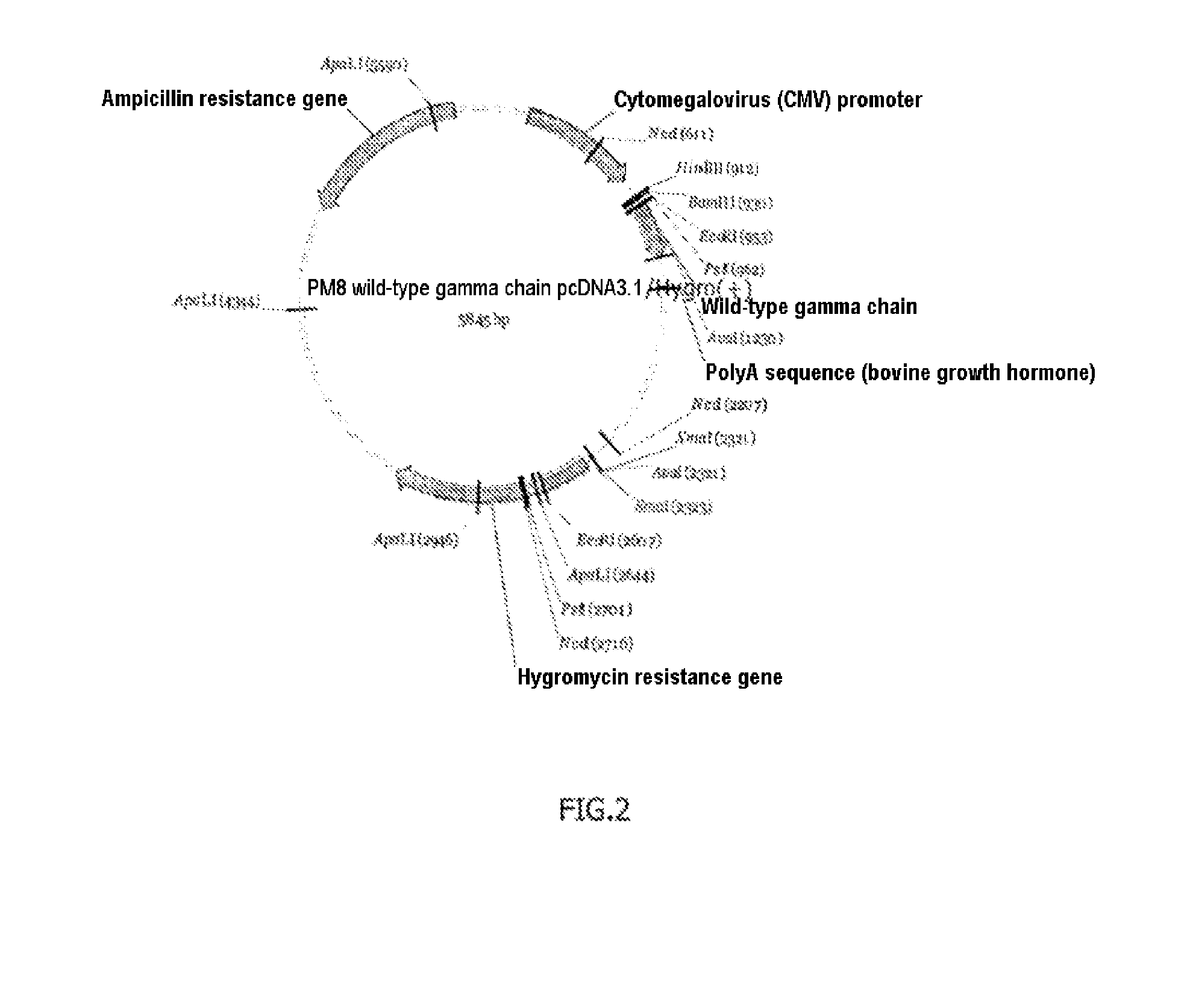
[0118]Plasmid gamma chain of the FcεRI receptor: The DNA sequence (SEQ ID No. 5) coding for the gamma chain of the FcεRI receptor was inserted in an expression plasmid by the classical techniques. The map of this plasmid is shown in FIG. 2. It is known that the CD16a receptor forms dimers with this gamma chain, and it is therefore recommended to coexpress the two proteins. The inventors nevertheless discovered that this coexpression was not necess...

example 2

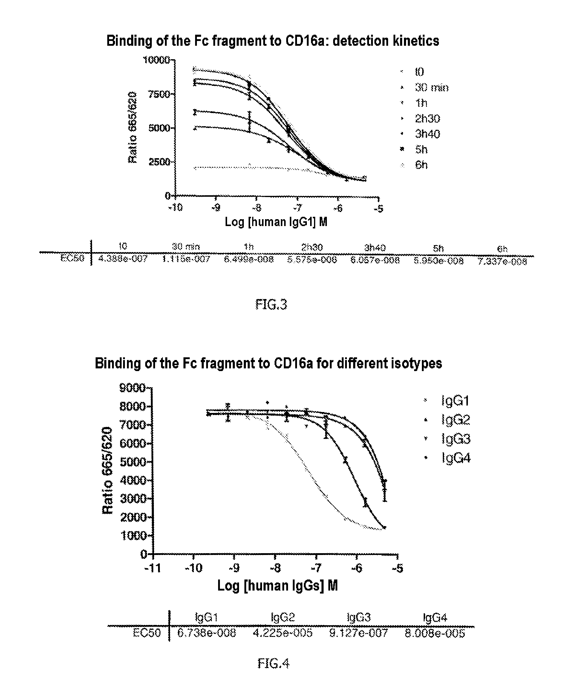
[0130]The cells prepared by the method described in example 1 were thawed at 37° C. and quickly mixed with 15 ml of PBS. The suspension obtained was centrifuged for 5 min at 1200 rpm and the supernatant was removed. The pellet was resuspended in Tag-lite® buffer to obtain a suspension for distributing HEK-CD16a-Tb cells in the wells of a 384 LV multiwell plate at a concentration of 10 000 cells per well under 10 μl.

[0131]An unlabeled human antibody of IgG1 isotype (its epitope specificity is unimportant) was added at different final concentrations from 0.3 nM to 5 μM under 5 μl.

[0132]The same antibody, labeled with the d2 fluorophore acceptor (d2 labeling kit Cisbio Bioassays, ref. 62D2DPEA), was added at 200 nM under 5 μl for a final concentration of 50 nM.

[0133]The FRET signals emitted by the 384 plate were measured immediately, then after 30 min, 1 h, 2 h30, 3 h40, 5 h and 6 h.

[0134]The results presented in FIG. 3 show that the method according to the invention allows effective m...

example 3

[0135]Example 2 was reproduced but this time the unlabeled IgG1 antibody (competing antibody) was replaced with human antibodies of isotypes IgG2, IgG3 and IgG4 (their epitope specificities are unimportant), which are known not to have as good affinity for the receptor CD16a as the antibodies of the IgG1 type. After adding different concentrations of these antibodies, the reaction mixture was incubated for 4 h20.

[0136]The results presented in FIG. 4 confirm what was known in terms of affinity of IgG2, IgG3 and IgG4 for the CD16a receptor and therefore validate the method according to the invention, which allows effective comparison of a reference antibody with other antibodies and which is sufficiently sensitive to allow visualization of differences in affinity of these antibodies, here for the CD16a receptor.


PUM

PropertyMeasurementUnit
incubation timeaaaaaaaaaa
Time Resolved FRETaaaaaaaaaa

Abstract

The invention relates to a method for detecting the binding of an antibody to an Fc receptor present on the surface of a cell as well as to a method for determining the level of glycosylation of an antibody. The invention also relates to a reagent kit for carrying out these methods.

Description

FIELD OF THE INVENTION[0001]The invention relates to a method for detecting the binding of an antibody to a receptor for the crystallizable Fc fragments (hereinafter “Fc receptors” or FcR) present on the surface of a cell as well as a method for determining the level of glycosylation of an antibody.PRIOR ART[0002]For some years now, recombinant monoclonal antibodies have permitted a therapeutic revolution. Although their clinical efficacy no longer requires any proof, still little is known about their manner of action in patients. The therapeutic effect of antibodies targeting membrane antigens notably involves the recruitment of effector cells expressing receptors for the crystallizable fragment of the antibodies (hereinafter, “Fc receptors”).[0003]The Fc receptors are proteins present on the surface of certain cells contributing to the functions of the immune system, in particular NK (“natural killer”) cells, macrophages, neutrophils and mastocytes. Their name comes from their cap...

Claims


Application Information

Patent Timeline
Patent Type & Authority Applications(United States)
IPC IPC(8): G01N33/68G01N33/58
CPCG01N33/6854G01N2440/38G01N2400/02G01N33/582
Inventor JAGA, DELPHINEMOKRANE, HAMEDMARTINEZ, STEPHANEFINK, MICHEL
Owner CISBIO BIOASSAYS
Why Eureka
Try Eureka